The Problems of Pulpotec Material
22/06/2024
Fellow
Warning: Undefined variable $post in /home/styleendo/htdocs/styleitaliano-endodontics.org/wp-content/plugins/oxygen/component-framework/components/classes/code-block.class.php(133) : eval()'d code on line 2
Warning: Attempt to read property "ID" on null in /home/styleendo/htdocs/styleitaliano-endodontics.org/wp-content/plugins/oxygen/component-framework/components/classes/code-block.class.php(133) : eval()'d code on line 2
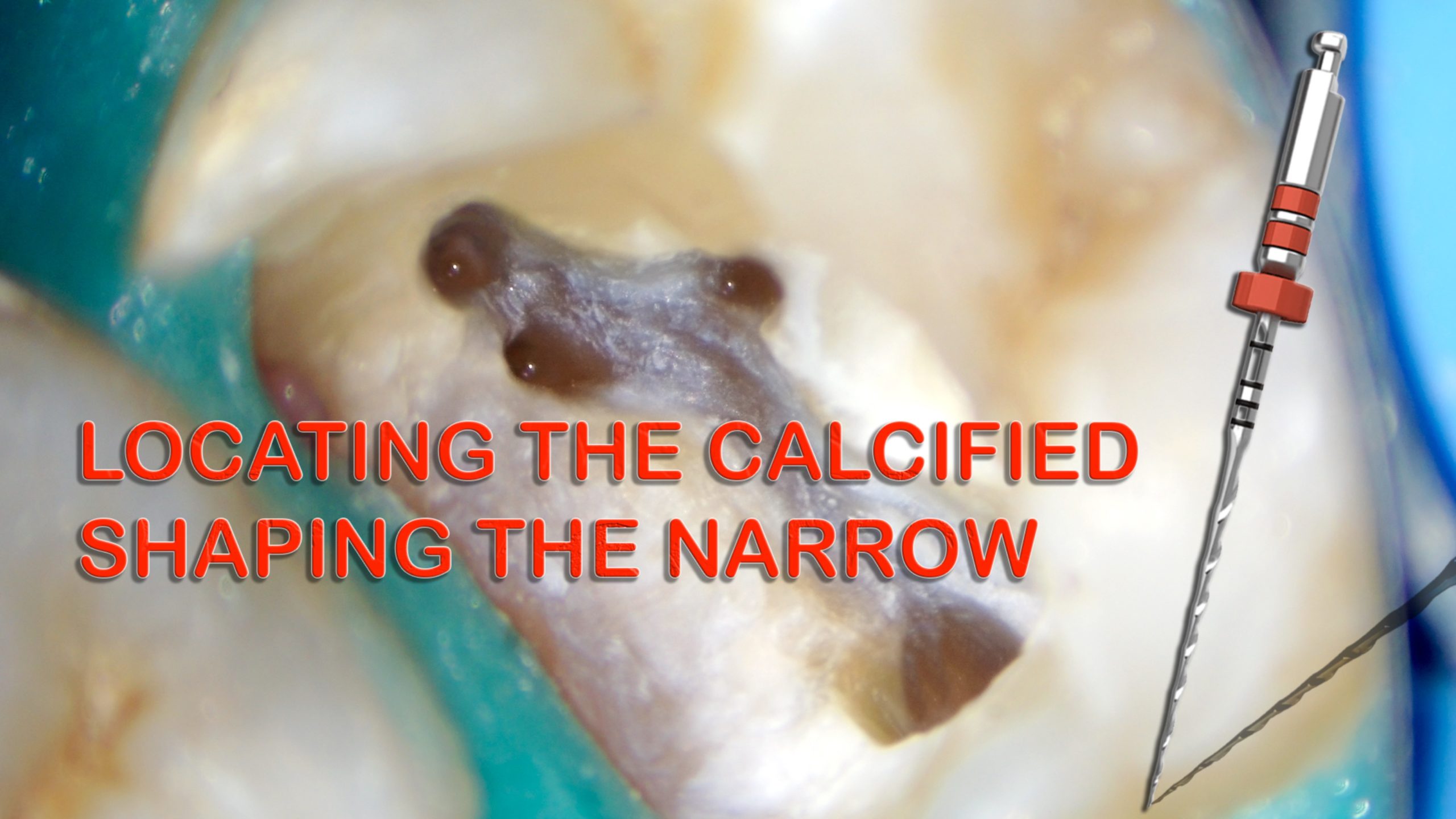
Narrow canal is a great challenge to Endodontist, When combined with calcified entrance the difficulty doubled.
Preoperative assessment to the periapical radiograph would help in avoiding many errors that could happen if case is underestimated.
CBCT has a great role in assessment and planning for management of canal calcification and blockage and it’s considered of great importance
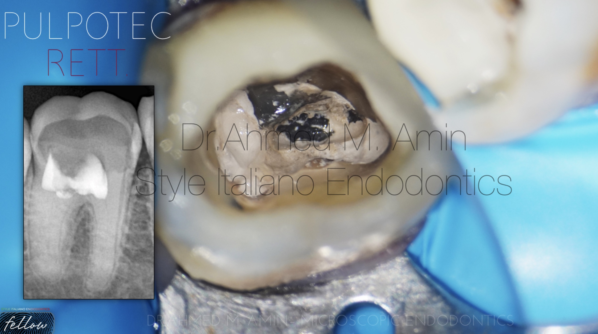
Fig. 1
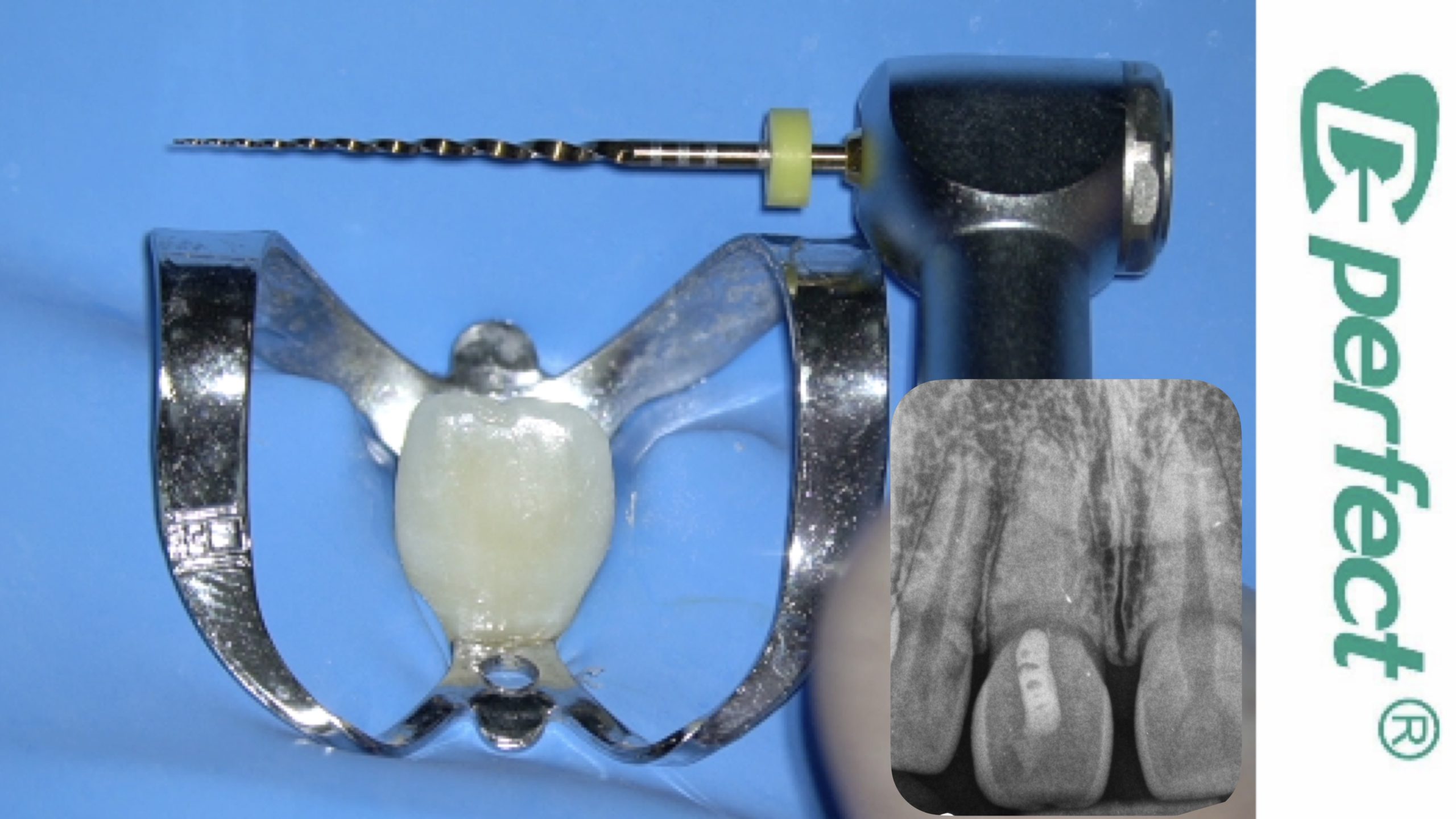
38-year female patient came to my clinic referred from a colleague to manage such a calcified canals as he saw at preop xray
Preop xray : Radio-opaque material covering the canals , very tight or nearly calcified canals
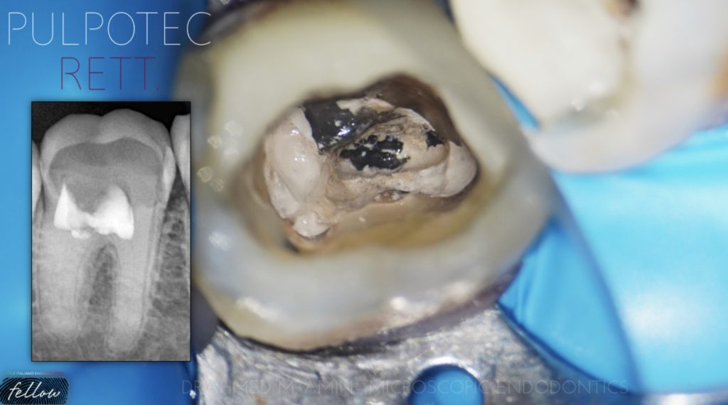
On clinical diagnosis : open tooth with a pulp champer full of pulpotec material
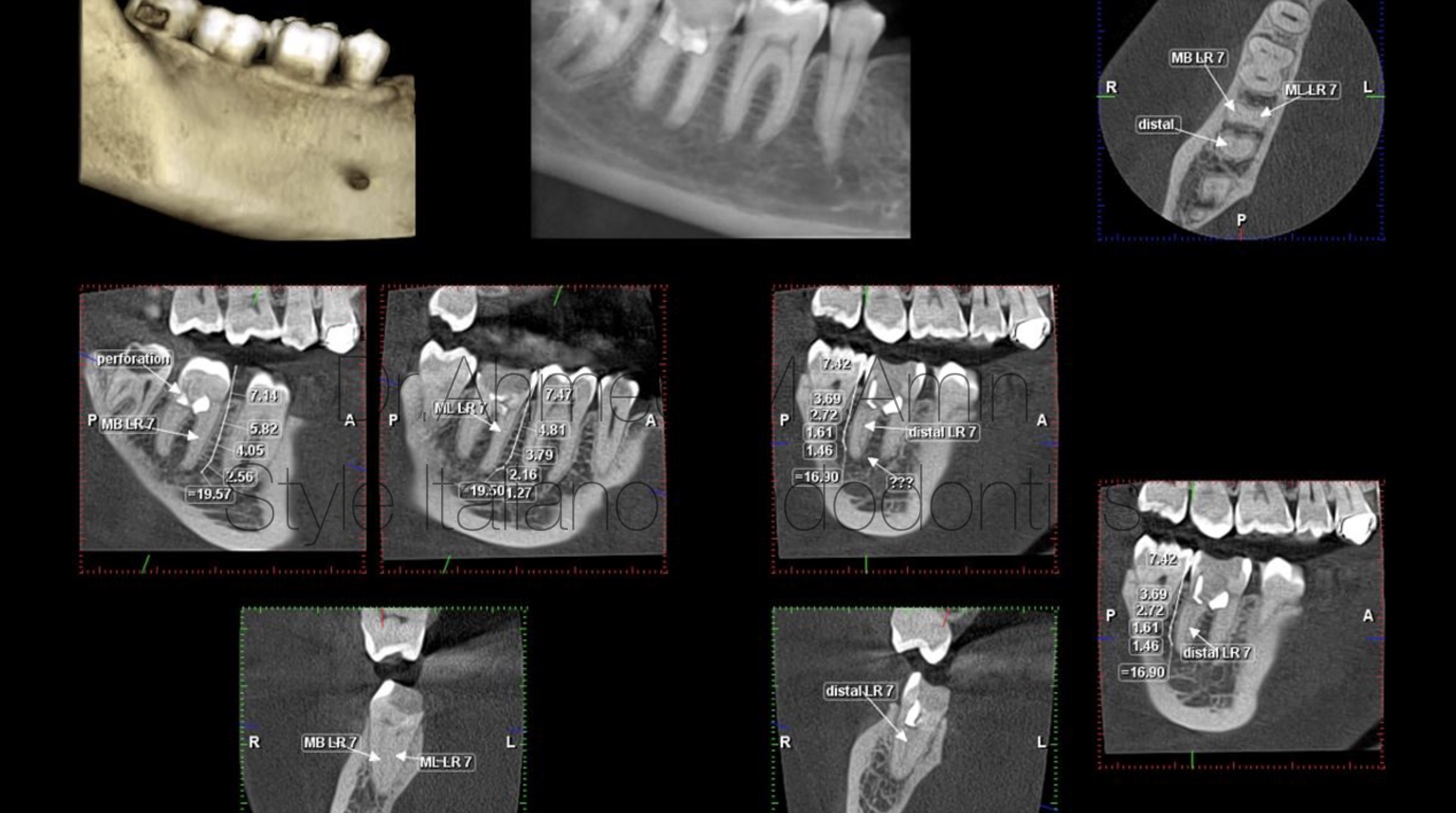
Fig. 2
CBCT has a great role in assessment and planning for management of canal calcification and blockage
On CBCT : we found very tight 3 canals , with lateral foramena related to distal canal
- possible of floor perforation sealed with the same pulpotec material
- Type II mesial canals
Removal of pulpotec from pulp champer using long shank round bur in a low speed , then removal of pulpotec from canals using ultrasonic tip to avoid any mishaps
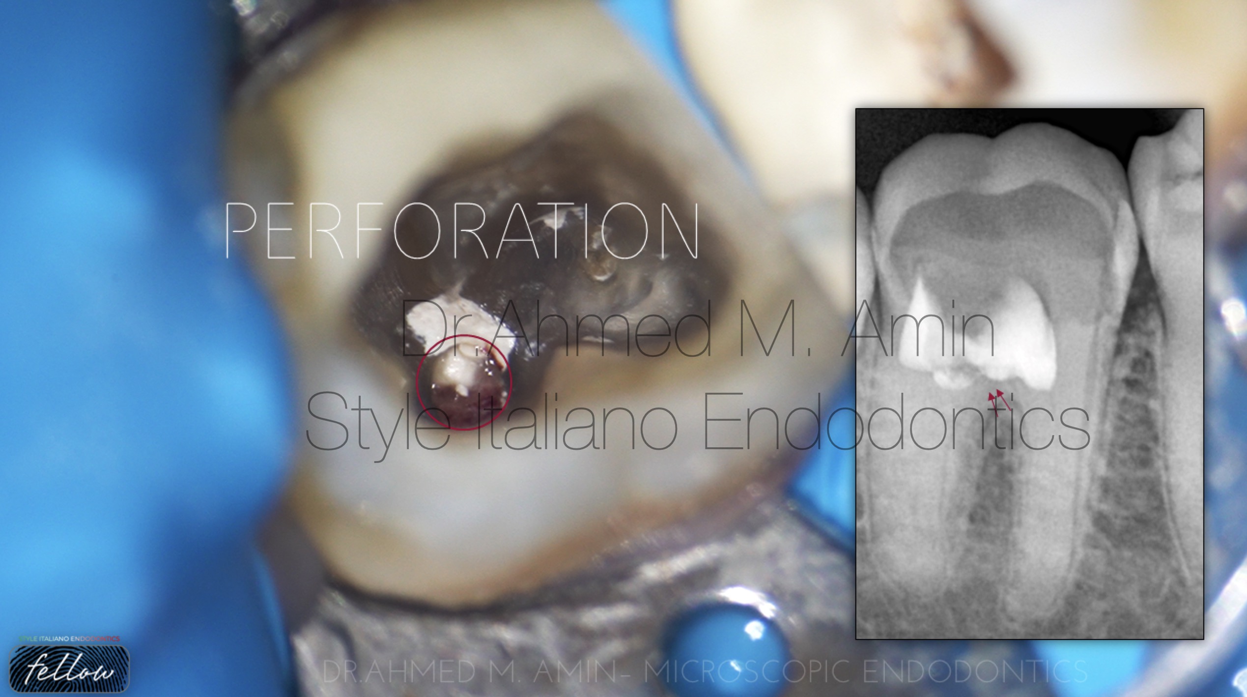
Fig. 3
After removal of Pulpotec , there was a perforation in the floor of pulp champer
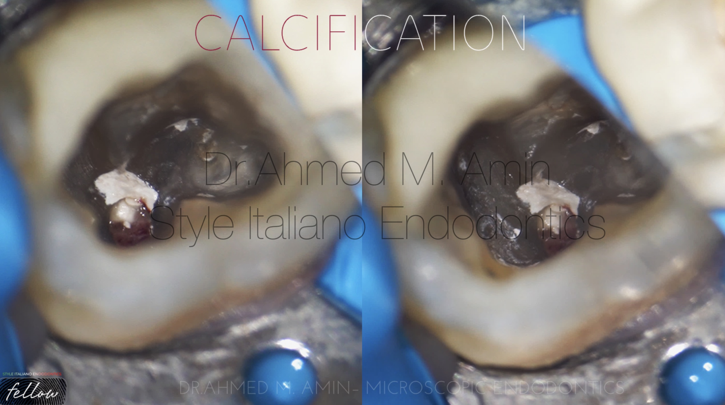
Fig. 4
calcified Canals entrance “ Mesials & Distal “
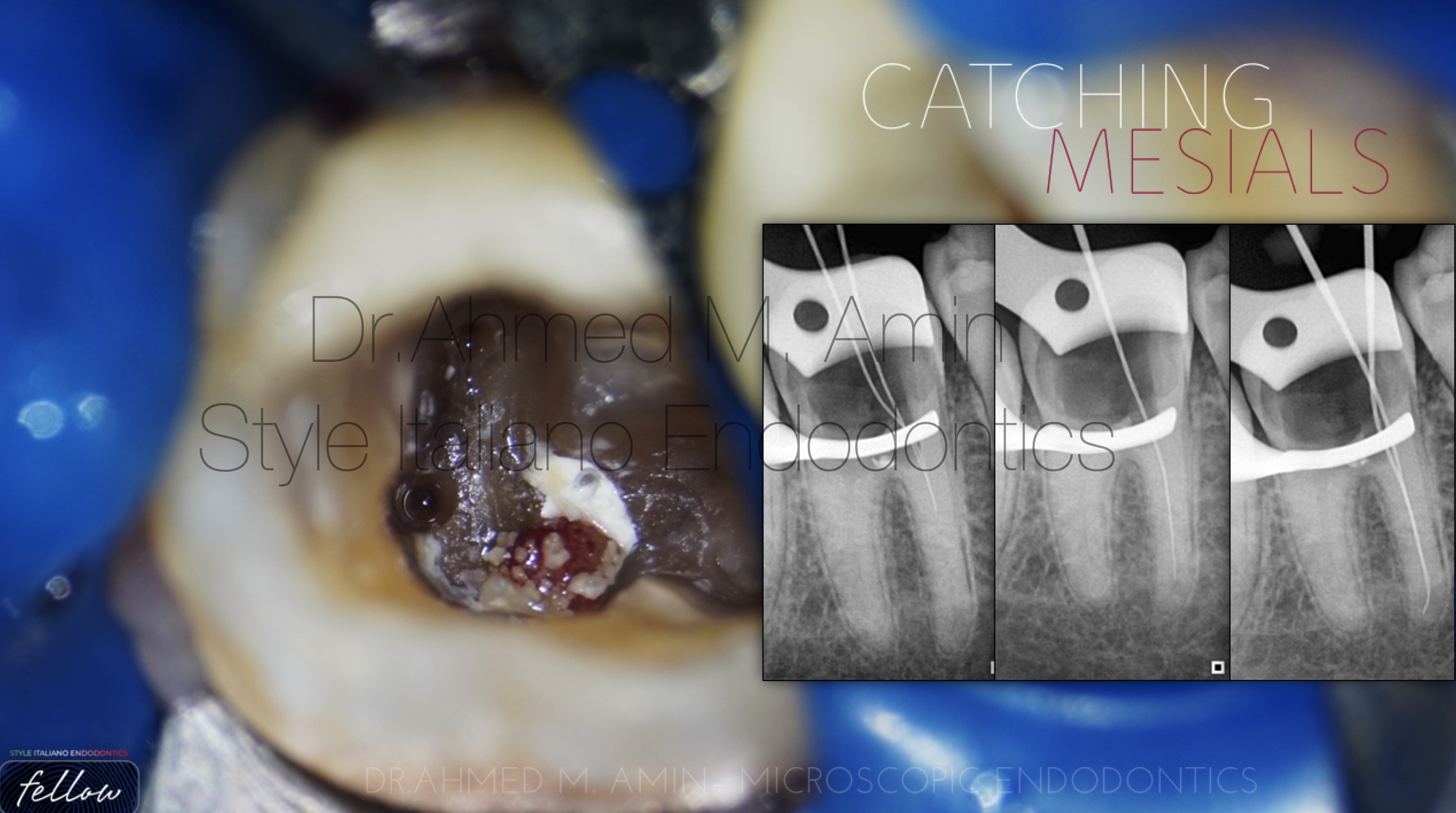
Fig. 5
The use of magnification and ultrasonic is considered a safe technique to locate calcified canal entrances , so we started with ED15D US Tip to locate the mesials and finally locating the tow mesials and catching them using D-Finder files from Mani 8,10,12,15 until the full wl
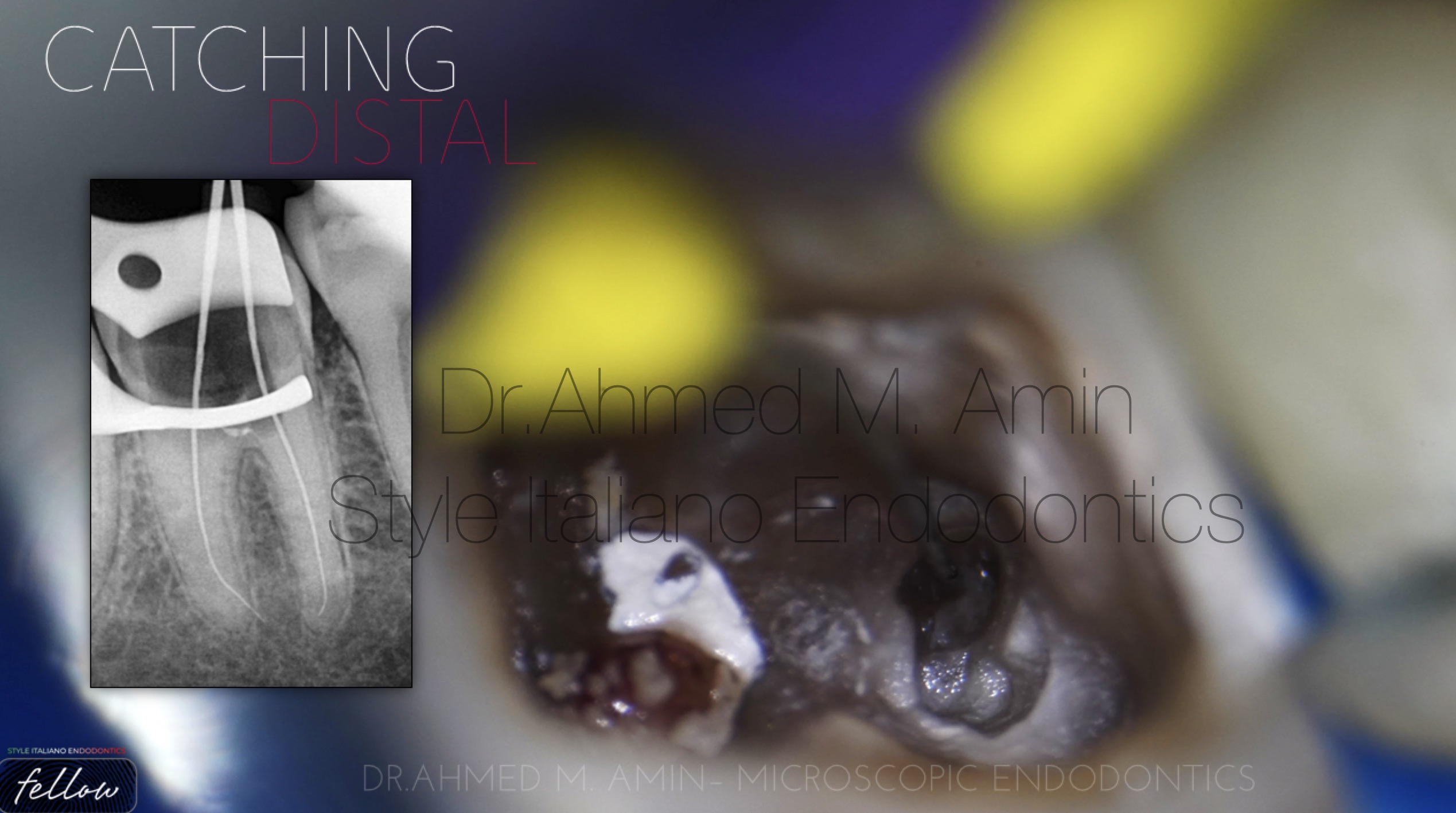
Fig. 6
With the same US tip we located & catched the d canal and prepared it manually before rotary files usage
25/4 mechanical Preparation for mesials & 30/4 for single distal canal
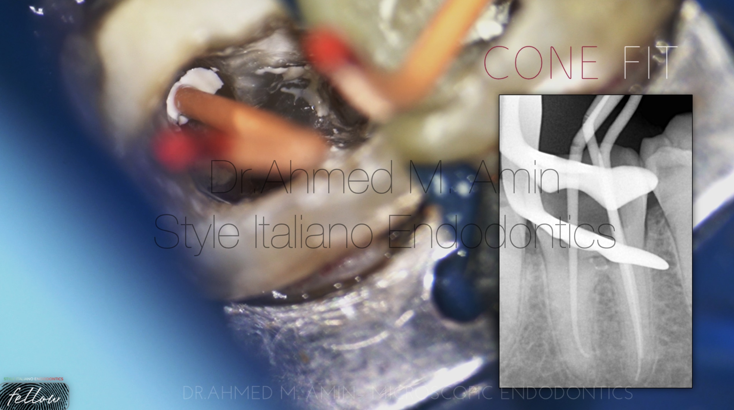
Fig. 7
Confirmation of Cone fit at the exact working length
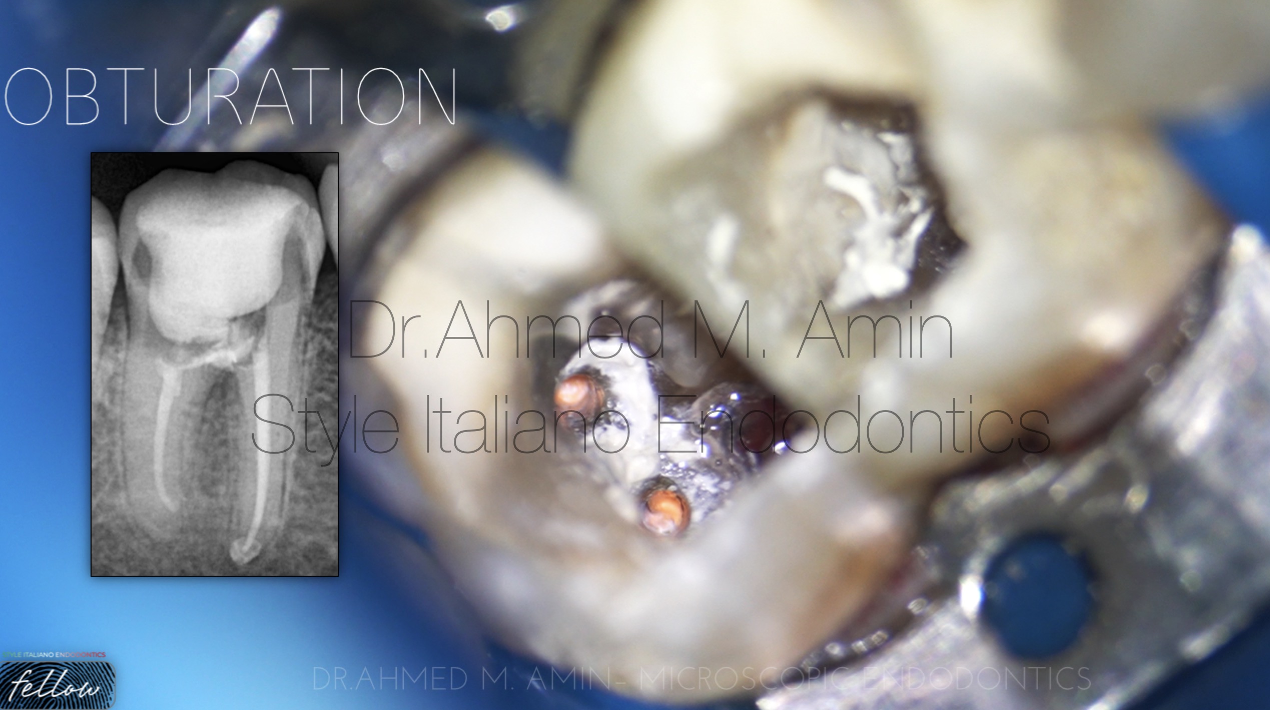
Fig. 8
Down packing then backfilling using eighteeth system with BIO C Sealer from angelus
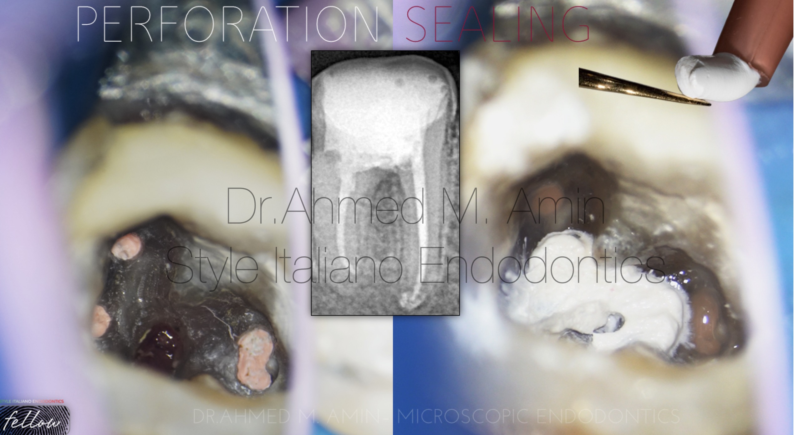
Fig. 9
Floor perforation was temporarily sealed with Intracanal Medication for 2 weeks to elevate the PH Level and act as antibacterial before permanently sealed with MTA
Then final restoration using bulkfill Composite
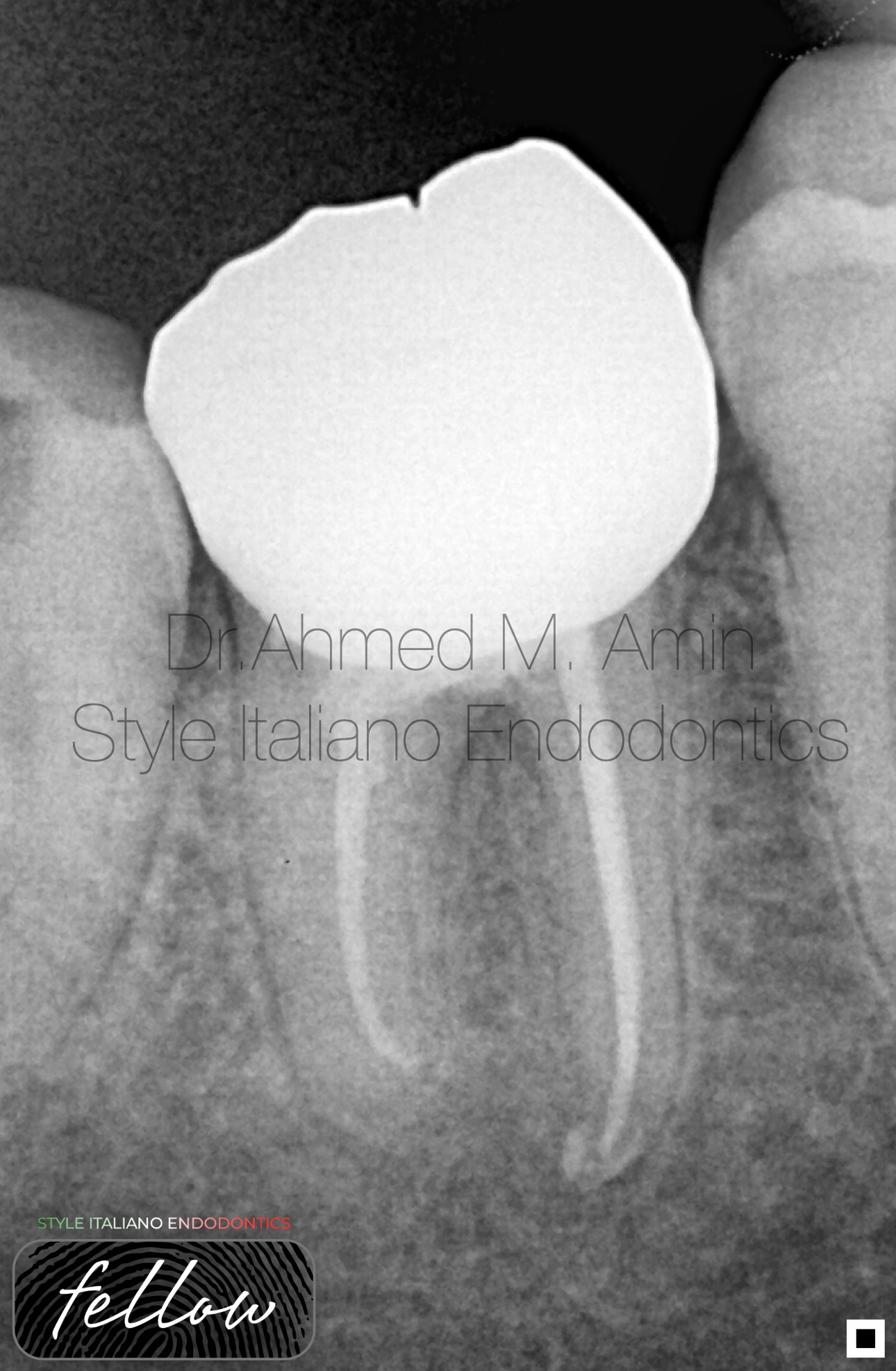
Fig. 10
Prosthetic part : Zircon Crown
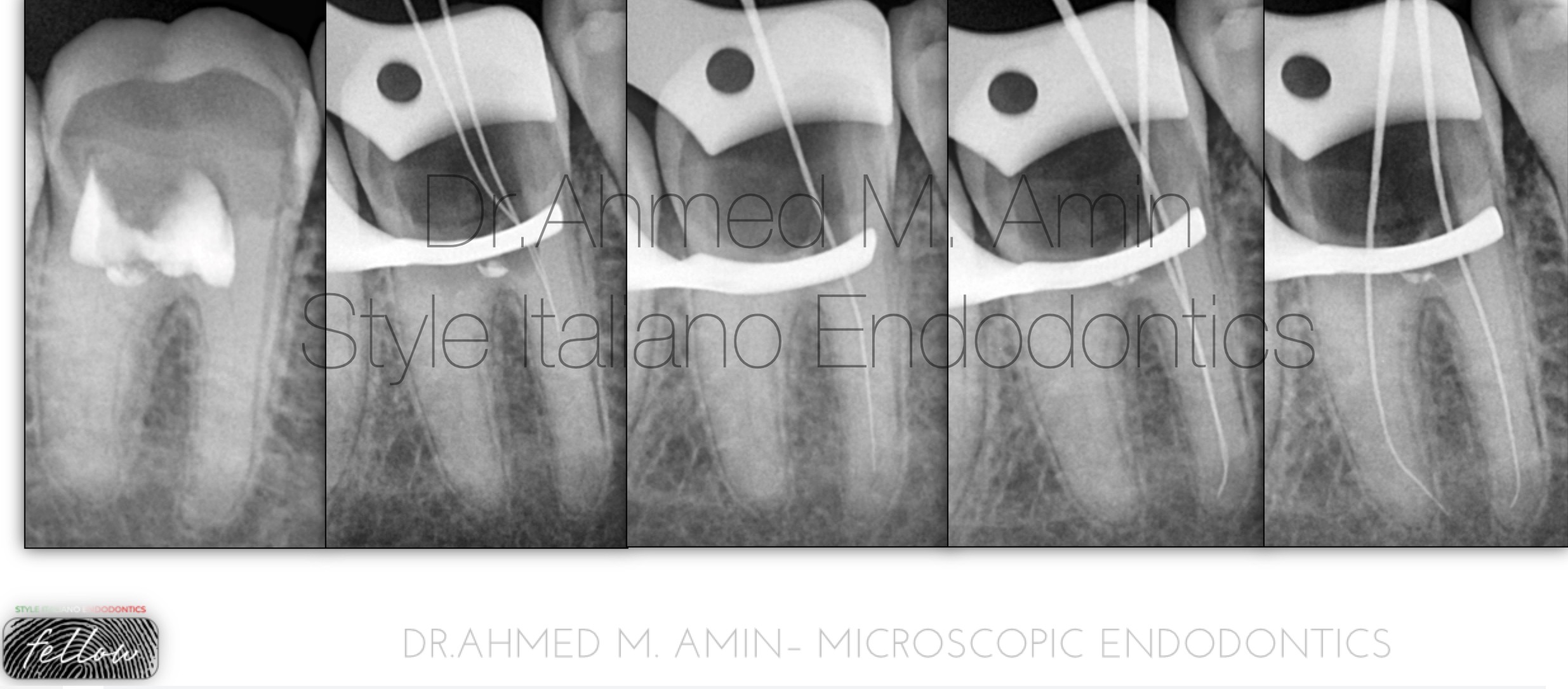
Fig. 11
Preoperative xray , and Scouting Trials Xrays
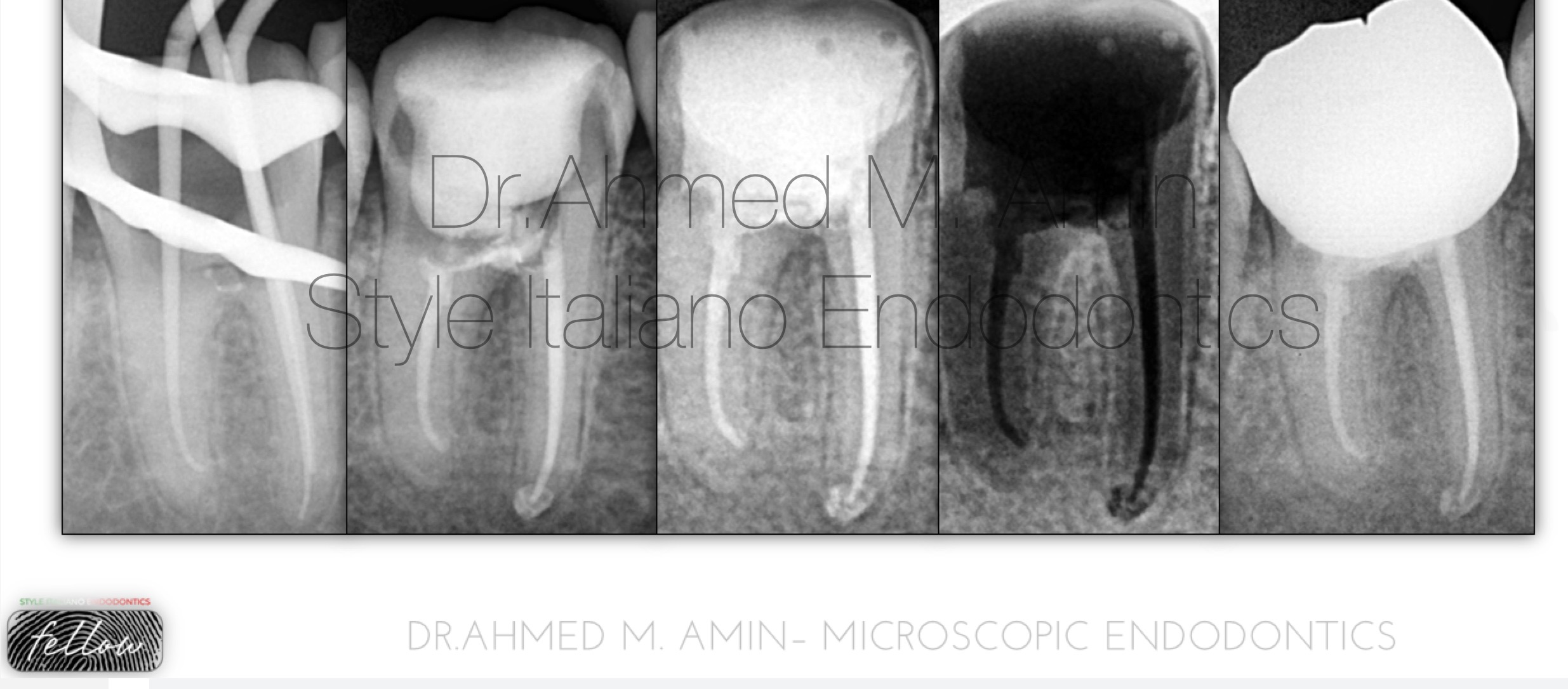
Fig. 12
Cone Fit , Obturation , Final Restoration and Finally Crown delivery Xray
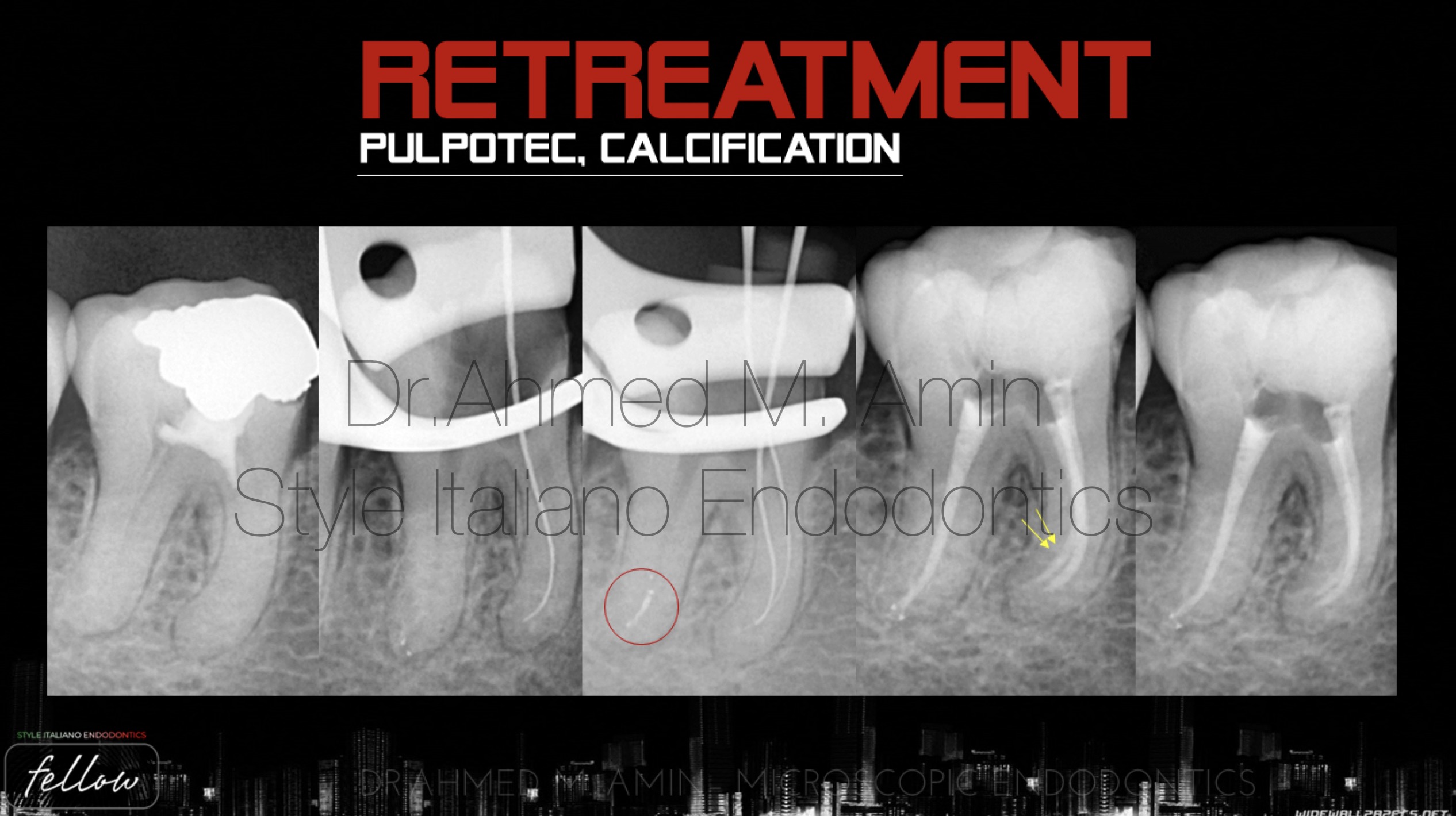
Fig. 13
Case no. 2
Tooth no. 46 with pulpotec material
Preop xray show calcified mesials and very tight distal canal
Many trials to locate and scout the mesials with the help of ultrasonic tips ‘ ED15D ‘ & Non-Active STST. D Finder Files
Broken Side vented needle in D Canal retrieved later
Preparation till 25/4
Obturation with wvc using eighteeth system
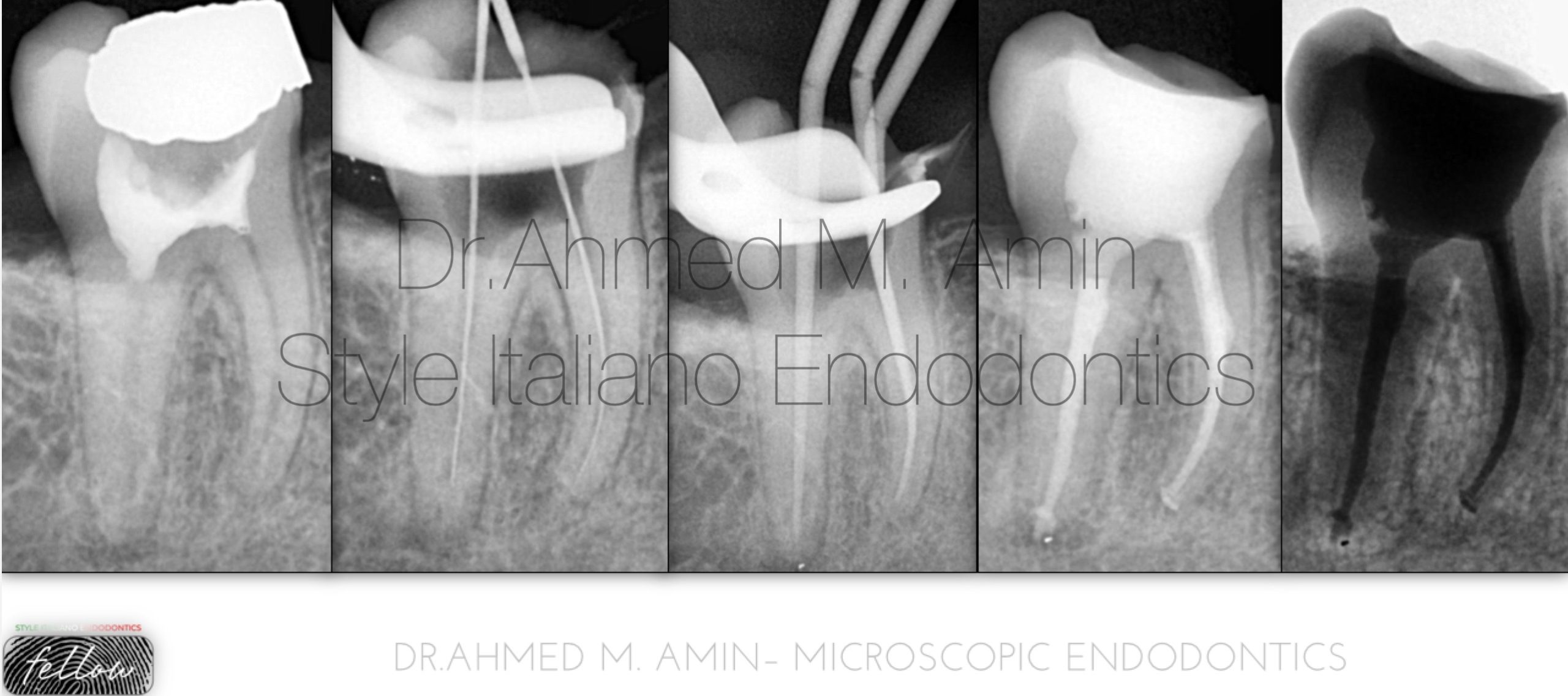
Fig. 14
Case No. 3
Another case of pulpotec retreatment starting from diagnosis , scouting , preparation , cone fit and finally obturation till the full working length
Step by step retreatment of pulpotec filling
 : eval()'d code</b> on line <b>19</b><br />
)
Fig. 15
Conclusions
- Preoperative radiograph is very important before starting endodontic treatment
- Case difficulty assessment will lead to successful treatment through good planning and selection of the approach technique and the right tools.
- The use of magnification and ultrasonic is considered a safe technique to locate calcified canal entrances.
- Implementation of CBCT, Magnification and ultrasonics is recommended in complex cases
- D Finder files from Mani is recommended for negotiation of narrow and tight canals.
Bibliography
Krasner P, Rankow HJ. Anatomy of the pulp chamber floor. JOE 2004; 30(1):5.
Cohen S, Hargreaves K. Pathways of the Pulp 9th ed. Mosby, St. Louis, MO, 2006.
Buhrley LJ, Barrows MJ, BeGole EA, Wenckus CS. Effect of magnification on locating the MB2 canal in maxillary molars. JOE 2002. doi: 10.1097/00004770-200204000-00016.
Nasiri K, Wrbas KT. Management of calcified root canal during root canal therapy J Dent Sci. 2023 Oct; 18(4): 1931–1932.
Falcon P.A., Falcon C.Y., Abbasi F., Hirschberg C.S. Chamberless endodontic access for treatment of calcified anterior central incisors. J Endod. 2021;47:322–326.
Rodríguez G, Patel S, Durán-Sindreu F, Roig M, Abella F. Influence of Cone-beam Computed Tomography on Endodontic Retreatment Strategies among General Dental Practitioners and Endodontists. J Endod. 2017 Sep;43(9):1433-1437. doi: 10.1016/j.joen.2017.04.004. Epub 2017 Jul 8. PMID: 28689702.