
Two Ways of Warm Vertical Compaction
07/07/2022
The Community
Warning: Undefined variable $post in /var/www/vhosts/styleitaliano-endodontics.org/endodontics.styleitaliano.org/wp-content/plugins/oxygen/component-framework/components/classes/code-block.class.php(133) : eval()'d code on line 2
Warning: Attempt to read property "ID" on null in /var/www/vhosts/styleitaliano-endodontics.org/endodontics.styleitaliano.org/wp-content/plugins/oxygen/component-framework/components/classes/code-block.class.php(133) : eval()'d code on line 2
Cleaning and shaping root canals without sealing is not enough so Warm Vertical Compaction technique is the most useful technique to seal the root canal system
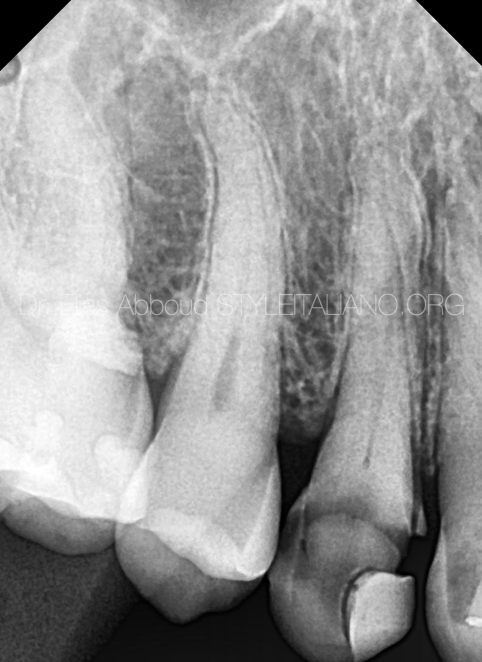
Fig. 1
A 35 years old male patient was referred because of severe, continuous and diffuse pain in the upper right jaw.
Clinical examination showed that there was very huge distruction on the first premolar.
Radiography examination showed that there were very deep caries.
Pulp pain has two type of inflammation:
1) Reversible pulpitis: we can treat the tooth without pulp extraction and root canal treatment. Partial caries removal , selective caries removal and vital pulp therapy are most favorable choices here.
2) irreversible pulpitis: hard, continuous and diffuse pain will be appear, the only choice is to do root canal treatment (pulp extraction).
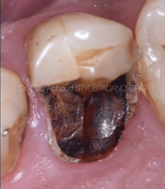
Fig. 2
Pre operative situation: a large decay is visible.
Rubber dam isolation is the most important step in all endodontic treatment because adds clearance, non contamination and perfect vision .

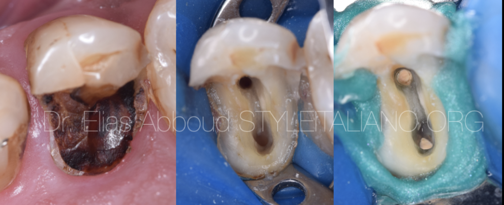
Fig. 3
After we removed all caries, it was noticed that tertiary dentin covered the orifices of the canals.
Ultrasonic diamond-coated tips were the best weapon in this phase for preventing any fault
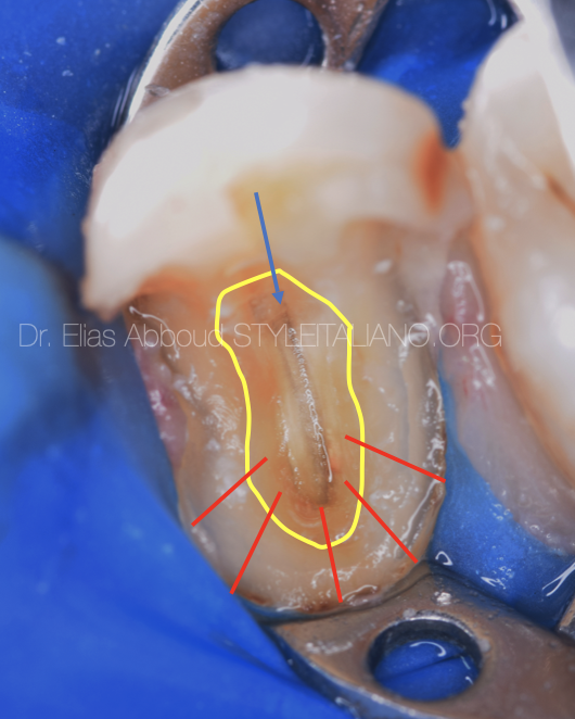
Fig. 4
All tertiary dentin was removed by T3 ultrasonic tip. Color of the dentin and pulp floor is the secret to find the canals so we have to remove all yellow & brown dentin
We have to follow the Anatomy of the Pulp-Chamber Floor principles (red arrows)
The distance from the external surface of the clinical crown to the wall of the pulp chamber was the same throughout the circumference of the tooth at the level of the CEJ
The orifices of the root canals are always locate at the junction of the walls and floors
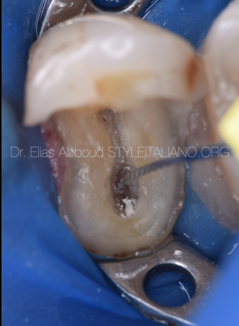
Fig. 5
The palatal canal was located in the center of the gray color as we notice before after removing dentin tissue
Then we started to search for the opposite canal ( buccal canal )
the mission here started to be easier
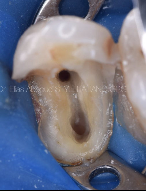
Fig. 6
After cleaning and shaping with
#17 2%
#20 4%
#25 4%
#30 4%
And irrigation with
Sodium hypochloride 5.25 %
Edta 17 %
Chlorhexidine 2%
And saline between each irrigant, the canals were ready for obturation
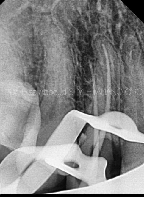
Fig. 7
Master cone radiography
This is the most important image in the whole treatment because here we can make an adjustment before obturation
Warm vertical compaction technique one of the standard technique in obturation of the root canals
There are two ways of obturation in WVC technique
The first one is cutting the gutta percha at the orifice of the canal, then condense it by coronal plugger; after that, down packing through the canal at the level of 4- 6 mm to the apical third
Then we condense the gutta percha in the apical third by apical plugger
The second way is to do down packing directly to 4 - 6 mm from the apex

Fig. 8
Down packing radiography confirm
Now we are ready for fast pack and obturate the middle and coronal third
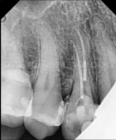
Fig. 9
Post operative X-ray
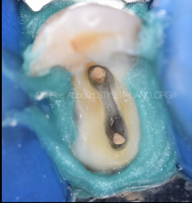
Fig. 10
Post operative picture
Fig. 11
The Author:
Dr . Elias Abboud
Damascus Syria
2018 : B.D.S ( Damascus university )
2019 : ENDODONTICS AND OPERATIVE DENTISTRY AT THE NATIONAL DENTAL CENTER
Conclusions
During the root canal treatment , regaining the anatomy and cleaning the entire endodontic system is necessary for a better long term prognosis of the tooth. We have always to follow the principle of preparation and access cavity steps. A long and sharp explorer such as DG16 will give us the possibility to detect the orifices
Bibliography
Deivanayagam Kandaswamy, Nagendrababu Venkateshbabu, Reddy Gopi Krishna, Rosaline Hannah, Ganesh Arathi, and Riaz Roohi. Comparison of laterally condensed, vertically compacted thermoplasticized, cold free-flow GP obturations – A volumetric analysis using spiral CT
Woolard RR, Brough SO, Maggio J, Seltzer S. Scanning electron microscope examination of root canal filling materials. J Endod. 1976;2:98–110
Cohen's Pathways of the Pulp Ha JH, Park SS. Influence of glide path on the screw-in effect and torque of nickel-titanium rotary files in simulated resin root canals, Restor Dent Endod 2012;37:4.

