
Thermafil Retreatment of a right upper first molar
01/07/2021
Marc Kaloustian
Warning: Undefined variable $post in /var/www/vhosts/styleitaliano-endodontics.org/endodontics.styleitaliano.org/wp-content/plugins/oxygen/component-framework/components/classes/code-block.class.php(133) : eval()'d code on line 2
Warning: Attempt to read property "ID" on null in /var/www/vhosts/styleitaliano-endodontics.org/endodontics.styleitaliano.org/wp-content/plugins/oxygen/component-framework/components/classes/code-block.class.php(133) : eval()'d code on line 2
The Thermafil Obturation technique was first introduced by Dr. W.B. Johnson in an article published in the Journal of Endodontics in 1978 as a simple method for the delivery of thermo-plasticized gutta-percha (GP) to the prepared root canal space.
Many studies proved that it was an efficient, easy and reproducible obturation technique with a high success rate (Gutmann et al.1993).
However, the presence of short root fillings and those with voids can happen, due to lack of operator expertise and skill in the application of this technique, therefore leading sometimes to a failure of the primary root canal treatment. In these cases, retreatment is indicated and it requires the complete removal of the carrier and the GP around it.
This case report will describe a detailed retreatment technique of an upper right first molar filled with the Thermafil technique.
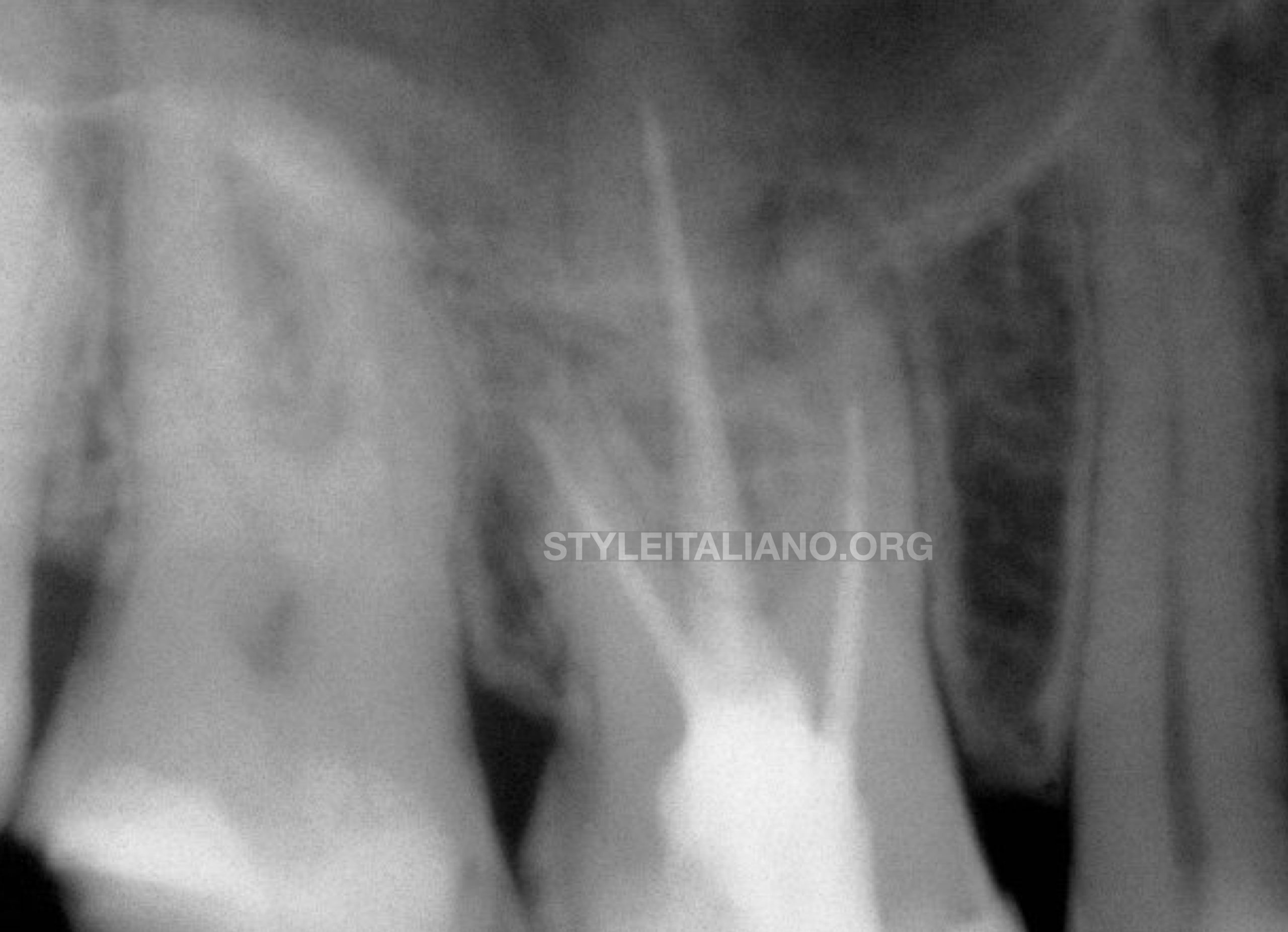
Fig. 1
Upon radiographic examination, the mesio-buccal 1 (MB1), disto-buccal (DB) and palatal (P) canals presented short fillings and the mesio-buccal 2 (MB2) was completely neglected.
The radiolucency detected in the apical region of the mesial root, and the absence of symptoms, suggested the diagnosis of an apical asymptomatic periodontitis.
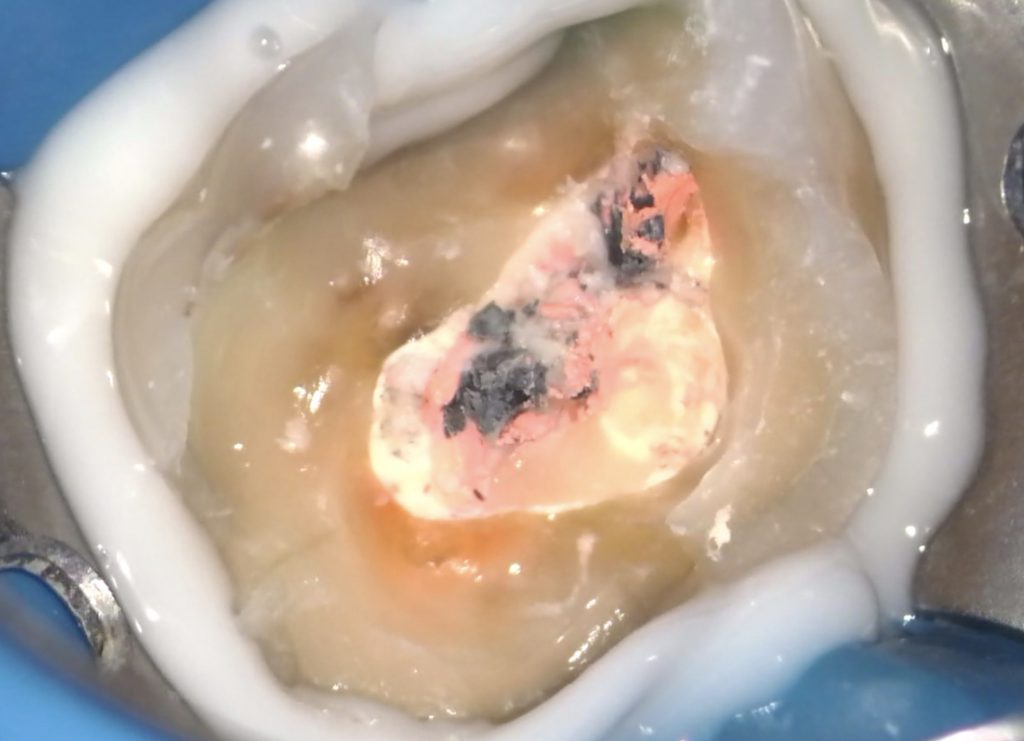
After the elimination of the composite build -up, the first step of the retreatment procedure was to remove safely the remnants of GP surrounding the head of the thermal carrier in the access cavity.
An Endodontic explorer was used for this purpose, and the 3 Thermafil grey carriers were clearly visualized.
A Stieglitz 45 degree angled forceps was used to retrieve in one piece the carrier, first in the MB1 canal, then in the DB canal and finally in the P canal.
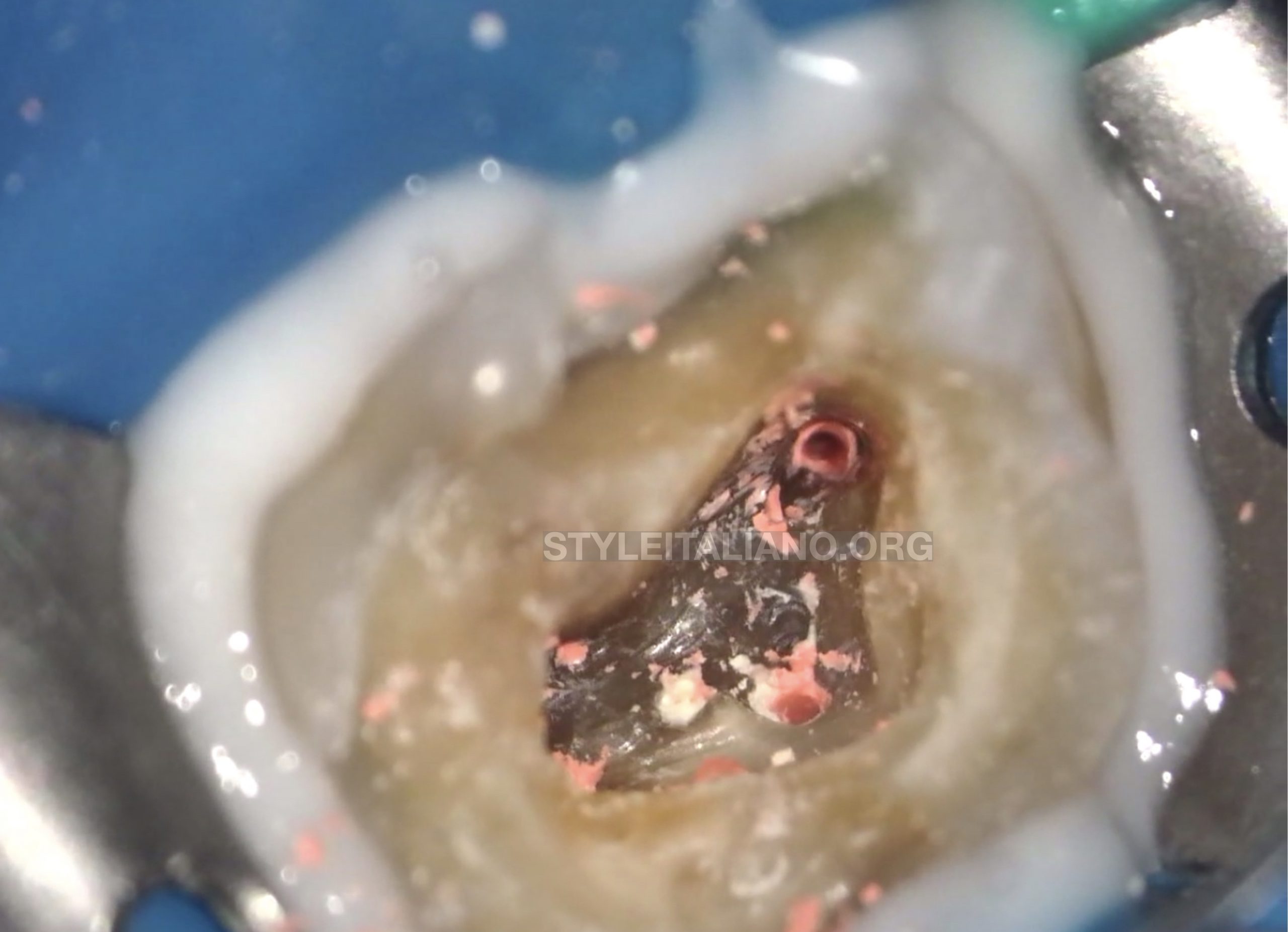
Fig. 2
Access cavity aspect after the retrieval of the plastic carriers, showing remnants of GP.
The access cavity was refined using a diamond coated ultrasonic tip ET18 (Satelec, Acteon Group, Merignac, France).
The GP was mechanically removed in all canals using heat treated files Endostar E3 Azure (Poldent, Warsaw, Poland) 30 /08 without using a solvant.
The orifice of the MB2 was detected, and the patency and scouting process were performed manually with a 08 Kfile (Dentsply Sirona, Ballaigues, Switzerland) using a watch winding motion followed by a push pull motion. Once this file was considered loose inside the canal, a mechanical glide path was done with the Easy Path (Poldent) 14/04 file. The same procedure was applied for the 3 other canals.
The working length was established with a 31 mm 10 K file (VDW, Munich, Germany) and an apex locator (Eighteeth, Changzhou, China).
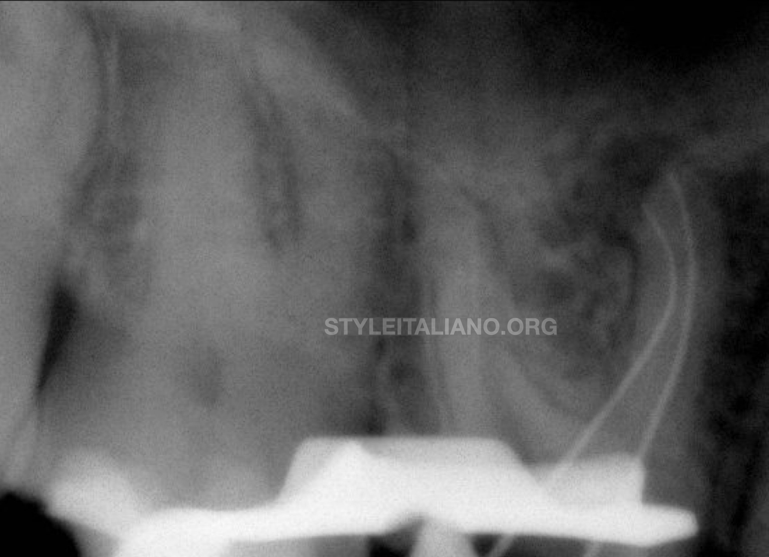
Fig. 3
A radiograph was taken showing the absence of filling material remnants in all canals. A 10 K files at the working length in both MB1 and MB2 canals confirmed the working length. The trajectory of the canals was preserved during the patency procedure.
The canals were first shaped with a 20/06 Endostar E3 Azure file (Poldent). An additional shaping with a 25/06 file was performed in the DB and P canal.
The irrigation was done with 12 ml of NaOCL 5.25 % using the IrriFlex (Produits Dentaires, Vevey, Switzerland) needle.
The canals were dried with a micro-canula and EDTA 17% was placed for 1 mn. A final rince with sterile water was done to eliminate the EDTA.
The irrigation was activated with a sonic device , the EndoActivator (Dentsply Sirona) with the 25/04 tip, for one minute in each canal and a push and pull motion up to 2 mm from the working length. The use of activation showed its effectiveness in removing the smear layer and also the filling material remnants (Volponi A et al. 2020)
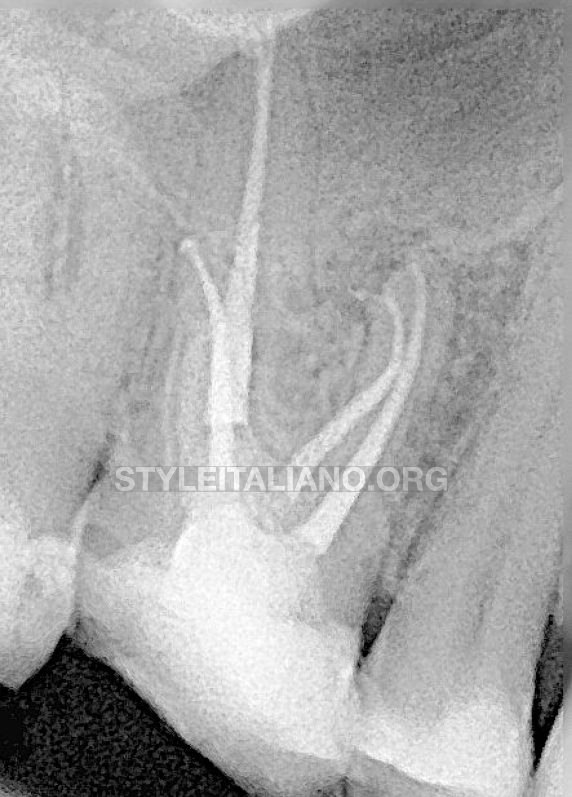
Fig. 4
The canals were filled with warm vertical compaction with 20/05 GP points (Coltène Whaledent, Langenau, Germany) and EssenSeal (Produits Dentaires).
Injecting technique with Obtura II gun and a 23 G tip (Obtura Spartan Endodontics, Algonquin,IL, USA) was used for back pack.

Fig. 5
The access cavity aspect after the final obturation.
Conclusions
Many techniques are described in the literature for the retreatment of carrier based obturation. Some authors recommended the usage of a solvent to soften the GP surrounding the carrier, heating the the material using the System B was also proposed (Wolcott et al. 1999). However most of the authors recommend the direct usage of mechanical Niti files in continuous rotation or reciprocation (Beasley et al. 2013, Rödig et al. 2018).
In this particular case, since the head of the carrier was kept intact in the access cavity, a direct retrieval was possible using a Steigliz plier, prior to the easy removal of the surrounding GP.
Bibliography
- Beasley RT, Williamson AE, Justman BC, Qian F. Time required to remove guttacore, thermafil plus, and thermoplasticized gutta-percha from moderately curved root canals with protaper files. J Endod 2013, 39:125-8.
- Gutmann JL, Saunders,WP, Saunders EM, Nguyen L. An assessment of the plastic Thermafil obturation technique. Part 2. Material adaptation and sealability. Int Endod J 1993, 26:179-83.
- Johnson WB. A new gutta-percha technique. J Ended 1978, 4:184-8.
- Rödig T, Wagner J, Wiegand A, Rick M. Efficacy of the ProTaper retreatment system in removing thermal, GuttaCore or vertically compacted gutta-percha from curved root canals assessed by micro-CT. Int Endod J 2018, 51:808-815.
- Volponi A, Pelegrine RA, Kato AS, Stringheta CP, Lopes RT, Silva ASS, Bueno CEDS. Micro-computed Tomographic Assessment of Supplementary Cleaning Techniques for Removing Bioceramic Sealer and Gutta-percha in Oval Canals. J Endod 2020, 46:1901-1906.
- Wolcott JF, Himel VT, Hicks ML. Thermafil retreatment using a new "System B" technique or a solvent. J Endod 1999, 25:761-4.


