The power of FLEX Endodontic Treatment of a Mandibular first molar with T- Flex files
17/02/2022
Viresh Chopra
Warning: Undefined variable $post in /var/www/vhosts/styleitaliano-endodontics.org/endodontics.styleitaliano.org/wp-content/plugins/oxygen/component-framework/components/classes/code-block.class.php(133) : eval()'d code on line 2
Warning: Attempt to read property "ID" on null in /var/www/vhosts/styleitaliano-endodontics.org/endodontics.styleitaliano.org/wp-content/plugins/oxygen/component-framework/components/classes/code-block.class.php(133) : eval()'d code on line 2
The development of various areas of dentistry requires precise study of morphology of human teeth so that better oral health can be provided. The mandibular molars play a principal role in mastication and help to maintain the vertical dimension of the face, continuity of the dental arch, maintaining the cheeks and tongue in their position and therefore maintaining the stomatognathic function. The conventional root canal anatomy indicates the location of the initial access. The knowledge of both the normal and abnormal anatomy of molars shows the parameters under which root canal therapy is to be performed and can directly modify the probability of success.Failure to treat a canal is an obvious reason for root canal treatment failure. Therefore, all clinicians must make every effort to locate and treat all existing canals during endodontic treatment.Success of Endodontic therapy depends upon thorough cleaning and shaping, disinfection & 3D obturation of the root canal system. Failure to follow the protocol in any of the above steps can lead to failure of the endodontic treatment.
This case report shows a Case of Irreversible pulpitis with Symptomatic Apical Periodontitis (SAP) due to deep distal carious lesion in a mandibular second molar.
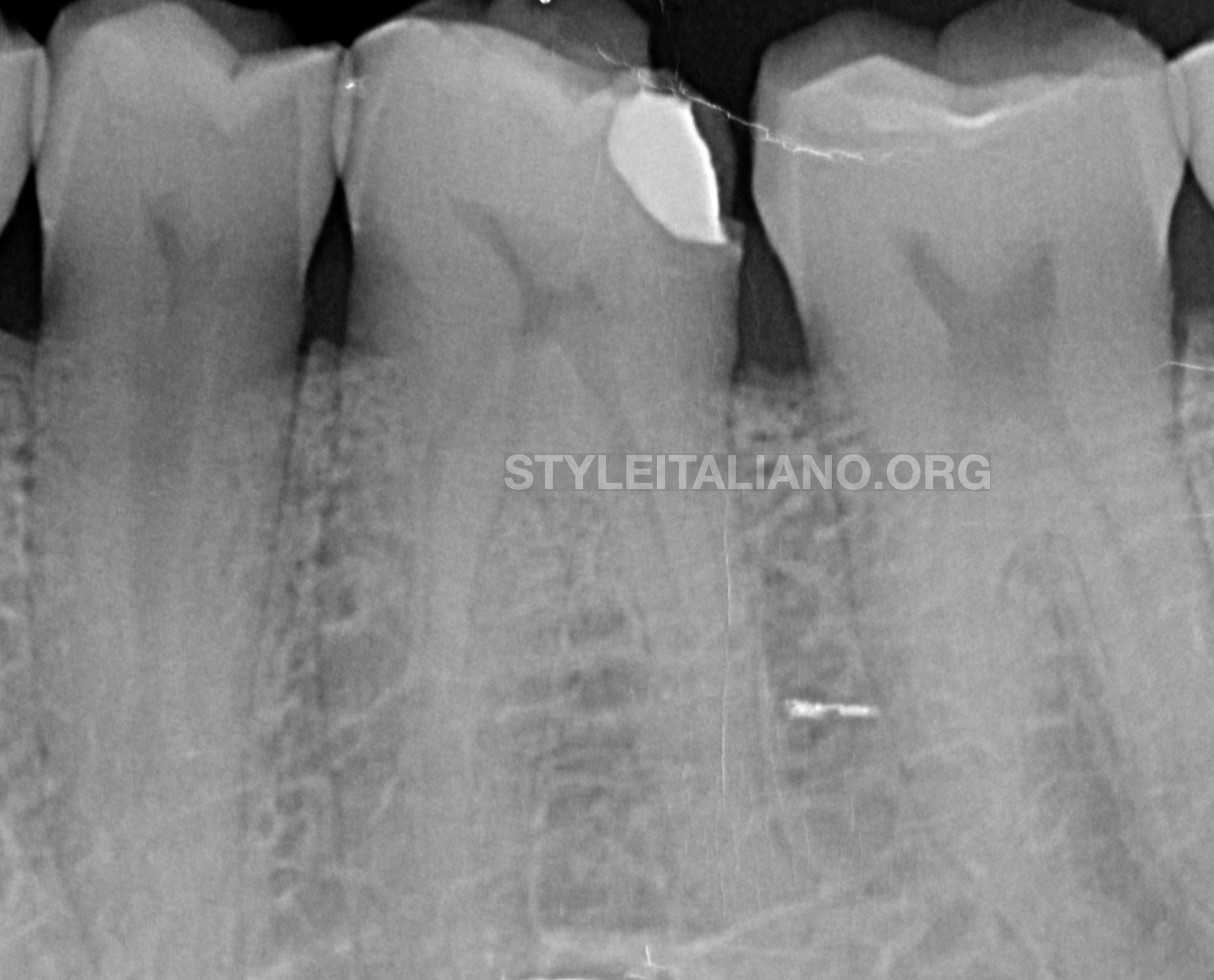
Fig. 1
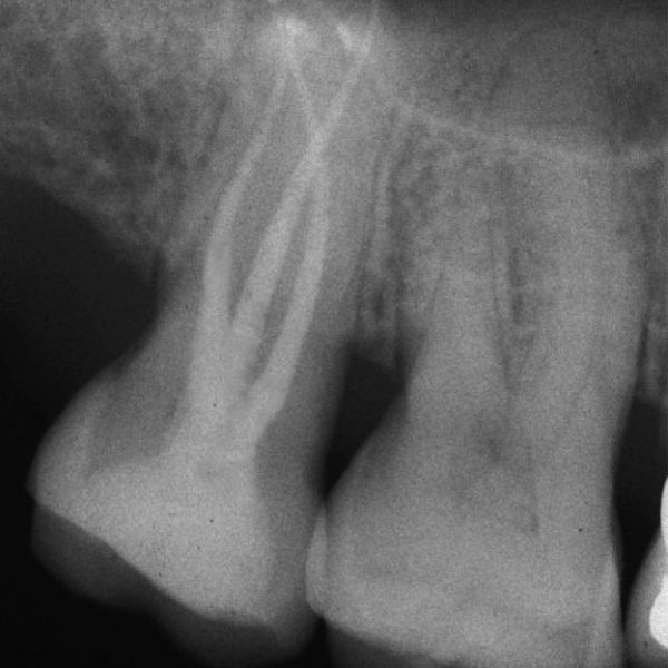
Preoperative radiological assessment:
- The periapical radiograph revealed a deep distocclusal temporary restoration. The root canal show signs of calcifications.
- Diagnosis (Pulpal and periapical):
- Irreversible Pulpitis with Symptomatic apical periodontitis.
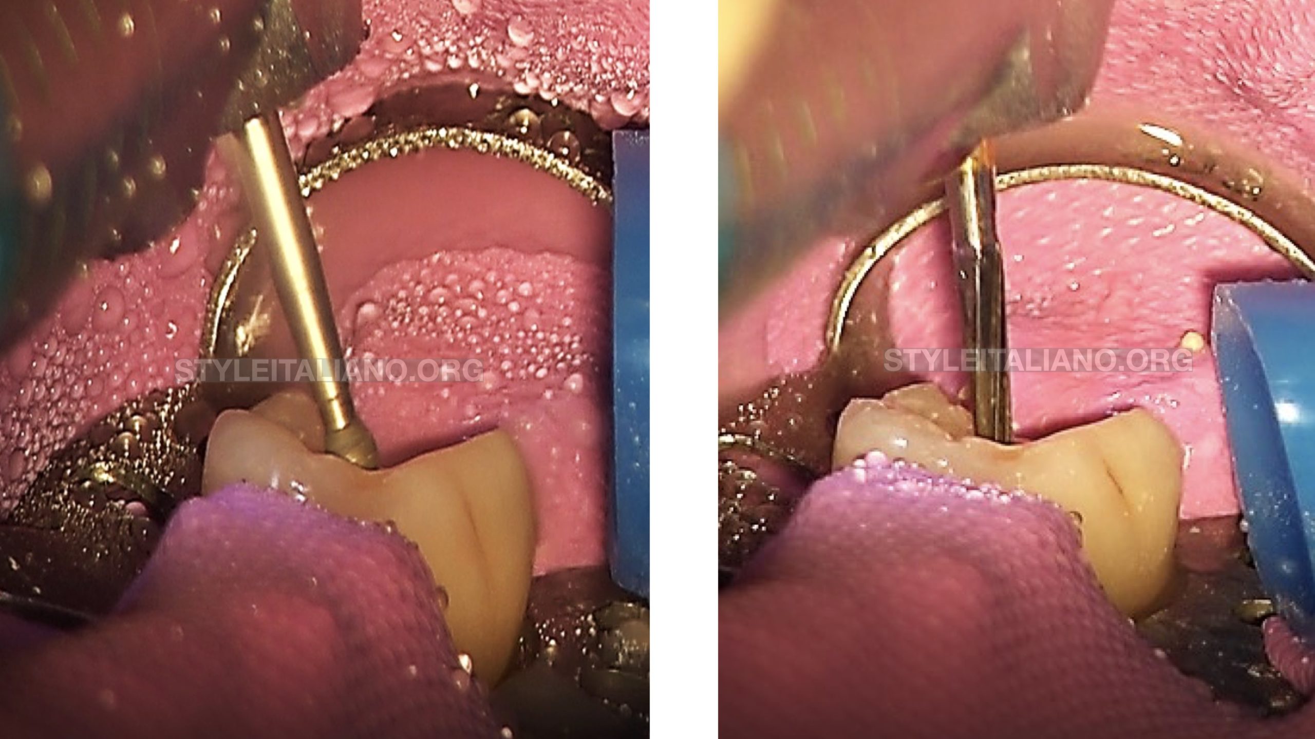
Fig. 2
The Endodontic access was initiated with a round bur and finally prepared with an Endo Z bur (Fig2a and 2b).
DG 16 explorer was used to locate the canals. The temporary restoration was not completely removed as it served as a proximal wall during the treatment protocol.
K files 10 and 15 were used to check the canal patency and estimate the working length.
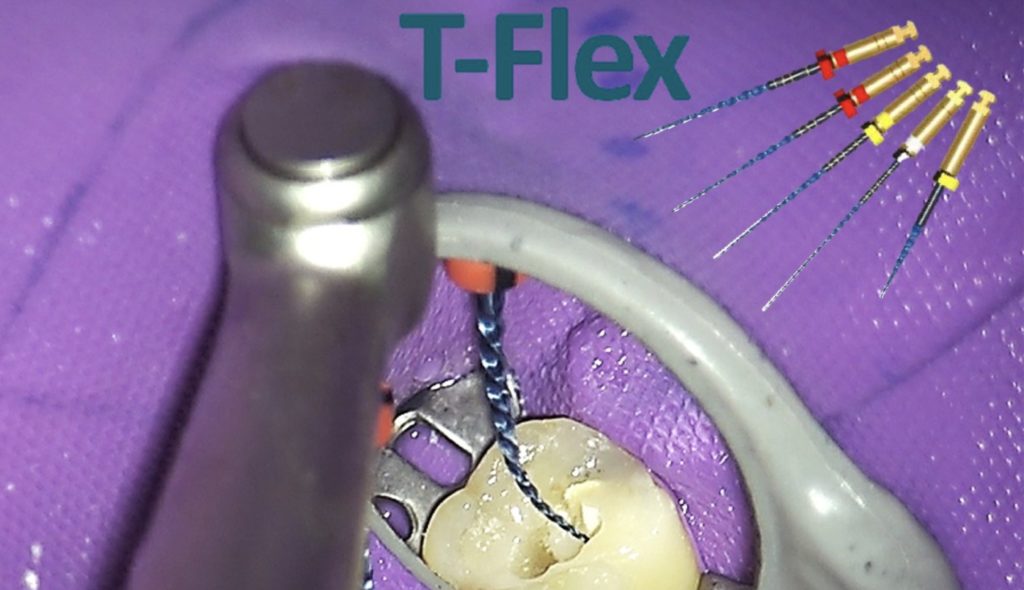
Once the working length was established, the canal orifices were enlarged with T flex orifice shaper 20/.10. (video 2)
Canals were thoroughly irrigated with 2.5% sodium hypochlorite.
Orifice shaping was followed by cleaning & shaping with 15/.03 file which reached the working length. It not only cleaned and shaped but also helped to achieve a glide path due to its narrow taper. (Video 3)
Profuse irrigation was carried on with a 27 gauge side vented needle.
20/.04 file was used next unto the working length for cleaning and shaping. (Video 4).
All the files were used in wet canals filled with the irrigant.
Finally the canal was cleaned and shaped unto the working length with 25/.04. Since the mouth opening of the patient was restricted, all the files were prebend prior to insertion inside the canal. (Video 5)
All the files were used in wet canals filled with the irrigant.
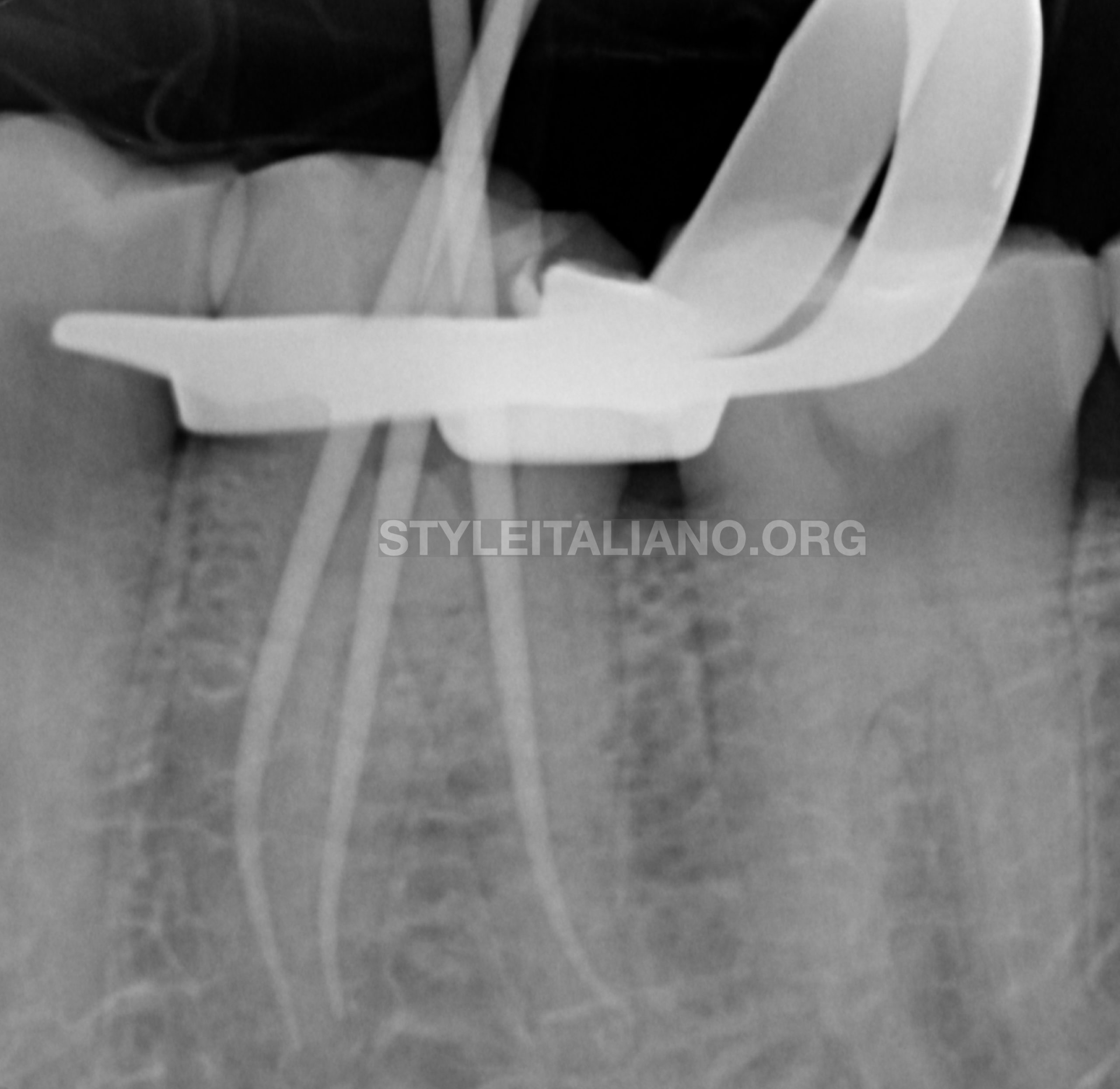
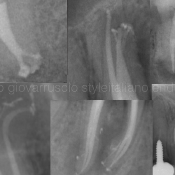
Fig. 3
Master cones were tried for a tug back fit. The master cone fit was also verified with a radiograph
Final drying of the canals prior to obturation were done with intracranial suction tips and paper points
Obturation was done using resin based sealer with single cone obturation technique.
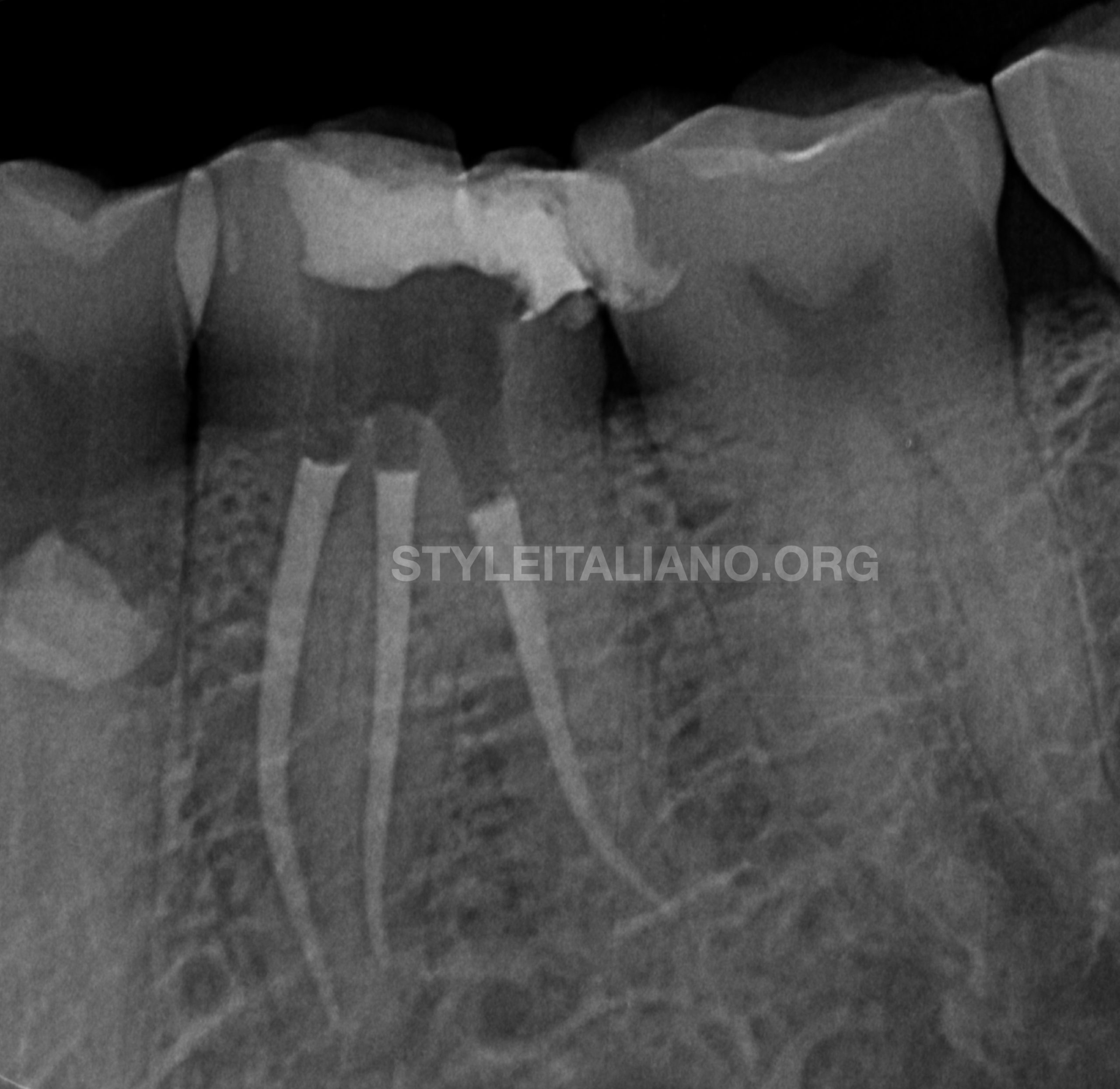
Fig. 4
Obturation was finally verified with an immediate postoperative periapical radiograph.
Conclusions
Learning Objectives: The reader should be able to understand:
- The importance of magnification in Endodontics.
- The significance of proper interpretation of the preoperative radiograph.
- The significance of a selection of correct rotary file system..
- The importance to use of efficient tools to make endodontic treatment easier and less messy.
- The importance of copious irrigation in endodontics.
- The need to plan/modify the treatment steps as per the clinical findings during the procedure.
- How to decide clinically, which instrument to be used for a particular step during an endodontic treatment.
- The concepts of understanding the prognosis of the tooth and giving a try to save the tooth instead of straight away extracting it.