Non surgical retreatment of a mandibular first molar with C-shaped canal system
27/05/2021
Riste Panajotu
Warning: Undefined variable $post in /var/www/vhosts/styleitaliano-endodontics.org/endodontics.styleitaliano.org/wp-content/plugins/oxygen/component-framework/components/classes/code-block.class.php(133) : eval()'d code on line 2
Warning: Attempt to read property "ID" on null in /var/www/vhosts/styleitaliano-endodontics.org/endodontics.styleitaliano.org/wp-content/plugins/oxygen/component-framework/components/classes/code-block.class.php(133) : eval()'d code on line 2
The C-shaped canal, which was first documented in endodontic literature by Cooke and Cox in 1979, is so named for the cross-sectional morphology of the root and root canal.
C shaped canals are mostly present in the mandibular second molars, while they are really rare in the first mandibular molars.
The cause of forming C shaped canals is the Hertwig’s epithelial root sheath which bends in a horizontal plane below the cemento-enamel junction and fuses in the center leaving openings for roots. Failure of the Hertwig's epithelial root sheath to fuse on the lingual or buccal root surface is the main cause of C-shaped roots, which always contain a C-shaped canal. The C-shaped root may also be formed by coalescence because of deposition of the cementum with time.
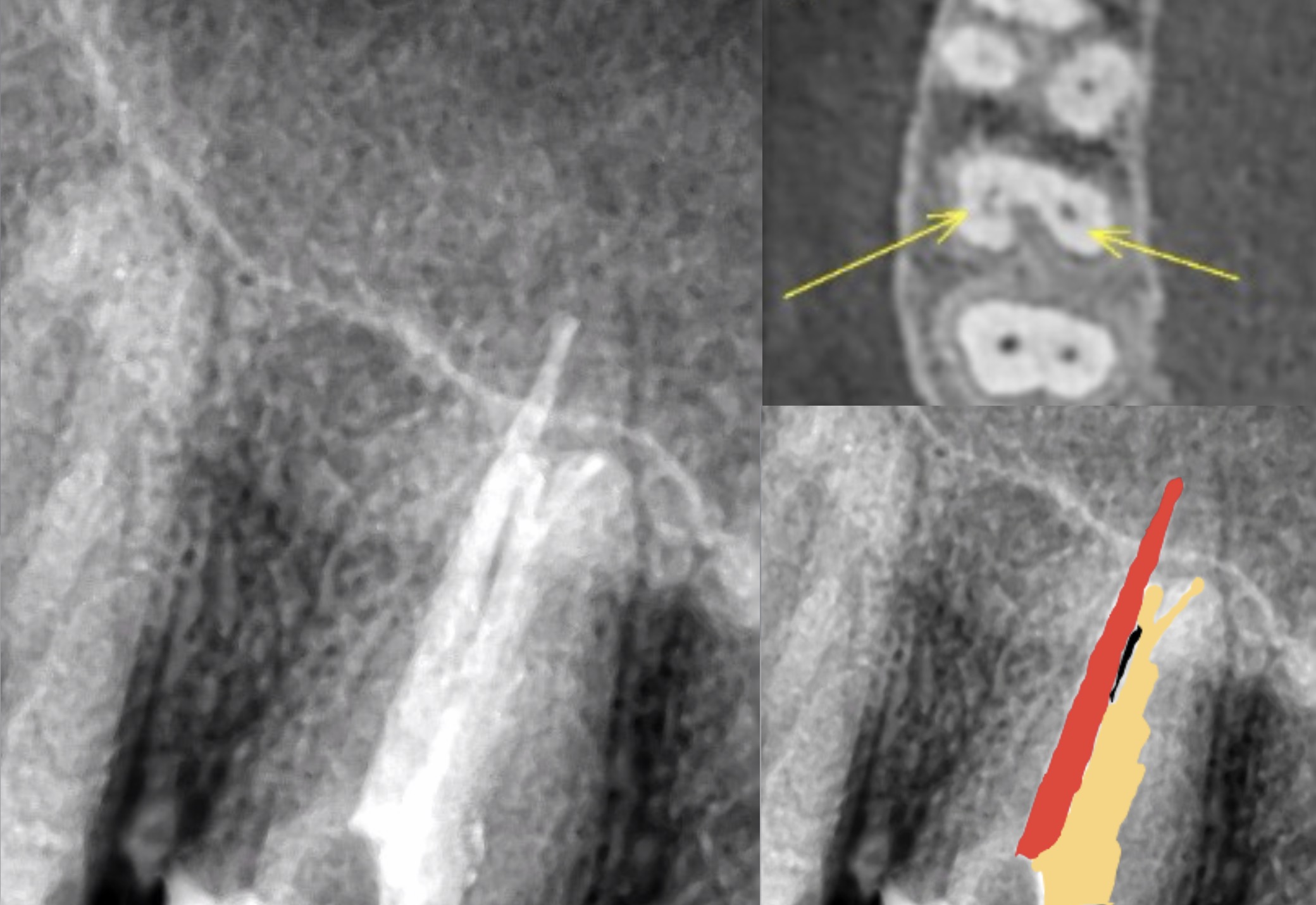
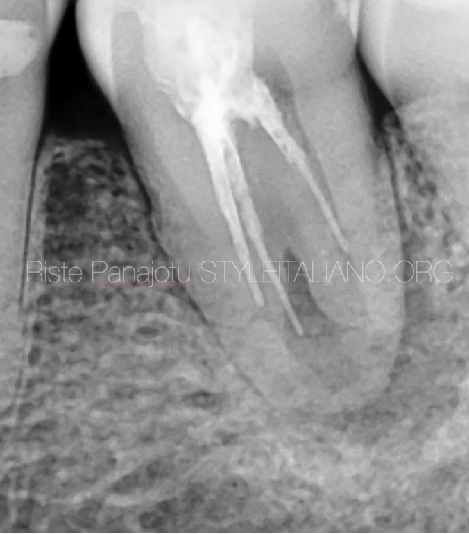
Fig. 1
65 year old woman
Complaining about pain while chewing in left mandible
On clinical examination big composite restoration was detected
Pain on precision and positive bite test.
No signs of crack or fracture
Probing dept of no more then 3 mm
x ray examination showed previously insufficient root canal treatment
Pulpal diagnosis : Previously treated 36
Periapical diagnosis:Symptomatic apical periodontitis
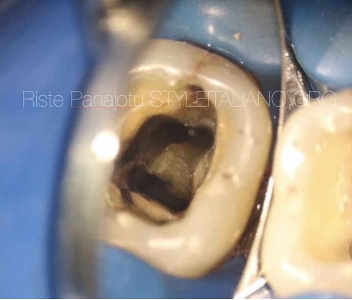
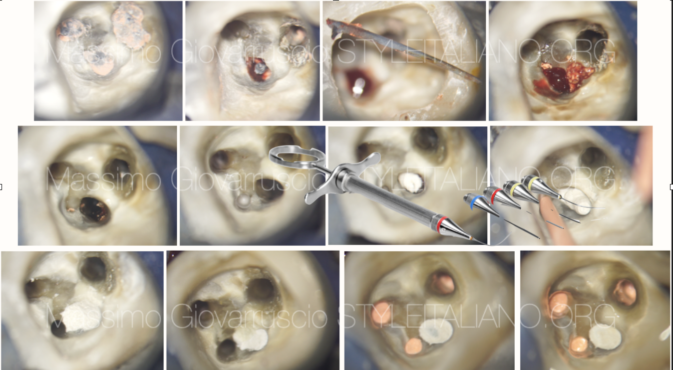
Fig. 2
After cleaning the pulp chamber from old filing material access to the root canal system was established.
It was obvious that the canal anatomy is challengin.
One oval mesial canal and one c shaped distal obturated with some gutta percha cones. The removal of gutta percha was easy .
TIP: Always try to remove the gutta percha mechanically from the canal: solvents make the gutta percha sticky and really is a challenge to remove completely the dissolved gutta from the canal walls
The shaping was done with CM files.
TIP: Irregular canal systems like oval canals and C shaped canals are impossible to be cleaned and shaped without brushing movements.The brushing movements have to be done away from the furcation.
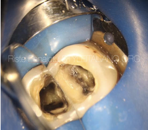
Fig. 3
The most crucial moment in such cases is the irrigation and activation of the irrigants. In this case I have used 5% Natrium Hypochlorite Sonically activated. Also I used EDTA 17% for dentin conditioning and better Hypochlorite penetration into dentin tubules.
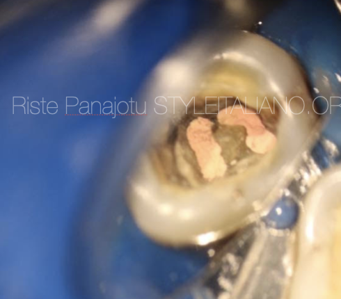
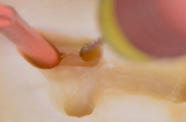
Fig. 4
The obturation was made with epoxy sealer and Squirt technique
Only warm gutta percha can fill all the gaps and adapt to the walls of this kind of irregular canal system anatomy.
TIP :Push the needle in the previously “painted” with sealer canal until it gets in contact with canal walls but avoid it to be wedged into the dentine. Start extruding warm gutta percha and wait for the needle to be pushed out of the canal from the back pressure of the gutta percha.
This tooth was finished with SDR composite over the orificiums then fiber reinforced composite and then nano composite on the top. Crown was advised.
TIP: Always cover the Short fiber reinforced composite with regular composite because of a possibility of microleakage in between the fibers.
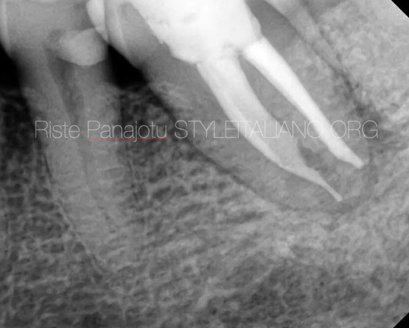
Fig. 5
post op
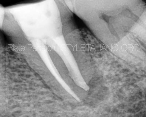
Fig. 6
Different shift
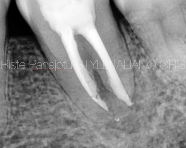
Fig. 7
6 months recall
Conclusions
The right diagnosis and the right technique of mechanical and chemical treatment as well as the right technique of obturation can lead us to a successful treatment of such irregular anatomies of the root canal system
Bibliography
Raisingani D, Gupta S, Mital P, Khullar P. Anatomic and Diagnostic Challenges of C-Shaped Root Canal System. Int J Clin Pediatr Dent 2014;7(1):35-39.
Jesús Alejandro Quiñones Pedraza (September 6th 2019). The C-Shaped Root Canal, Human Teeth. Key Skills and Clinical Illustrations, Zühre Akarslan and Farid Bourzgui, IntechOpen