Non surgical retreatment after surgical treatment of a maxillary central incisor
22/08/2020
Riste Panajotu
Warning: Undefined variable $post in /var/www/vhosts/styleitaliano-endodontics.org/endodontics.styleitaliano.org/wp-content/plugins/oxygen/component-framework/components/classes/code-block.class.php(133) : eval()'d code on line 2
Warning: Attempt to read property "ID" on null in /var/www/vhosts/styleitaliano-endodontics.org/endodontics.styleitaliano.org/wp-content/plugins/oxygen/component-framework/components/classes/code-block.class.php(133) : eval()'d code on line 2
Nowadays in our practices we can witness a lot of unsuccessful RCT. Sometimes we also can see surgical treatments which are failing . When planning the treatment we should keep in mind that not always surgical treatment means more aggressive treatment and that also surgical treatment can fail.
Very often in our practices we have requests for solution of endodontic problem from our referral coleagues and they ask sometimes for a quick solution. We should take the right approach that suits the best our needs as clinicians, to give the best treatment for our patient, and the needs of our referral doctor to fit in his treatment plan.
In the following case I will try to explain my approach, the referral needs and the benefit for the patient.
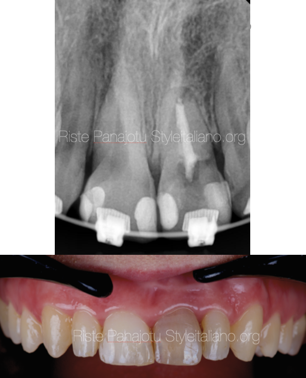
Fig. 1
This is the first X-ray that the referral doctor, a friend of mine, sent to me to consult me about the left central. The patient is a 27 year old woman.The orthodontic treatment was finished the same day the x-ray was taken and the braces were removed.
This patient is going to receive 1 crown and 3 veneers. The referral is upset how the left central eddodontic treatment looks . There is no time for a second surgery, so he asks me for an orthograde retreatment .
I agreed and really think that a good retreatment on this previously surgical treated tooth will be a good choice in the time frame that they have (they had 2 weeks to finish the treatment).
On this x-ray we can see a surgically treated tooth with no retro filing, insufficient canal filling and a bad coronal seal.
The treatment plan is to remove the existing filling material, to a good desinfection, perform an MTA plug followed by a warm gutta percha back fill and a custom core made from a fiber reinforced composite due to the size of the canal.
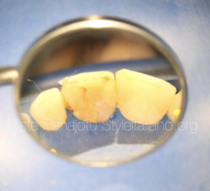
Fig. 2
Rubber dam isolation
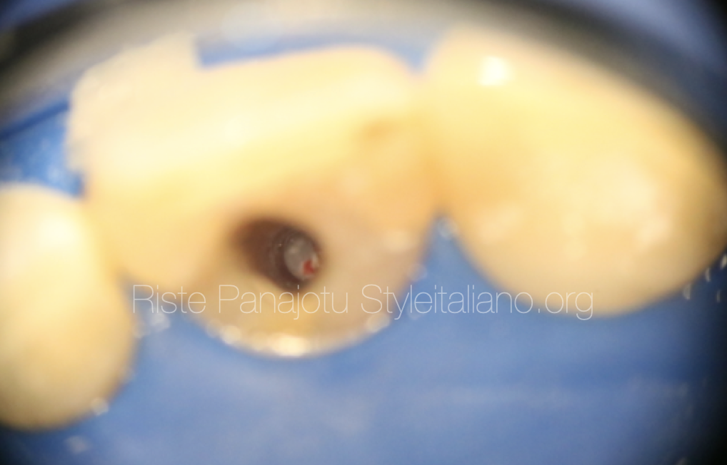
Fig. 3
After rubber dam isolation the access cavity was done from the palatal side of the tooth.
The old composite was removed, then the canal filling was removed mechanically, without using any solvent.
This microscopic photography allows to see directly the apical foramen.
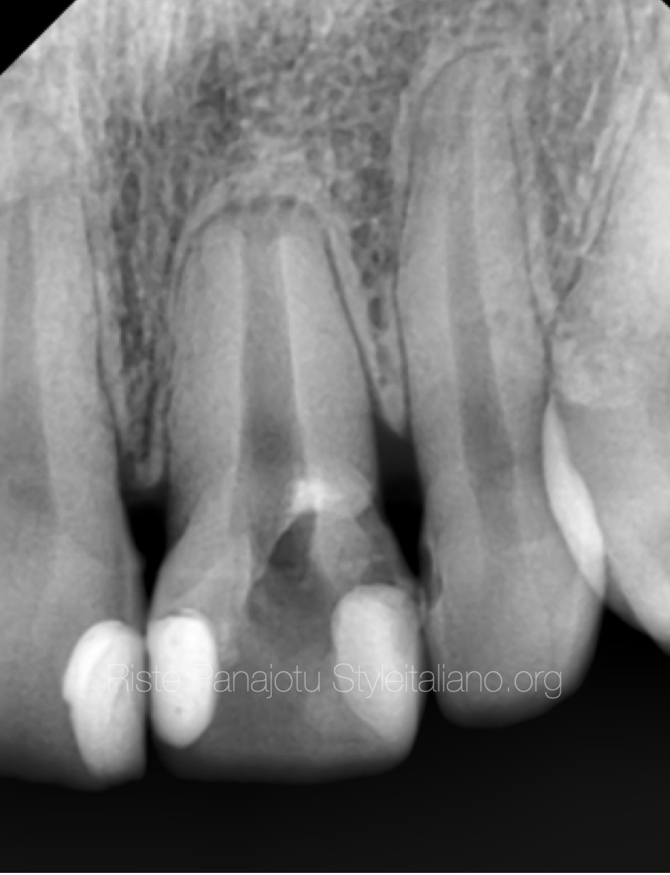
Fig. 4
An intraoperative x ray was taken to check for gutta-percha residuals
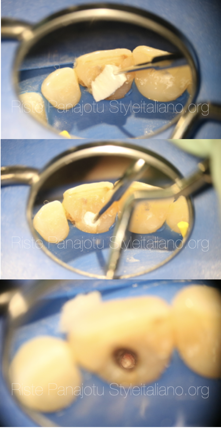
Fig. 5
After disinfection the apical collagen barrier was placed using a small size plugger with apex locator control
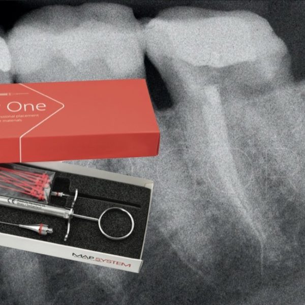
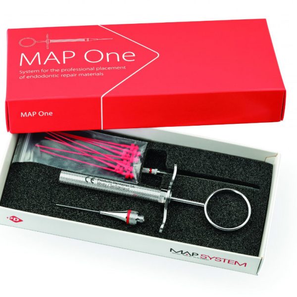
White mta properly mixed on the Ergo tool was then placed using the MAP system
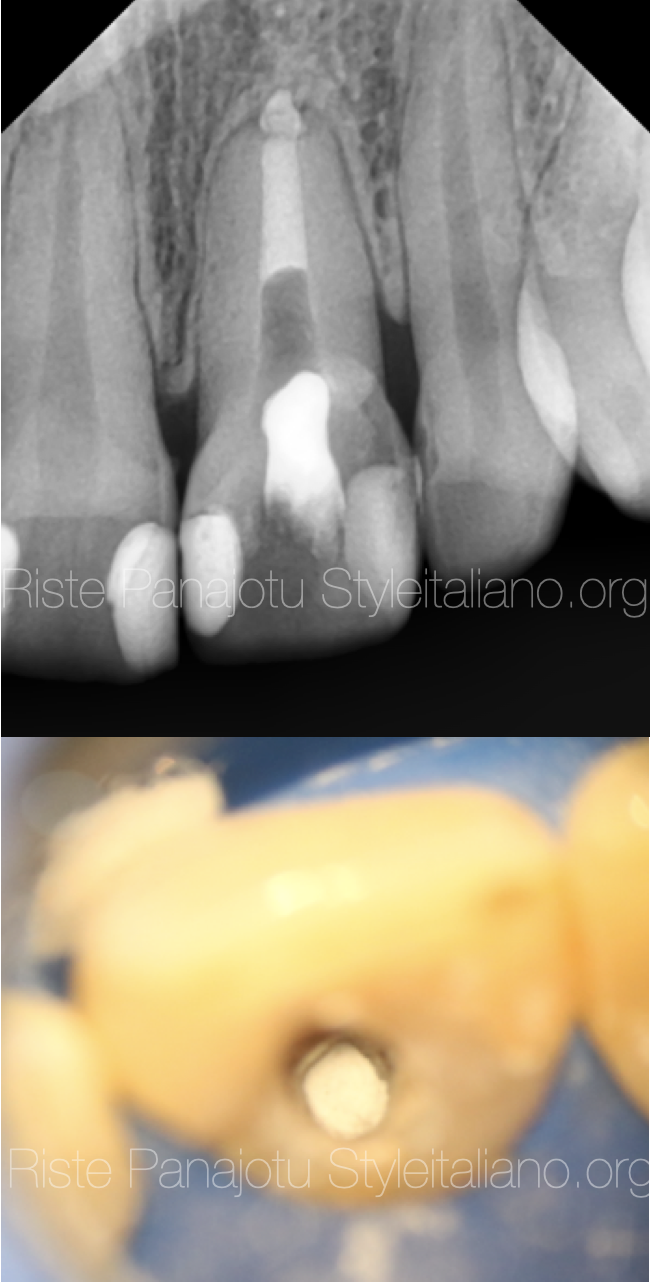
Fig. 6
The mta plug in place
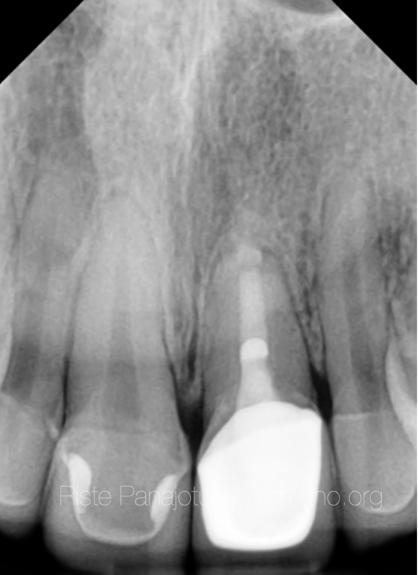
Fig. 7
6 months after the treatment
The tooth received a custom made post from fiber reinforced composite and a full crown
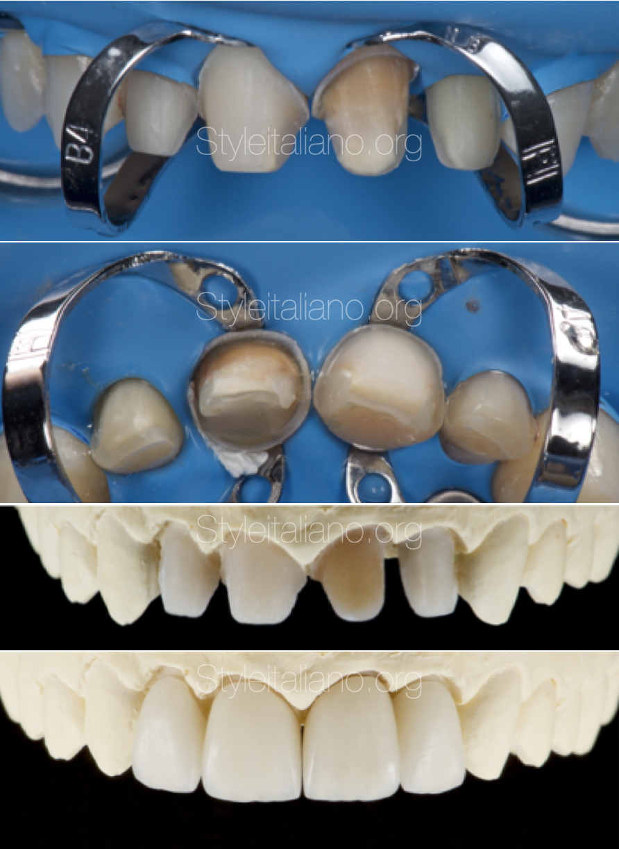
Fig. 8
Here are some photos from the prosthetic part dome by Dr.Aleksandar Trajanoski
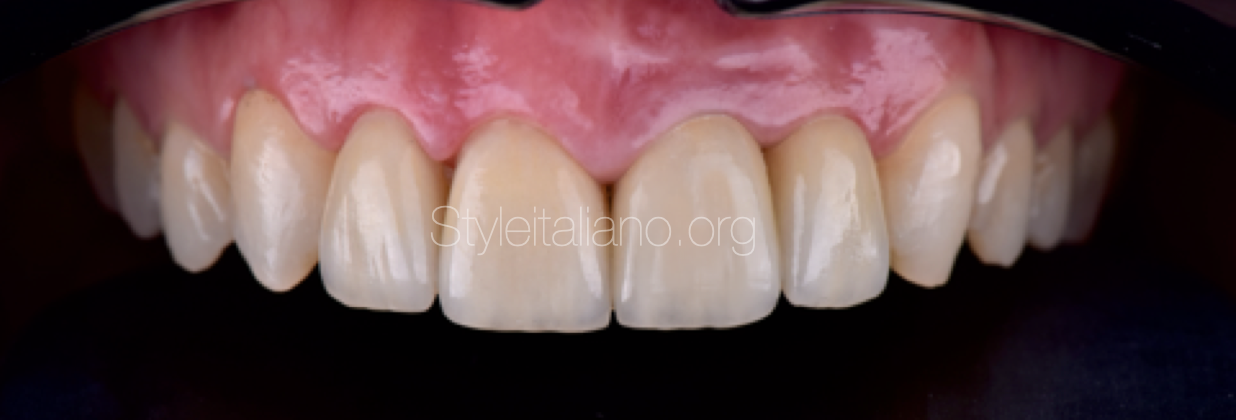
Fig. 9
The final result
Conclusions
Non surgical retreatment after surgical treatment sometimes can be a challenging task because of the large size of the apical terminus after root resection.
In cases where the time is important with proper equipment a well established treatment plan this type of treatment is possible without lowering the success rate.
Bibliography
Pace R, Giuliani V, Pini Prato L, Baccetti T, Pagavino G. Apical plug technique using mineral trioxide aggregate: results from a case series. Int Endod J. 2007;40(6):478-84.
Fridland M, Rosado R. Mineral trioxide aggregate (MTA) solubility and porosity with different water-to-powder ratios. J Endod. 2003;29(12):814-7.
Aminoshariae A, Hartwell GR, Moon PC. Placement of mineral trioxide aggregate using two different techniques. J Endod. 2003;29(10):679-82.
Hydration mechanisms of mineral trioxide aggregate Int. Endod. J., 40 (6) (2007), pp. 462-470