Magnification in Modern Endodontics
06/04/2023
Ahmed Shawky
Warning: Undefined variable $post in /var/www/vhosts/styleitaliano-endodontics.org/endodontics.styleitaliano.org/wp-content/plugins/oxygen/component-framework/components/classes/code-block.class.php(133) : eval()'d code on line 2
Warning: Attempt to read property "ID" on null in /var/www/vhosts/styleitaliano-endodontics.org/endodontics.styleitaliano.org/wp-content/plugins/oxygen/component-framework/components/classes/code-block.class.php(133) : eval()'d code on line 2
The introduction of the dental operating microscope and its ergonomics date back to mid to late 80s, being introduced by the famous endodontist Dr. Gary Carr.
Dr. Carr often quoted, “ YOU CAN NOT TREAT WHAT YOU CAN NOT SEE” reflecting the importance of augmented vision and illumination to impose precision to endodontic treatments.
This article will discuss how the use of magnification and coaxial illumination is considered an essentiality in modern endodontic treatments from the diagnostic procedures and decision making till the treatment finalization.
This article discuss some, and not all, of the benefits of the dental operating microscope.
1- Magnification in Diagnosis and decision making:
Restorative evaluation:
To make a decision whether to retain or remove a restoration, examination of the tooth/restoration interface can not be precisely done with naked eye vision.
In this video, a decision was taken to remove the restoration during endodontic retreatment due to the grossly defective margins as examined under high magnification.
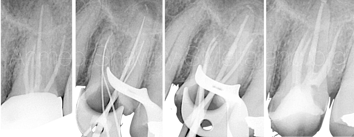
Fig. 1
Although it is an apparently good endo, the decision to do surgical retreatment was not taken. This is because restorative evaluation under magnification revealed compromised integrity of the existing restoration as well as CBCT scan data confirming a technically-deficient canal treatment
Fig. 2
2- Magnification in access preparations:
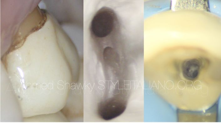
Calcified pulp chambers (pulp stones)
In the presence of long term, low-grade irritation (long-standing decay) the pulp may recede or undergo calcific metamorphosis.
Access cavity preparations in teeth with calcified pulp chambers (pulp stones) may lead to severe mutilation of the coronal tooth structure if committed without the use of magnification.
Image: Pretreatment radiograph of a maxillary first molar with huge carious lesion and massive reactive calcification in the pulp chamber and the root canals.
This video shows how working under magnification is safer for management of a calcified root canal system from access preparation to treatment finalization.
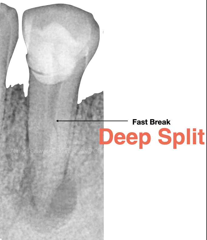
Fig. 3
3- Management of complex anatomy:
Deep splits represent an anatomical complexity which can impact the access cavity design, especially in teeth like mandibular premolars.
Using the dental operating microscope can allow the clinician to design an ASAP access cavity “As Small As Practical” in order to balance between the mechanical and biological treatment objectives
This video shows how pretreatment evaluation of anatomy, and execution of the access under high magnification provided a practical way for management of the splitting system without excessive removal of sound tooth structure.
Visibility is always enhanced by the use of ultrasonics in cavity refinement, providing unobstructed visual convenience during the procedure
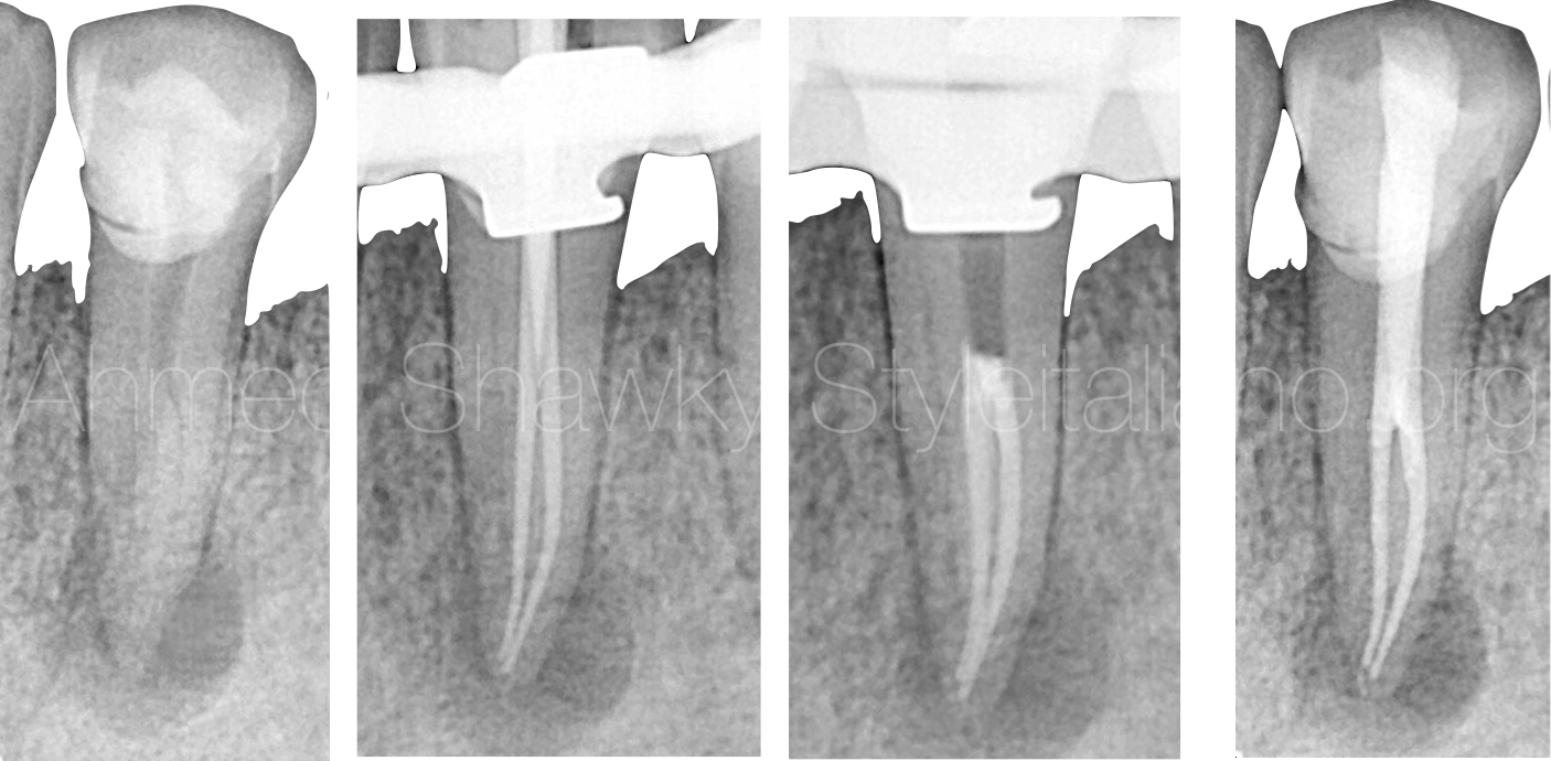
Fig. 4
Synergistic relationship exists between ultrasonics and magnification leading to a repeatable and predictable treatment approach
In This video, C-shaped anatomy represents disinfection challenge. Under magnification, it is possible for the clinician to expose the C-shape using medium power ultrasonics with intermittent cooling.
This will make the complex system accessible for the disinfection protocol and in the end, will be predictably sealed
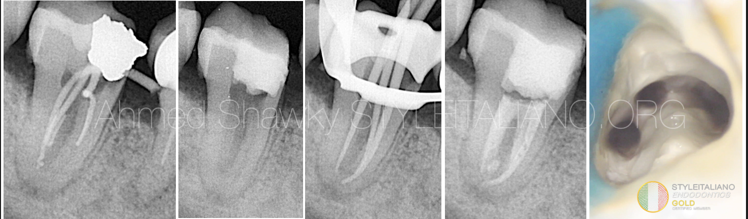
Fig. 5
The treatment of this C-shaped anatomy was performed over two visits. Obturation was done using warm vertical compaction and heat-friendly bioceramic sealer
4- Non-Surgical Retreatments:
Removal of retention elements:
In this video, you can see how removal of a metallic screw post is easy under high magnification, with selective cutting to conserve as much coronal structure as possible
In cases having fiber posts, the situation is more complex due to the presence of the resin inside the root canal
Sometimes it is difficult for the clinician to differentiate the boundaries of the fiber post without magnification and powerful illumination
The use of magnification can avoid transportations, perforations or excessive removal of dentin during fiber post disassembly
This video (Video 6) shows the steps of fiber post disassembly under magnification
Locating Missed canals:
Locating hidden canals is more predictable and safe when the procedure is performed under magnification
One of the clues for locating hidden canals, is the observation of color differences in the pulp chamber (e.g. white line test for locating missed MB2s)
This requires adequate and clear vision to make the procedure quick and safe
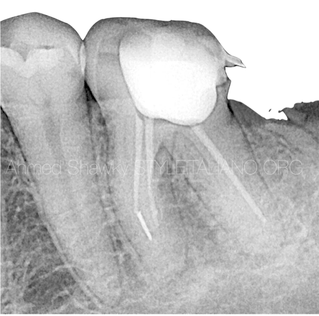
Fig. 6
Conservative retrieval of Separated instruments
During the procedure of separated instrument retrieval, care should be taken not to remove a lot of dentin.
Ultrasonic preparation beside broken files requires enhanced vision for precise and minimally invasive preps leading to successful retrieval.
Also, the use of grasping tools such as; loop techniques, requires visual control for successful placement of the grasping wire around the fragment (Video)
Instrument removal with loop technique
Isthmus Cleaning in retreatments
In the same case, inspection of the plural floor under magnification revealed the presence of a deep isthmus between the MB and the ML root canals
troughing of the isthmus was done using ultrasonics, followed by using thinner ultrasonic tip to remove the biofilm residing in that isthmus
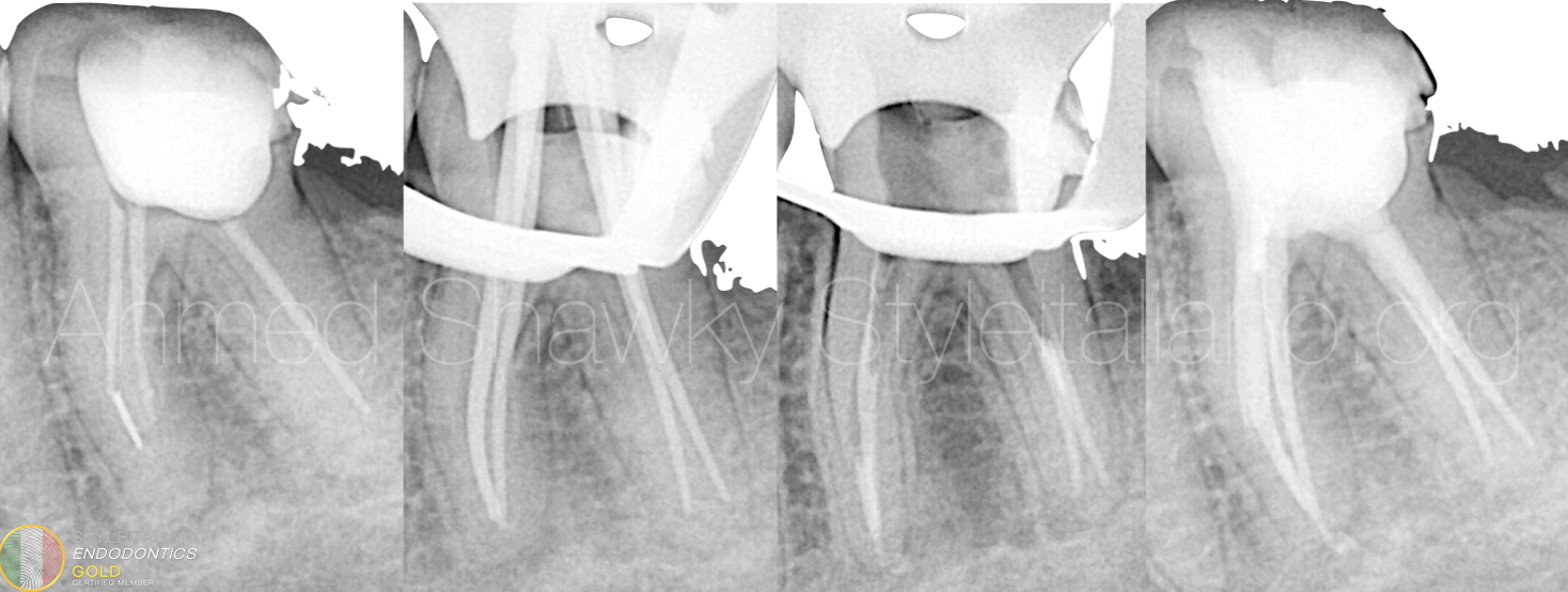
Fig. 7
With the help of magnification, successful retrieval of the fragment occurred, all missed anatomies treated and the root canal system was sealed tridimensionally
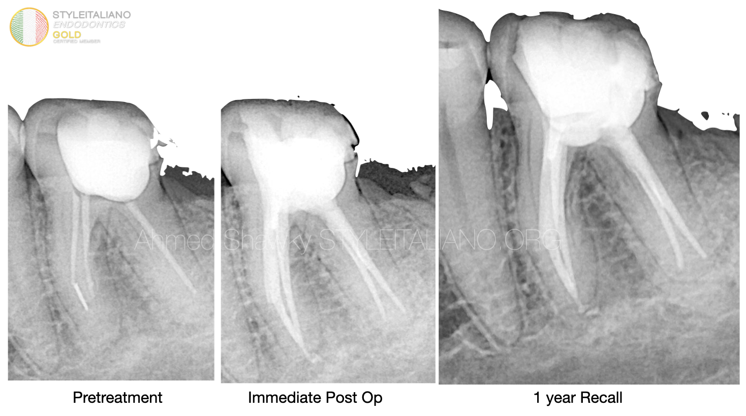
Fig. 8
Pre treatment, immediate post op and 1 year follow up X-rays
5- Apical plug placement:
The use of magnification allows precise and predictable placement of apical plugs in teeth with apical foramina larger Than #55 ISO size.
In this video non surgical retreatment was initiated across an existing fixed restorations (according to the criteria of Abusteit et al 2021). The procedure was performed on two visits and the obturation involved apical plug followed by backfilling of thermoplasticized GP injection
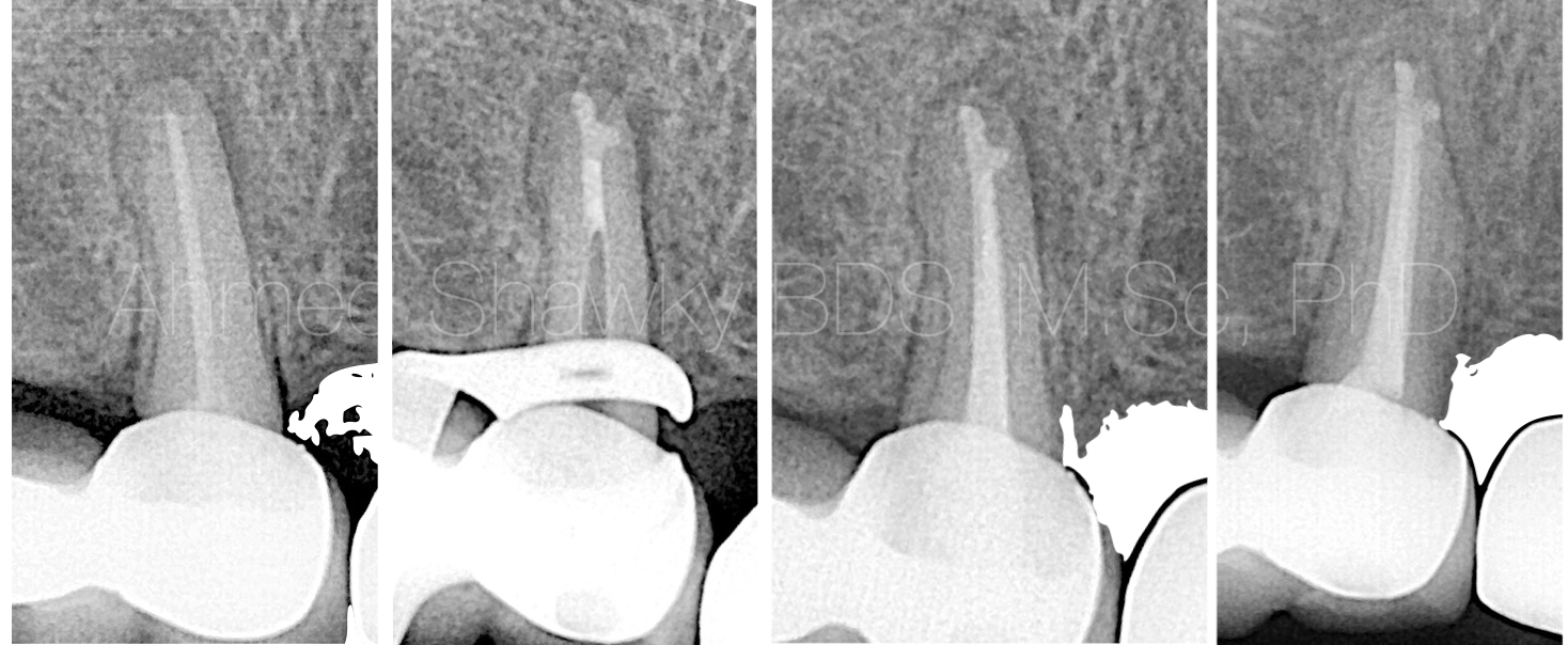
Fig. 9
One year recall shows complete resolution of apical periodontitis
A major role in the precision and predictability of apical plugs placement is played by the use of the dental operating microscope
Conclusions
- The use of the dental operating microscope is nowadays established as an important corner stone in Modern Endodontics
- There is a synergistic relationship between the use of magnification and clinical application of ultrasonics in endodontics
- The clinician must see well, in order to treat well.
Bibliography
1- Paul Krasner and Henry J. Rankow, Anatomy of the Pulp-Chamber Floor, Journal of Endodontics, VOL. 30 )1( 2004
2- Silva EJNL, Pinto KP, Ferreira CM, Belladonna FG, De-Deus G, Dummer PMH, Versiani MA. Current status on minimal access cavity preparations: a critical analysis and a proposal for a universal nomenclature. International Endodontic Journal. doi:10.1111/iej.13391
3- Outcome of Endodontic Treatment through Existing Full Coverage Restorations: An Endodontic Practice Case Series. https://doi.org/10.1016/j.joen.2021.11.008