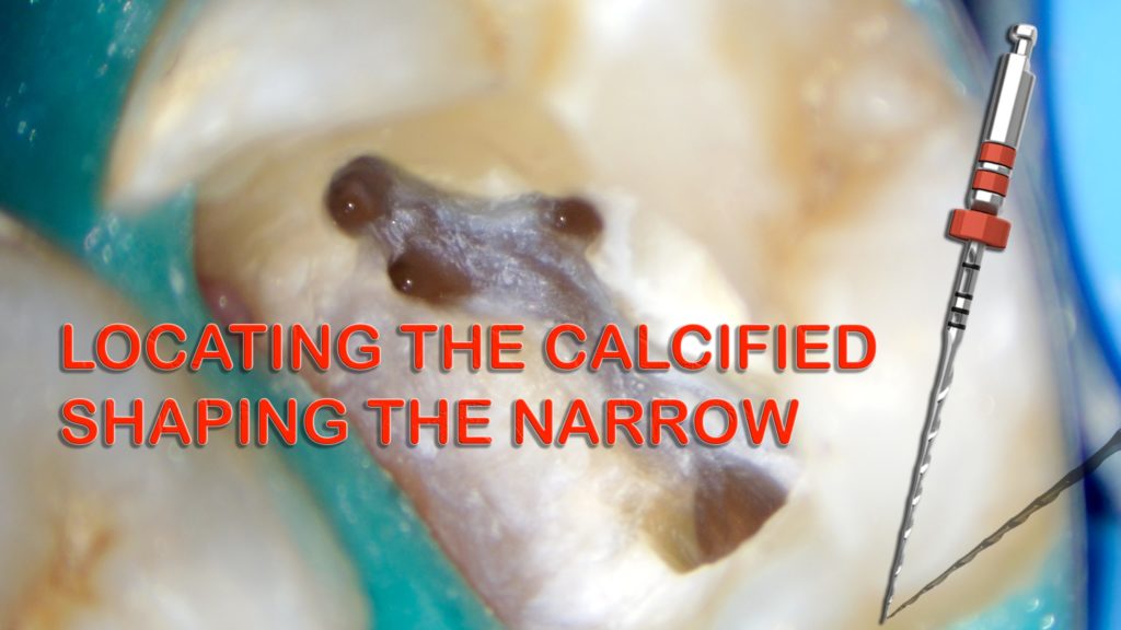
Locating the calcified - Shaping the narrow
17/03/2022
Mohamad Zaafrany
Warning: Undefined variable $post in /var/www/vhosts/styleitaliano-endodontics.org/endodontics.styleitaliano.org/wp-content/plugins/oxygen/component-framework/components/classes/code-block.class.php(133) : eval()'d code on line 2
Warning: Attempt to read property "ID" on null in /var/www/vhosts/styleitaliano-endodontics.org/endodontics.styleitaliano.org/wp-content/plugins/oxygen/component-framework/components/classes/code-block.class.php(133) : eval()'d code on line 2
Narrow canal is a great challenge to Endodontist, When combined with calcified entrance the difficulty doubled.
Preoperative assessment to the periapical radiograph would help in avoiding many errors that could happen if case is underestimated.
In this article we’ll describe how we overcome the challenge using the correct technique and the right tools.
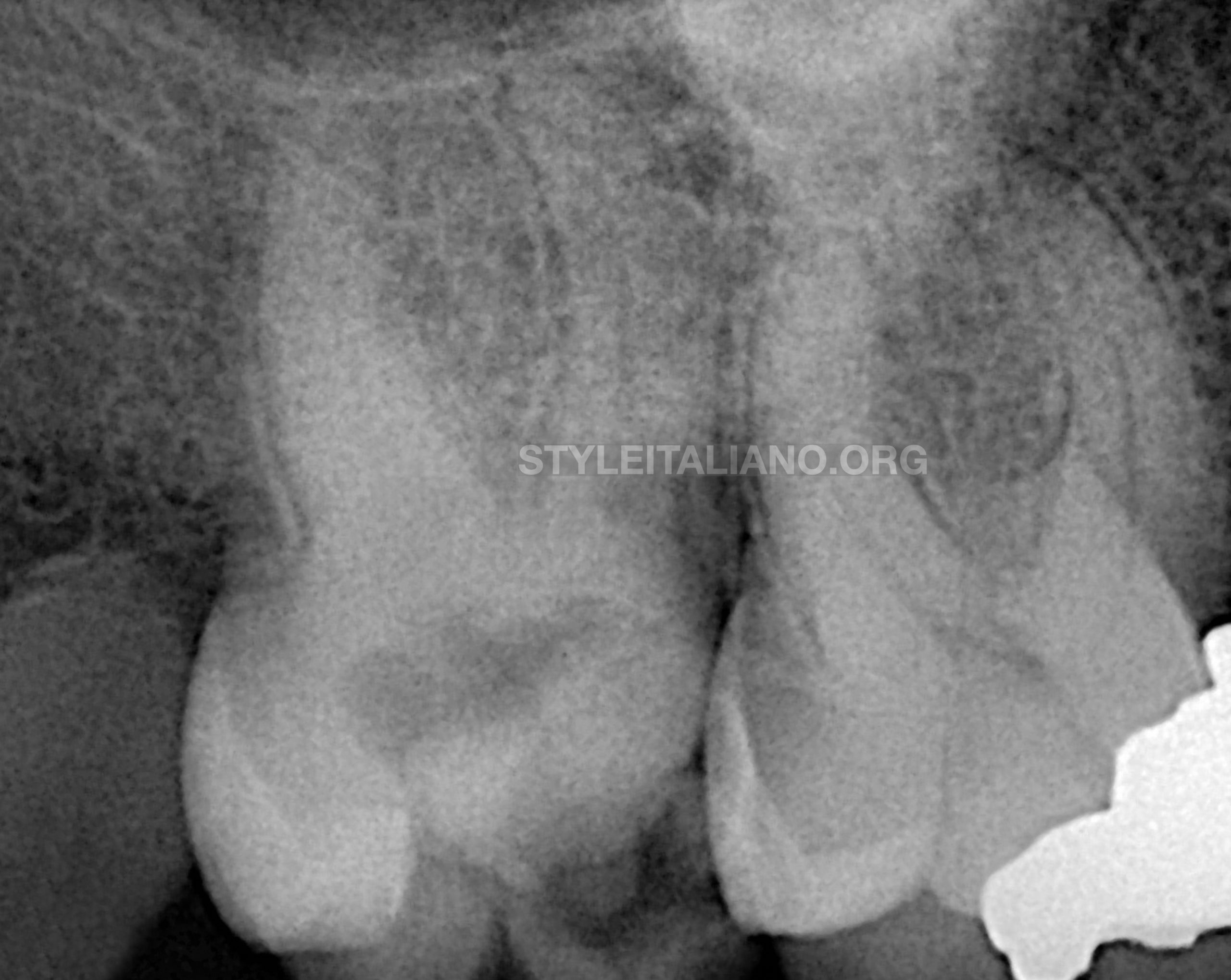
Fig. 1
Preoperative radiographic analysis
Referred case of tooth no. 17 with previously initiated therapy.
Inability of locating the orifices of the canals was the reason of referral.
From preoperative radiograph it was noticeable the iatrogenic overextension of the access cavity distally as a trial to locate entrance to the pulp space.
It was evident also the possibility of dealing with narrow canals specially with the absence of canals shade in the radiograph.
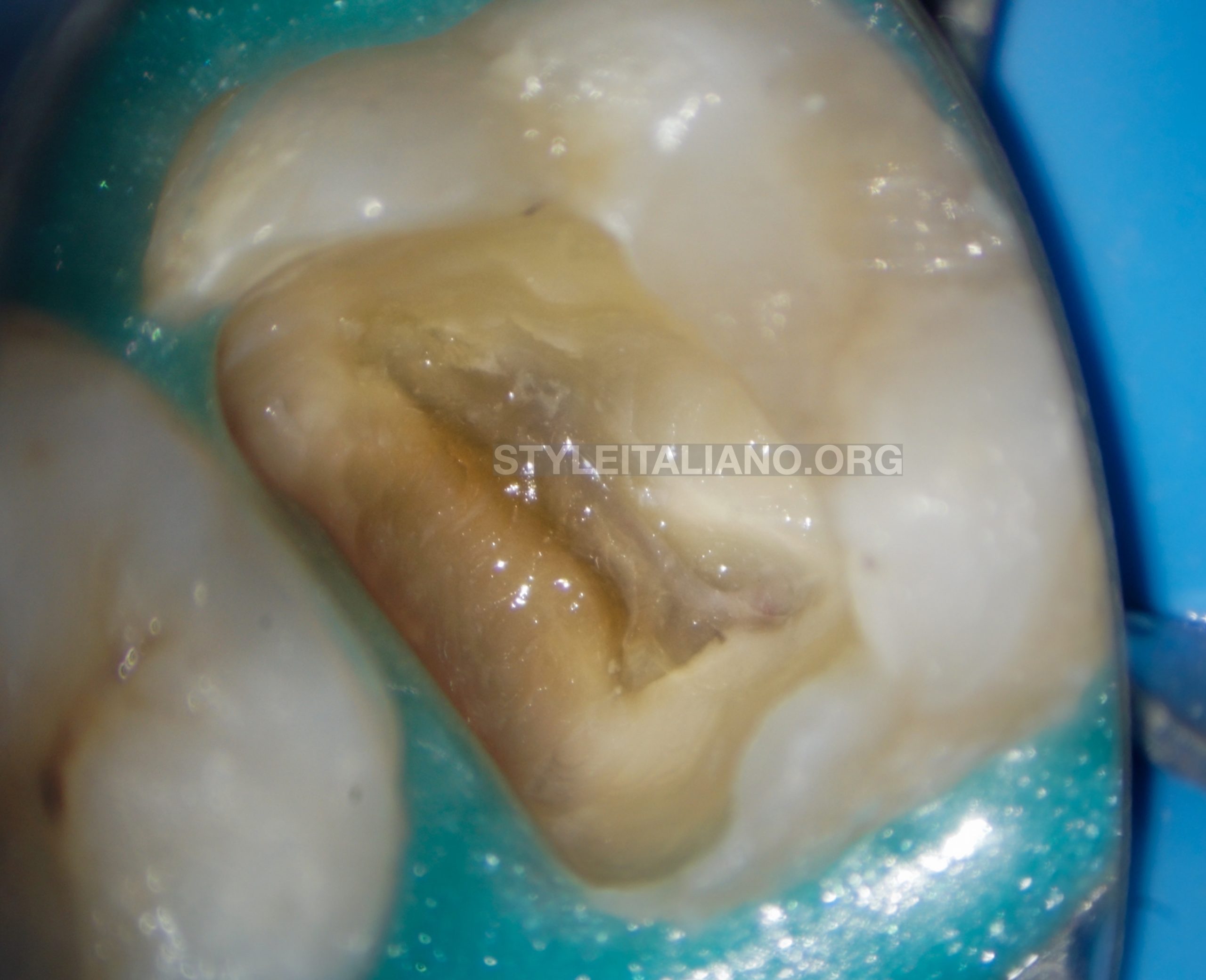
Fig. 2
After temporary filling removal. It was noticeable the calcification of the pulp tissue in the pulp chamber.
With the presence of color difference and in the pulp chamber, just under magnification and with the use of ultrasonic tips on the calcified tissue, locating the canals would be easy.
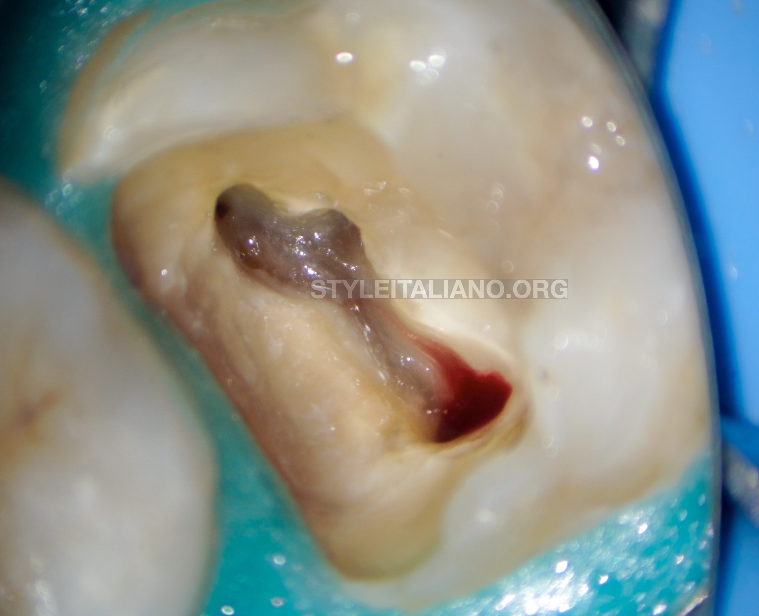
Fig. 3
After ultrasonic removal to the calcified tissue in the pulp chamber canals entrances located. But they were very narrow.
Note the conservative relocation of the floor of pulp chamber, Thanks to magnification and ultrasonics.
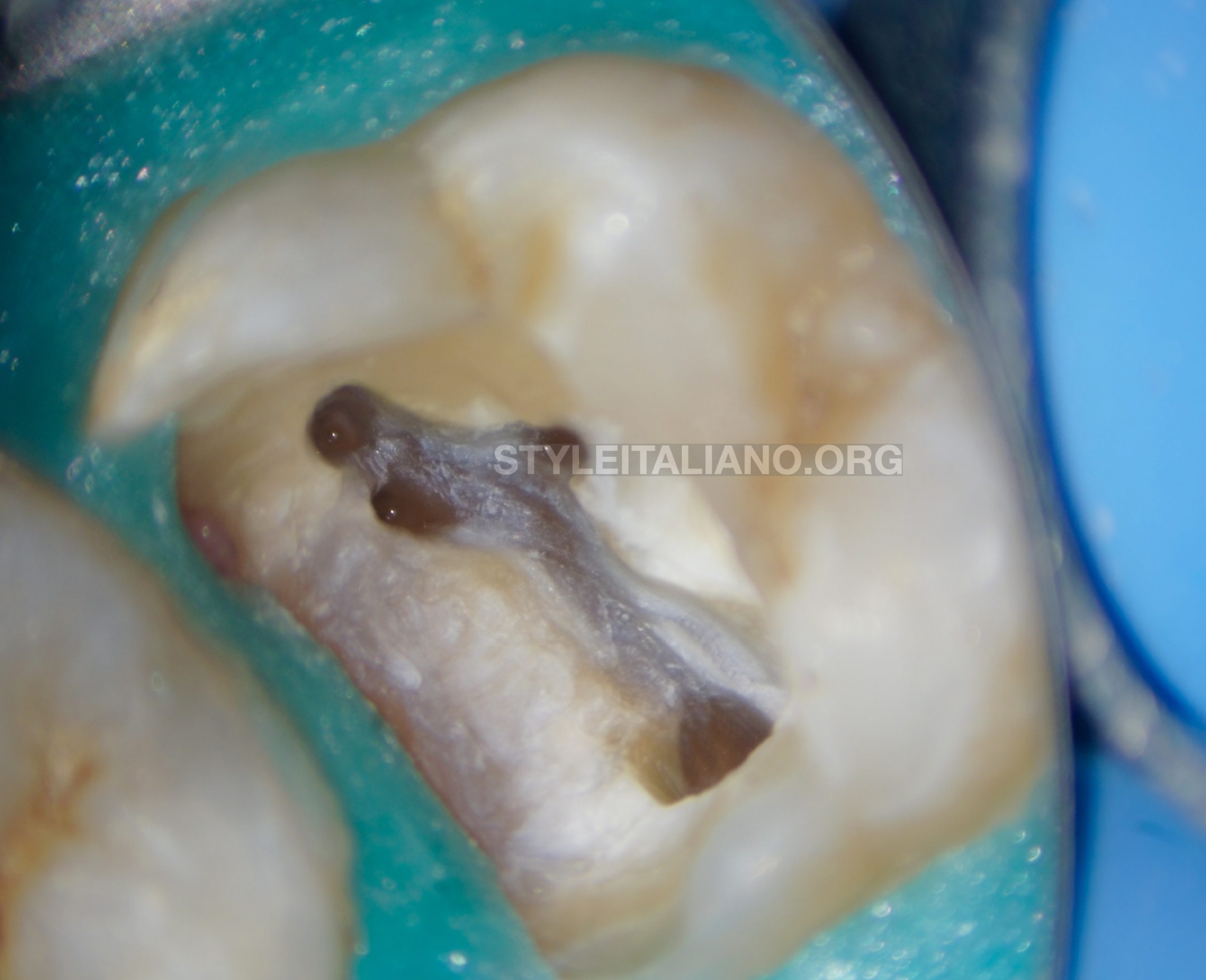
Fig. 4
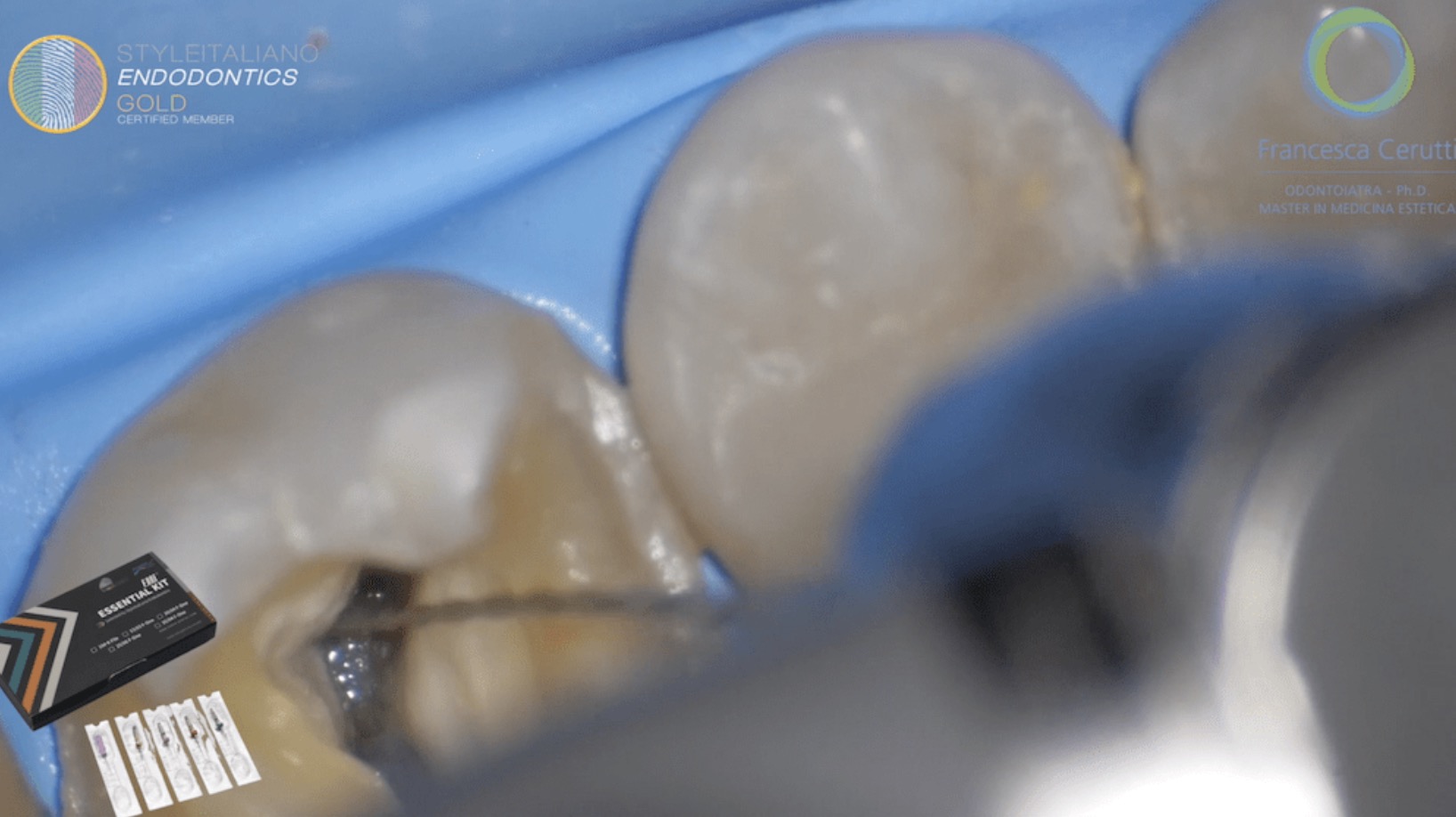
Using Fanta AF 13 - 03 followed by F1 25 - 06 for preflaring the canals only in the coronal portion till resistance, canals were sufficiently enlarged coronally for safer manual glide path before going to mechanical instrumentation to the canals.
Manual glide path done using, 8 and 10 K files till the WL in a watch wind motion.
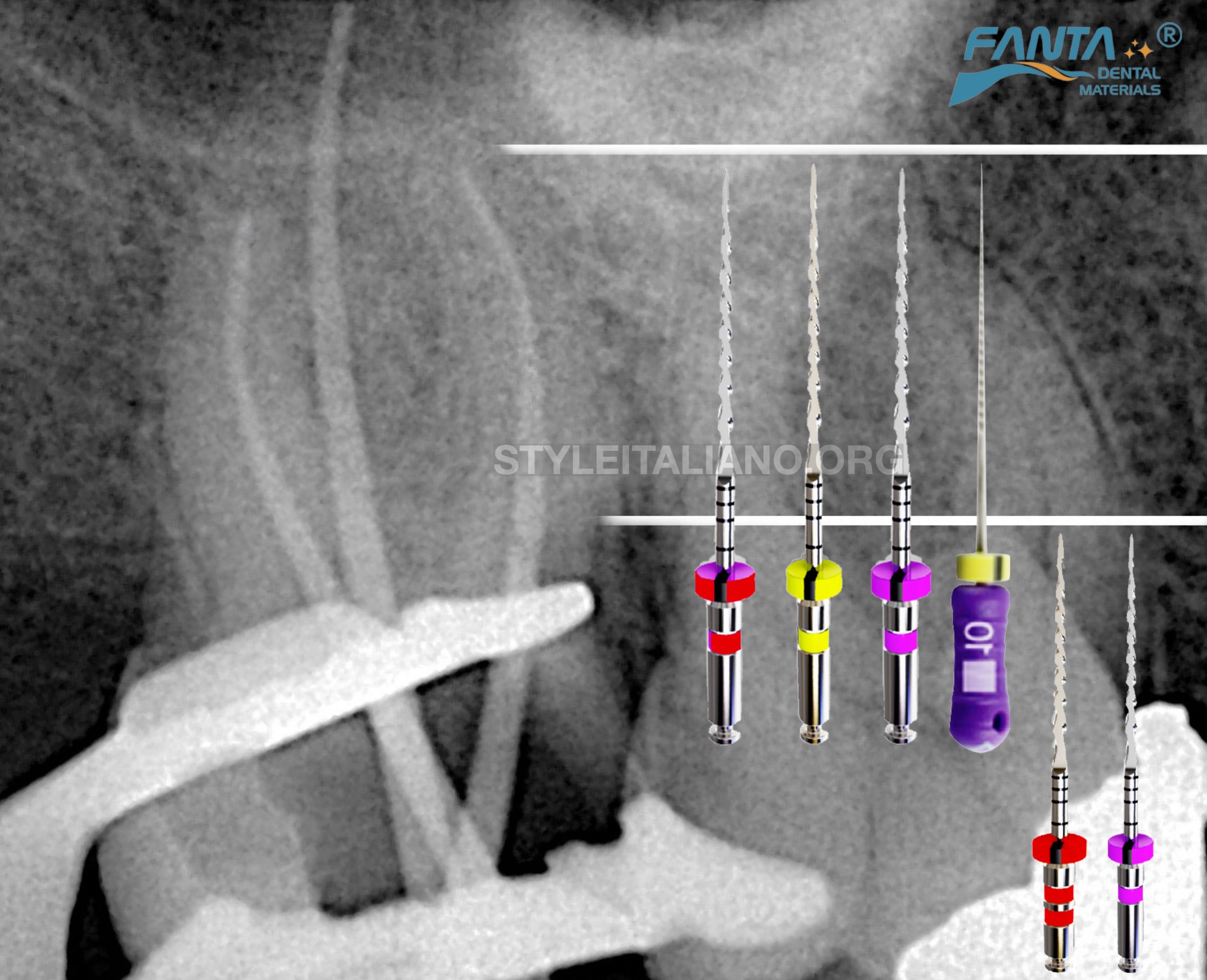
Fig. 5
After manual glide path, Mechanical glide path done using 13 - 03 to full length, followed by 20 - 04 and apical finishing in the buccal canals using 25 - 04.
Apical finishing in the palatial canal up to Fanta 35 - 04 AF F1.
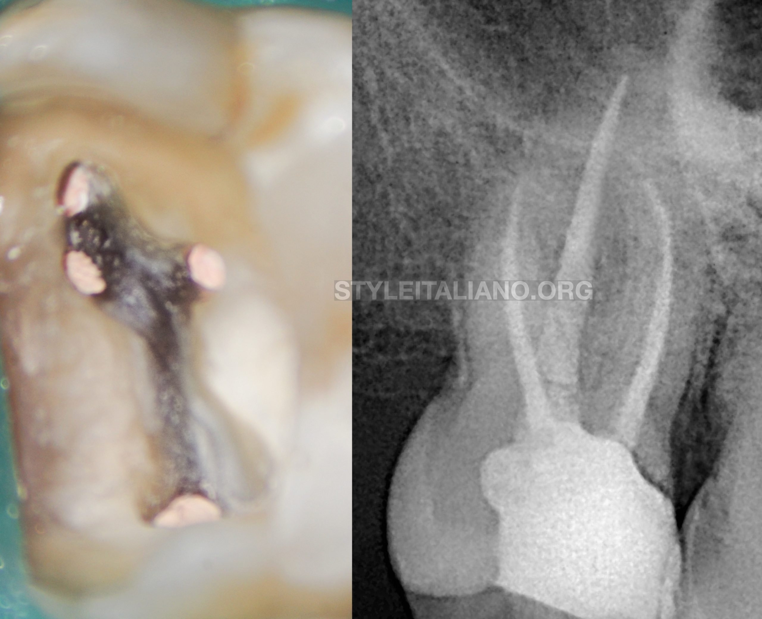
Fig. 6
Final radiograph with obturation done in using Single cone and BC sealer in buccal canals and modified hot technique in the palatal canal.
Conclusions
- Preoperative radiograph is very important before starting endodontic treatment
- Case difficulty assessment will lead to successful treatment through good planning and selection of the approach technique and sequence of shaping files.
- Planning the sequence of shaping files is important to avoid errors during shaping phase.
- Fanta AF F1 files is considered safe shaping of very narrow canals, thanks to the hybrid heat treatment and flat surface design
- The use of magnification and ultrasonic is considered a safe technique to locate calcified canal entrances.
Bibliography
Krasner P, Rankow HJ. Anatomy of the pulp chamber floor. JOE 2004; 30(1):5.
Cohen S, Hargreaves K. Pathways of the Pulp 9th ed. Mosby, St. Louis, MO, 2006.
Vertucci, F. J., & Williams, R. G. (1974). Furcation canals in the human mandibular first molar. Oral Surgery, Oral Medicine, Oral Pathology, 38(2), 308–314. doi:10.1016/0030-4220(74)90073-5
Louis J Buhrley 1 , Michael J Barrows, Ellen A BeGole, Christopher S Wenckus Effect of magnification on locating the MB2 canal in maxillary molars.
JOE 2002. doi: 10.1097/00004770-200204000-00016.
Hot modified technique with a new biosealer. Drs Alfredo Iandolo, Massimo Calapaj & Dina Abdellatif, Italy