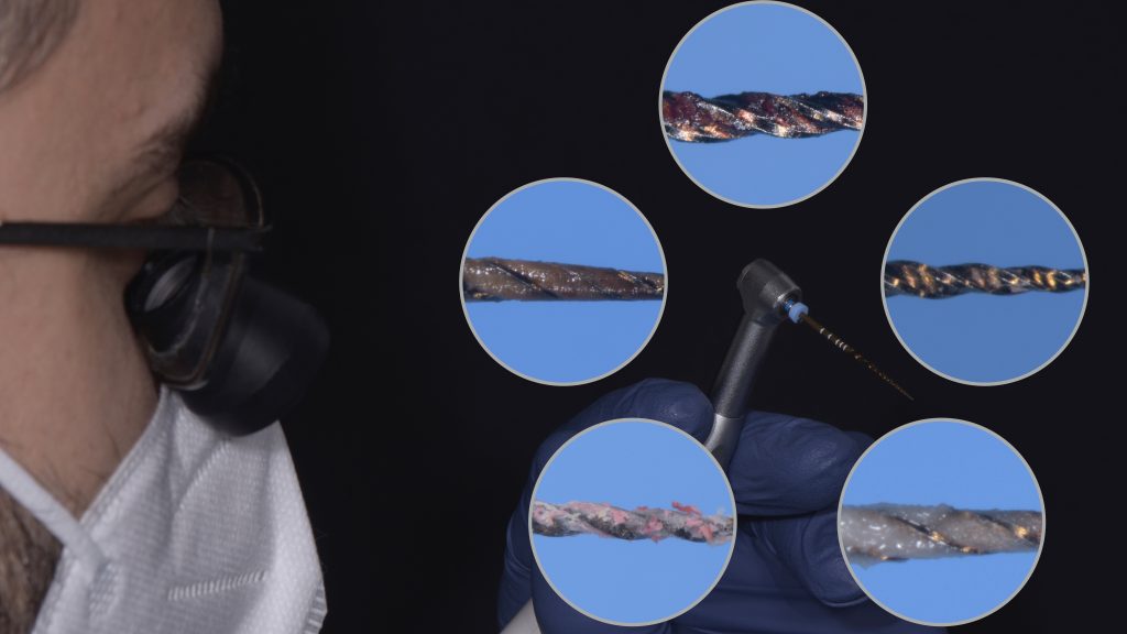
How to evaluate the debris on flutes
28/04/2021
Calogero Bugea
Warning: Undefined variable $post in /var/www/vhosts/styleitaliano-endodontics.org/endodontics.styleitaliano.org/wp-content/plugins/oxygen/component-framework/components/classes/code-block.class.php(133) : eval()'d code on line 2
Warning: Attempt to read property "ID" on null in /var/www/vhosts/styleitaliano-endodontics.org/endodontics.styleitaliano.org/wp-content/plugins/oxygen/component-framework/components/classes/code-block.class.php(133) : eval()'d code on line 2
The primary objective of the entire root canal treatment procedure is to eliminate microorganisms and pathogenic debris from the root canal system and to prevent its reinfection: mechanical instrumentation accompanied by irrigation could be considered as the most essential component that aids in reaching this goal.
To understand if the instrumentation of the endodontic space has been carried out in the right way it is mandatory to check the debris on the instrument flutes.
Debris fill the flutes of the instrument. An accurate analysis of the flutes may help the endodontist in deciding about the eternal dilemma: to stop the shaping or continue the shaping ?
During the shaping we can face different situations. An analysis of the flutes determines the choice of the endodontist that, obviously, has to be confirmed in the following phases of cone fit or paper point placement.
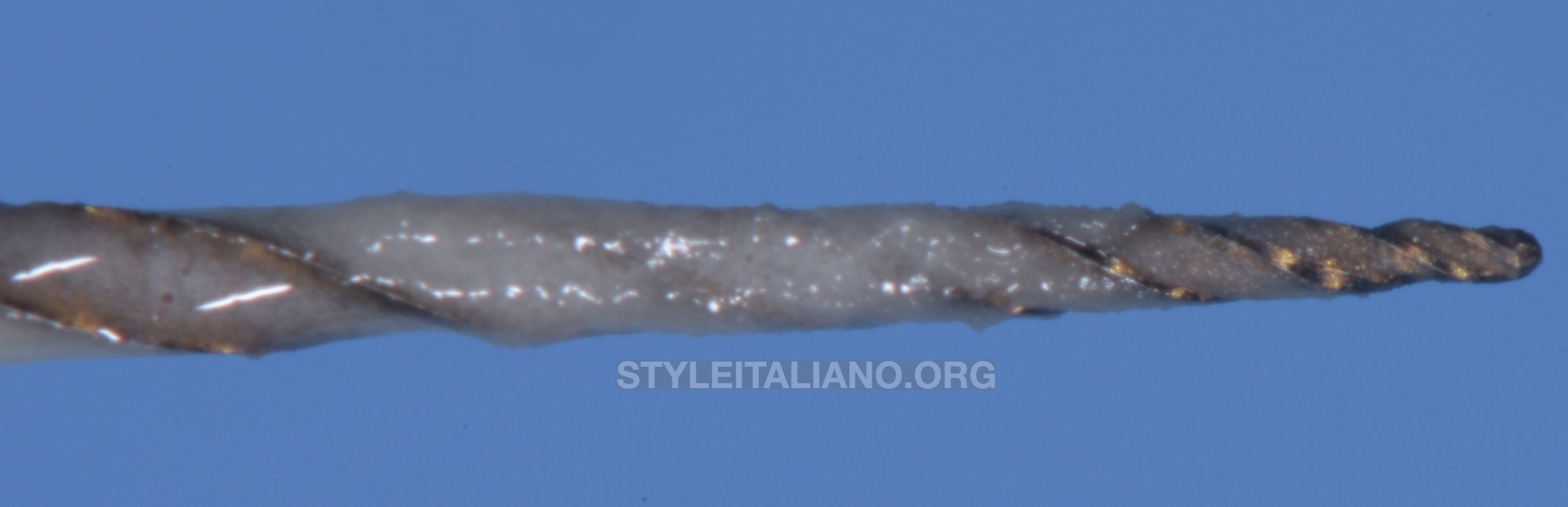
Fig. 1
White flutes:
Ideal preparation, the flutes are fill of "white" debris. This means that the instrument touched an wide area of the canal and the dentine is sound.
This is the ideal condition. In general we can stop the preparation because the instrument touches a high percentage of sound dentine.
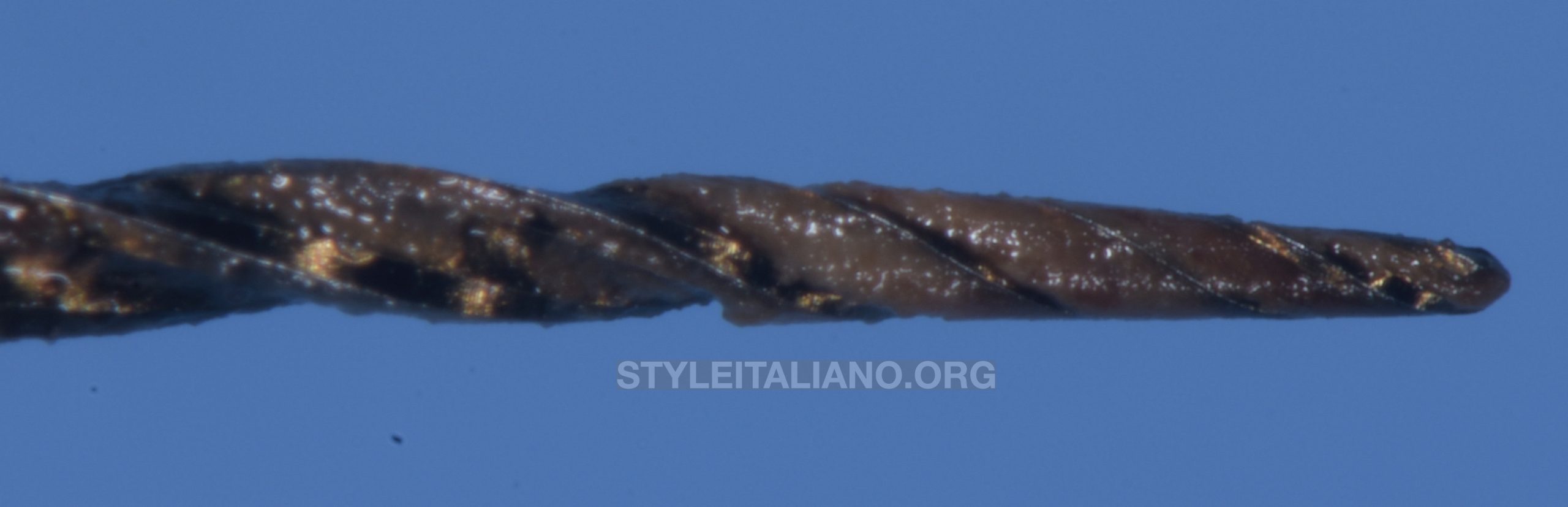
Fig. 2
Black flutes:
Even if the instrument is full of debris, the color means that the instrument touches only dirty dentin or residual endodontic pastes like endomethasone, thus the apical dentin is probably infected.
In this case it is advised to enlarge the taper until sound dentine can be seen on the instrument. This is mandatory for achieving the success.
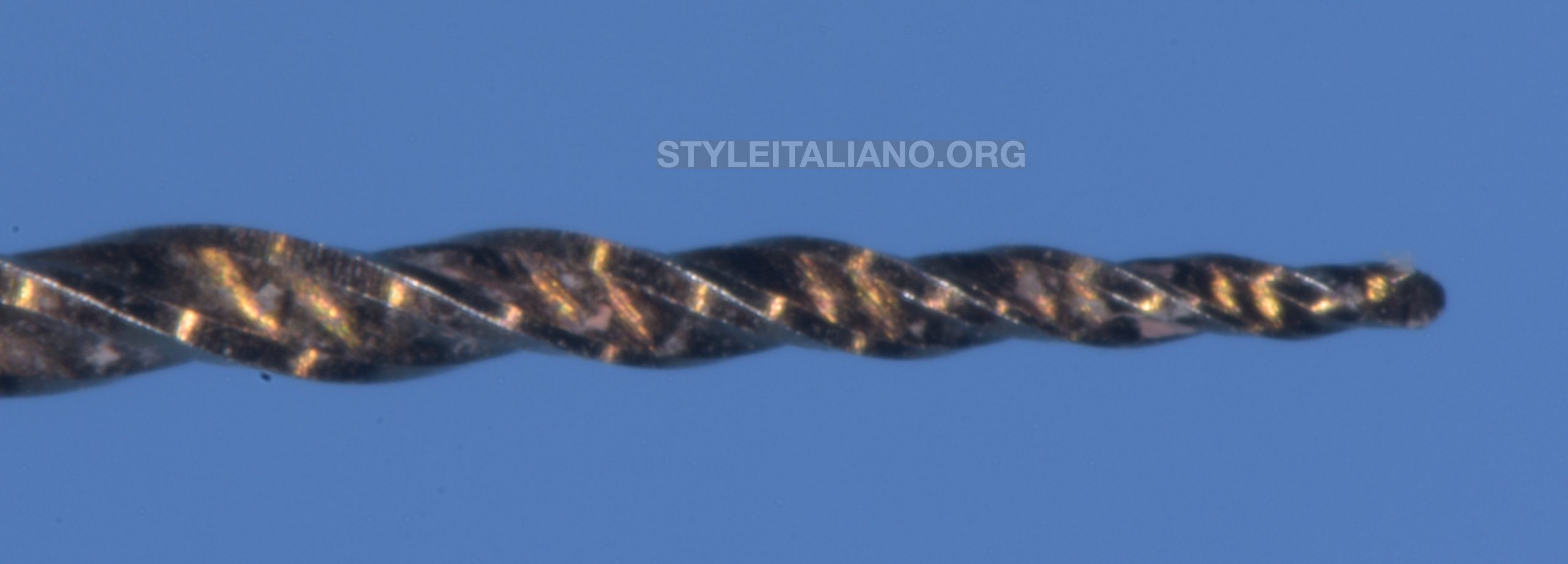
Fig. 3
Visible Flutes:
This situation happens when the canal is much larger than the instrument.
In this case, if it is possible, it necessary to increase taper and diameter of the endodontic instrument.
In case of wide canals, even a big instrument doesn't keep contact whit the dentine and it is impossible to appreciate the quality of the dentine. In this case copious irrigation has to be done, preferably using ultrasonic activation of the irrigants, in order to remove the debris from the root canal walls.
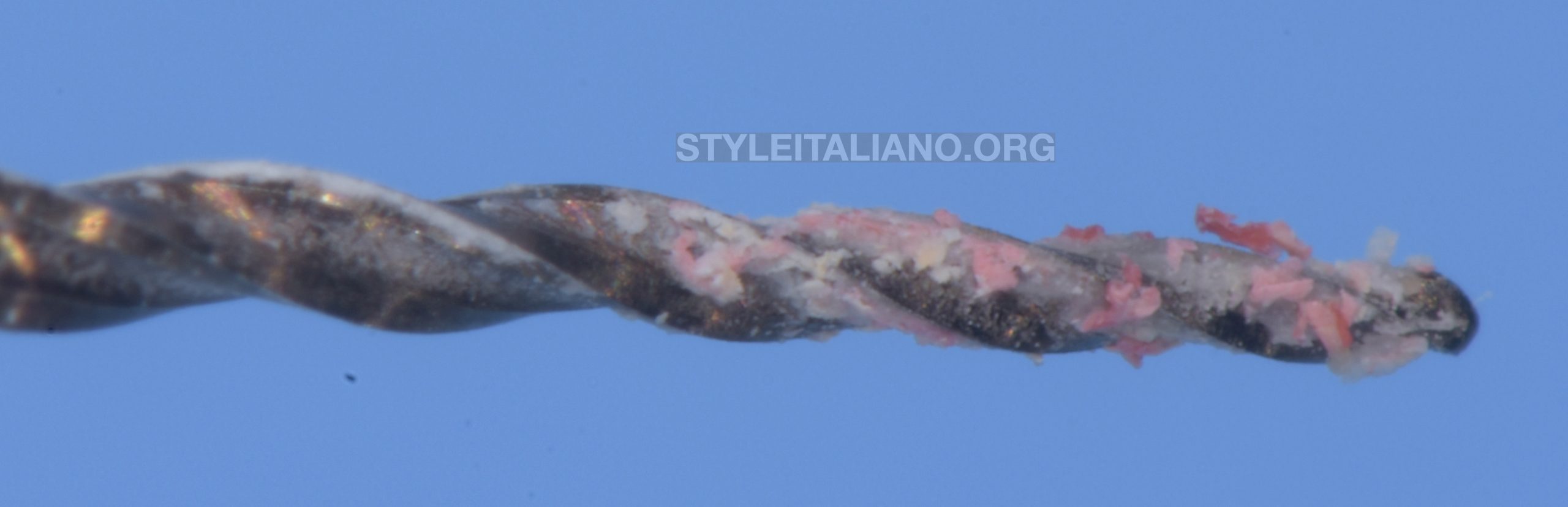
Fig. 4
Pink flutes:
This happens in case of retreatments and it means that in the apical area the walls are still filled with gutta-percha.
Ultrasonic tips help the clinicians in these case to clean the area from the gutta-percha remnants.
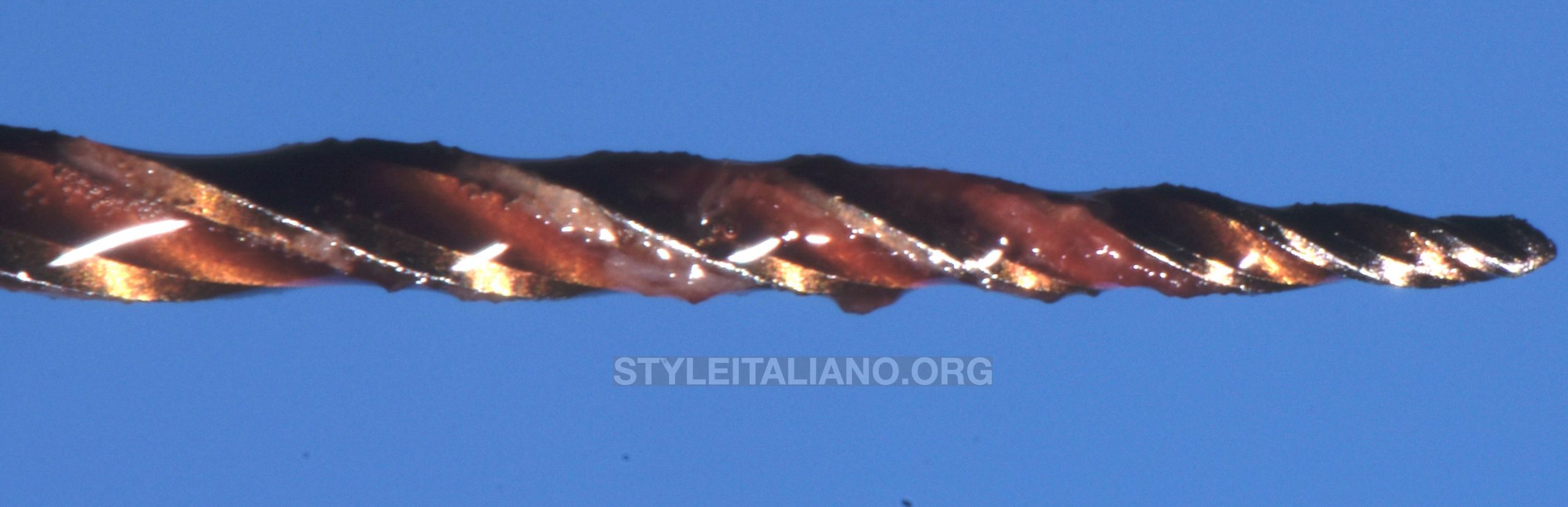
Fig. 5
Red flutes:
The presence of bloody flutes indicates the presence of pulp tissue in the apical area.
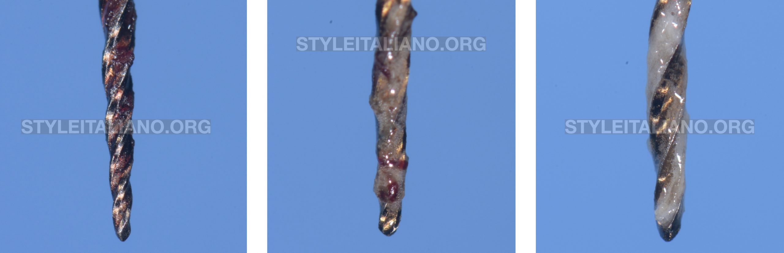
Fig. 6
Clinical example:
We have to increase the taper or the diameter preparation or both to eliminate the pulp tissue. In the picture, the shaping was conducted from 25.08 (full of blood); followed by 30.09 (partially full of blood); 40.06 (white debris).
In some cases, expecially with particulary infammed pulp, the presence of blood can origine from the periapical tissue. In these cases it is important to understand where the blood comes from, because in the last case increasing the diameter and taper might play a negative role in the fracture resistance of the tooth.
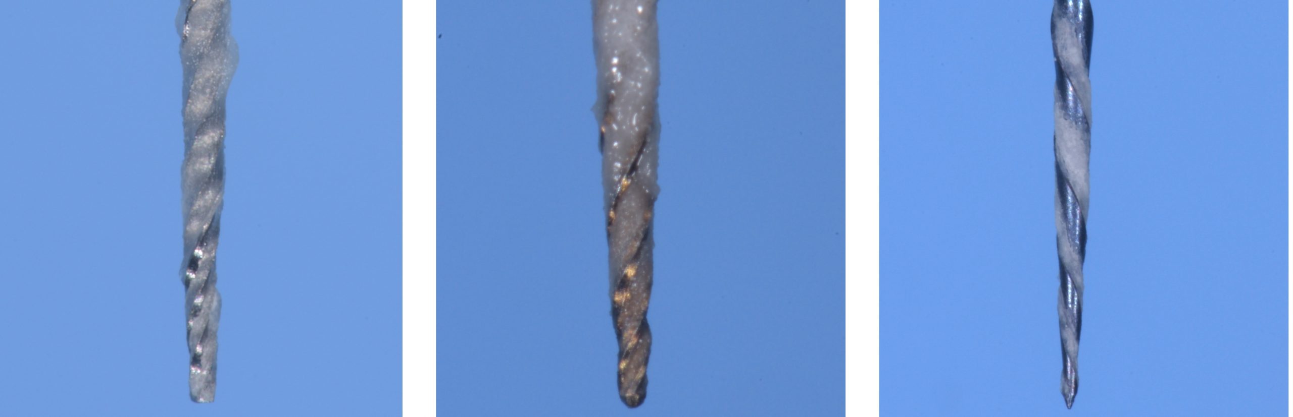
Fig. 7
Different flutes or different instruments may facilitate the vision of these debris.
Conclusions
The cause of the endodontic failure is the permanence of bacteria in the canals. Cleaning and shaping procedures act in synergy to eliminate bacteria.
The comprehension of the anatomy of the lasts millimiters of the endodontic space plays an important role for the success of the therapy.
Bibliography
T Mizutani , N Ohno, H Nakamura Anatomical study of the root apex in the maxillary anterior teeth J Endod 1992 Jul;18(7):344-7.
J Martos , C M Ferrer-Luque, M P González-Rodríguez, L A S Castro Topographical evaluation of the major apical foramen in permanent human teeth Int Endod J 2009 Apr;42(4):329-34.
Ariane Cassia Salustiano Marinho , Frederico Canato Martinho, Alexandre Augusto Zaia, Caio Cezar Randi Ferraz, Brenda Paula Figueiredo de Almeida Gomes. Influence of the apical enlargement size on the endotoxin level reduction of dental root canals Appl Oral Sci Nov-Dec 2012;20(6):661-6
A A Azim , J A Griggs , G T-J Huang The Tennessee study: factors affecting treatment outcome and healing time following nonsurgical root canal treatment Int Endod J 2016 Jan;49(1):6-16.
Dent Clin North Am. 2004 Jan;48(1):323-35