Endodontic surgery of an inferior central incisor
02/03/2023
Gabriele Ragucci
Warning: Undefined variable $post in /var/www/vhosts/styleitaliano-endodontics.org/endodontics.styleitaliano.org/wp-content/plugins/oxygen/component-framework/components/classes/code-block.class.php(133) : eval()'d code on line 2
Warning: Attempt to read property "ID" on null in /var/www/vhosts/styleitaliano-endodontics.org/endodontics.styleitaliano.org/wp-content/plugins/oxygen/component-framework/components/classes/code-block.class.php(133) : eval()'d code on line 2
Apical surgery is an option for the management of endodontically-treated tooth with persistent periapical lesions or symptom/sign.
Several epidemiological studies have suggested that 33–60% of endodontically-treated teeth still presented the pictures of apical periodontitis.
The possible causes may be persistent primary infection, secondary infection after endodontic therapy, vertical root fracture or cemental tears. The success rate of apical surgery was reported to range from 37% to 91%. Complete apical healing had been observed in 37%–96% of the endodontically-treated teeth after apical surgery.
The wide range and inconsistency of the results may be attributed to the variation in treatment planning, surgical technique, methodology, and follow-up period
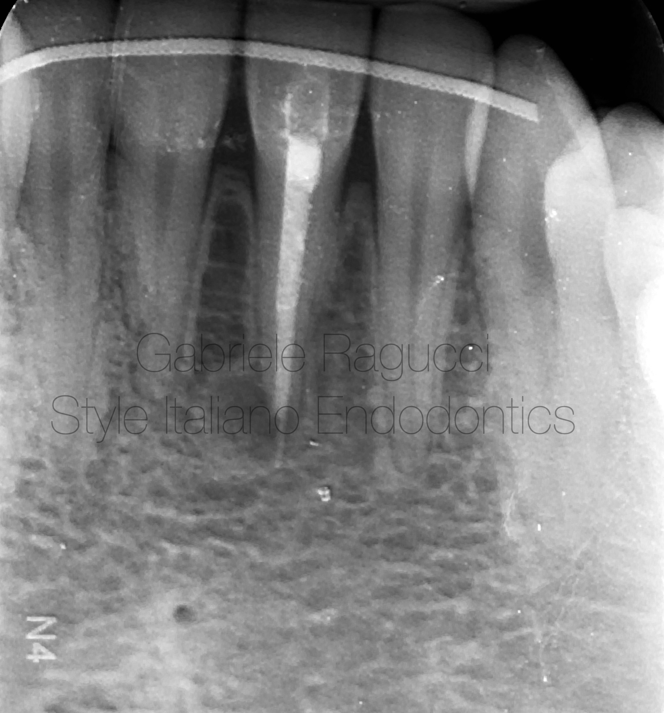
Fig. 1
Preoperative X-ray
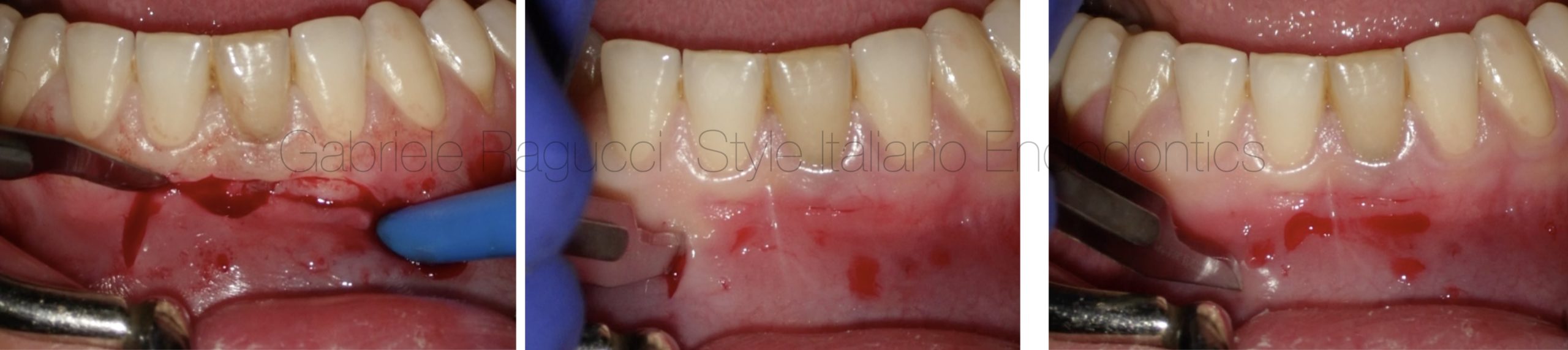
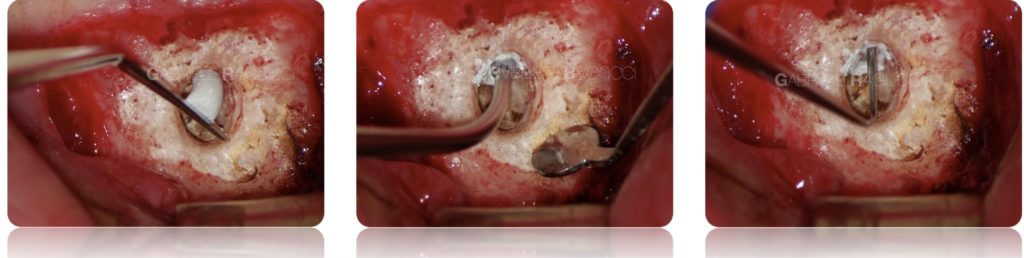
Fig. 2
Flap steps
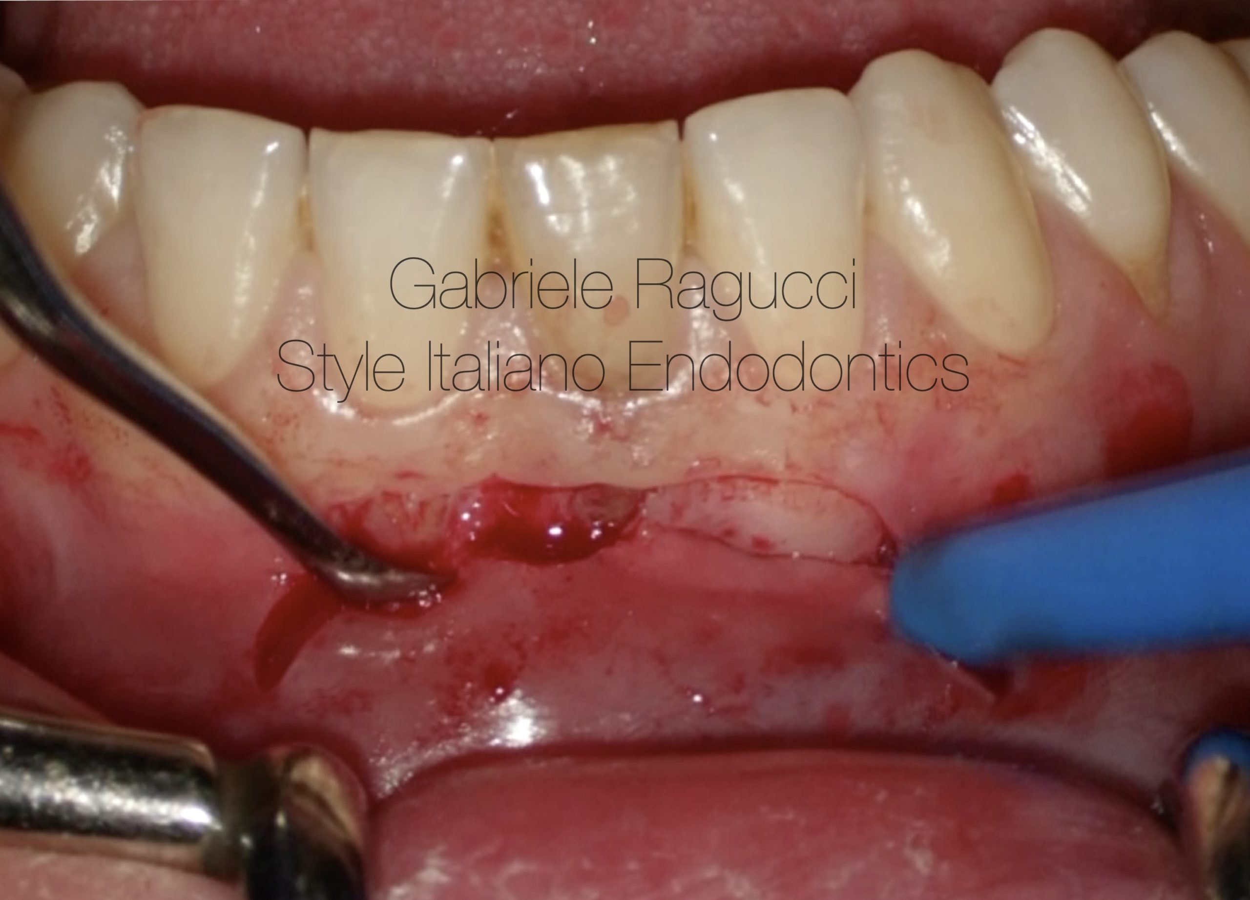
Fig. 3
Periosteal elevator (SIE3+FG V).
Double-curved periosteal elevator – two different sizes on each endused for a sharp dissection of periosteal tissue, without any tissue damage.
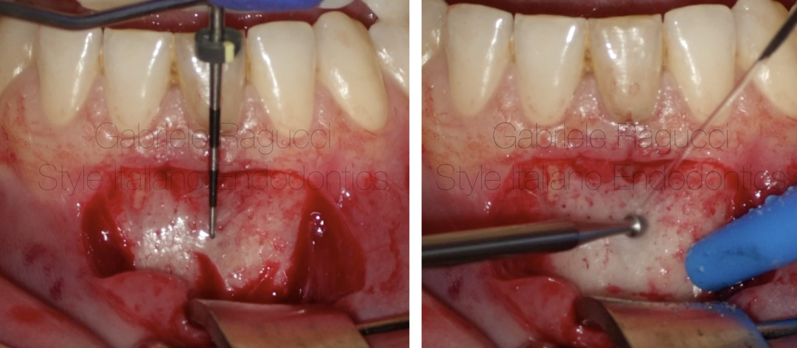
Fig. 4
apex identification
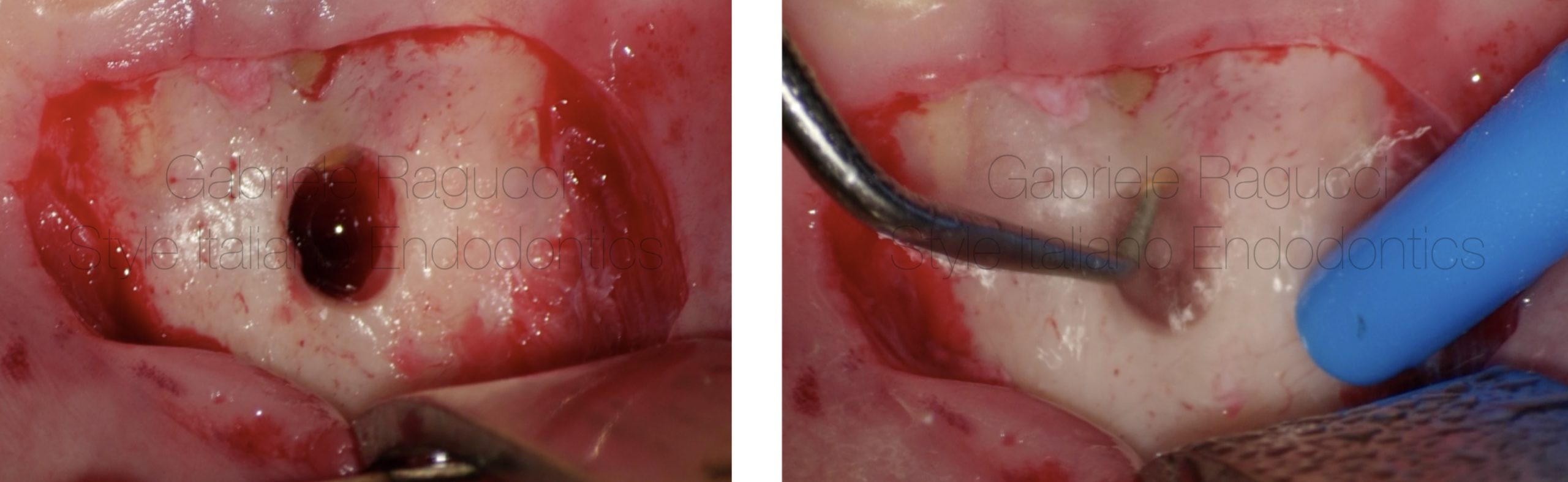
Fig. 5
Surgery phases retrogade ultrasonic preparation
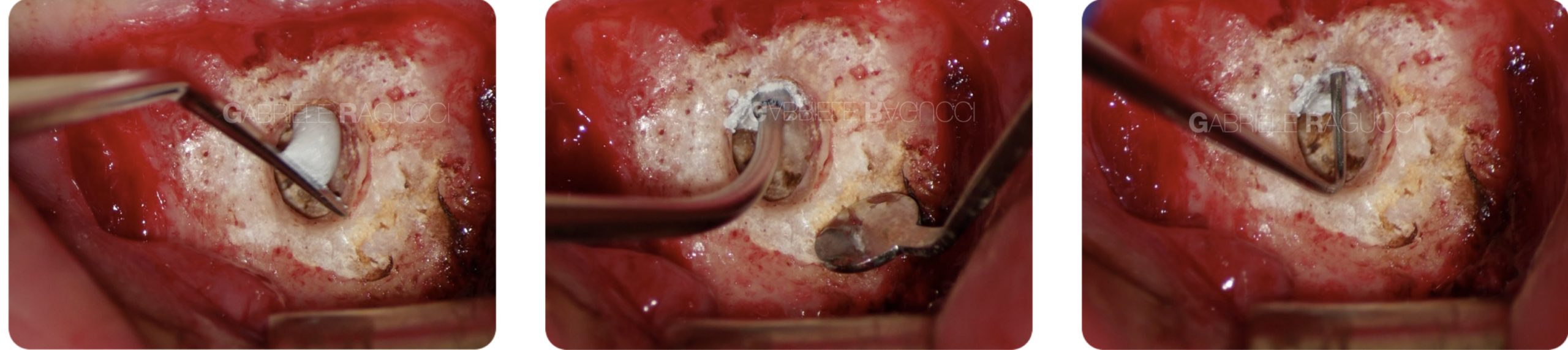
Fig. 6
Obturation
Obturation video
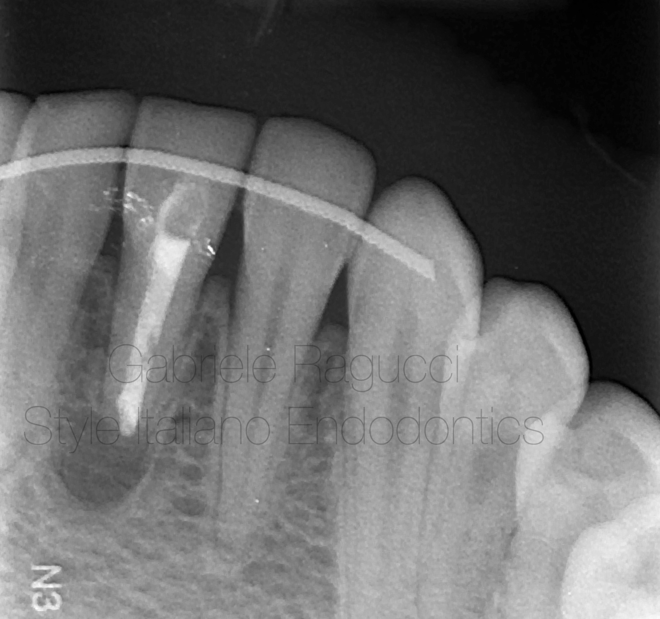
Fig. 7
Postoperative X-ray
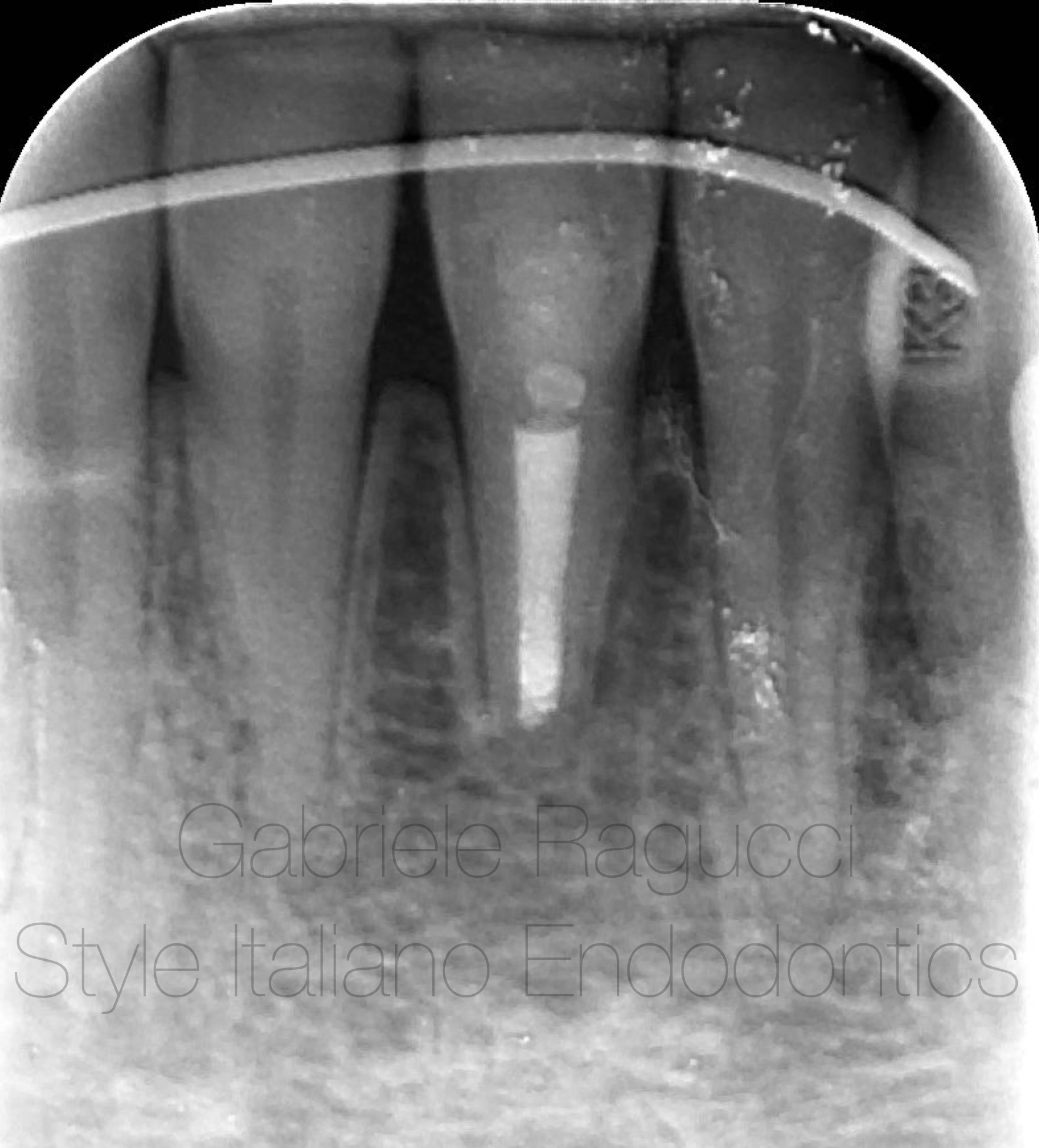
Fig. 8
4 years control X-ray
Conclusions
Micro apical surgery could be more conservative in many cases when we have heavy coronal restorations ,
The procedure should be done with high magnification under microscope and right tools for precise and better outcome .
Bibliography
1-H.M. Eriksen, L.L. Kirkevang, K. Petersson. Endodontic epidemiology and treatment outcome: general considerations. Endod Topics, 2 (2002), pp. 1-9
2-C. Barone, T.T. Dao, B.B. Basrani, N. Wang, S. Friedman. Treatment outcome in endodontics: the Toronto study—phases 3, 4, and 5: apical surgery. J Endod, 36 (2010), pp. 28-35
3- S. Friedman. The prognosis and expected outcome of apical surgery. Endod Topics, 11 (2005), pp. 219-262
4- T. von Arx, M. Penarrocha, S. Jensen. Prognostic factors in apical surgery with root-end filling: a meta-analysis. J Endod, 36 (2010), pp. 957-973