Endodontic management of a Traumatic Maxillary Central Incisor with wide open apex and evident resorption
08/12/2022
Viresh Chopra
Warning: Undefined variable $post in /var/www/vhosts/styleitaliano-endodontics.org/endodontics.styleitaliano.org/wp-content/plugins/oxygen/component-framework/components/classes/code-block.class.php(133) : eval()'d code on line 2
Warning: Attempt to read property "ID" on null in /var/www/vhosts/styleitaliano-endodontics.org/endodontics.styleitaliano.org/wp-content/plugins/oxygen/component-framework/components/classes/code-block.class.php(133) : eval()'d code on line 2
Pulp necrosis(mostly due to trauma) in teeth with incomplete roots can arrest root development resulting in open apices. he main challenge in performing root-canal treatment in teeth with necrotic pulps and wide-open apices is to obtain an optimal apical seal. The wide foramen requires a large volume of filling material that may extrude from the root canal into the periapical tissues creating foreign-body responses and compromising the apical seal.
With traditional means, induction of an apical barrier, regardless of the material used, takes at least 3–4 months and requires multiple appointments. Patient’s compliance with this regimen may be poor and they may fail to return for scheduled visits. Even the temporary seal may fail resulting in reinfection and prolongation or failure of treatment. For all these reasons, one-visit apexification has been suggested.
The rationale behind this is to establish an apical stop that would enable the root canal to be filled immediately. There is no attempt to induce root-end closure rather an artificial apical stop is created at the apex.
A number of materials have been proposed including tricalcium phosphate, calcium hydroxide, freeze-dried bone and freeze-dried dentine, MTA, and biodentine for this purpose
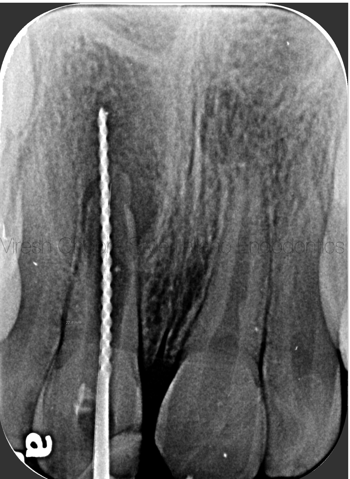
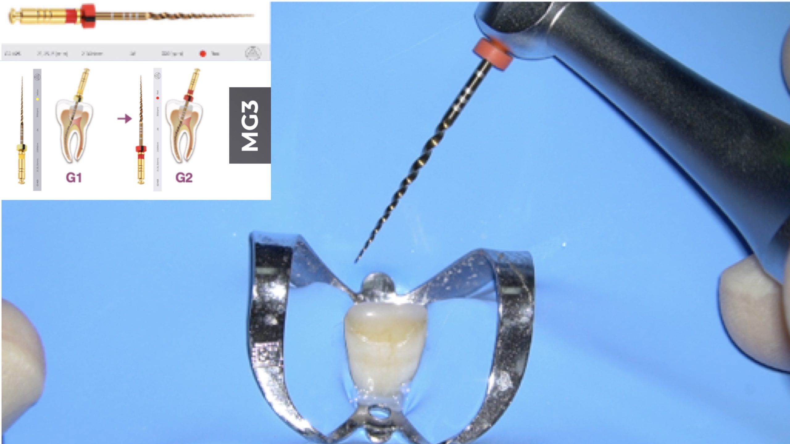
Fig. 1
Patient information
- Age:19-year old
- Gender: Male
- Medical history: non-contributory
- Identification: Right maxillary Central Incisor (Tooth 11)
- Dental history :
Chief complaint: I am having swelling and pain on biting
Clinical examination findings:
The tooth 11 is tender to percussion. Uncomplicated Mesio-incisal crown fracture present. History of trauma few years back while playing football.
The preoperative radiograph sent by the referring dentist showed wide open apex with evident root resorption along the walls of the root canal wall.
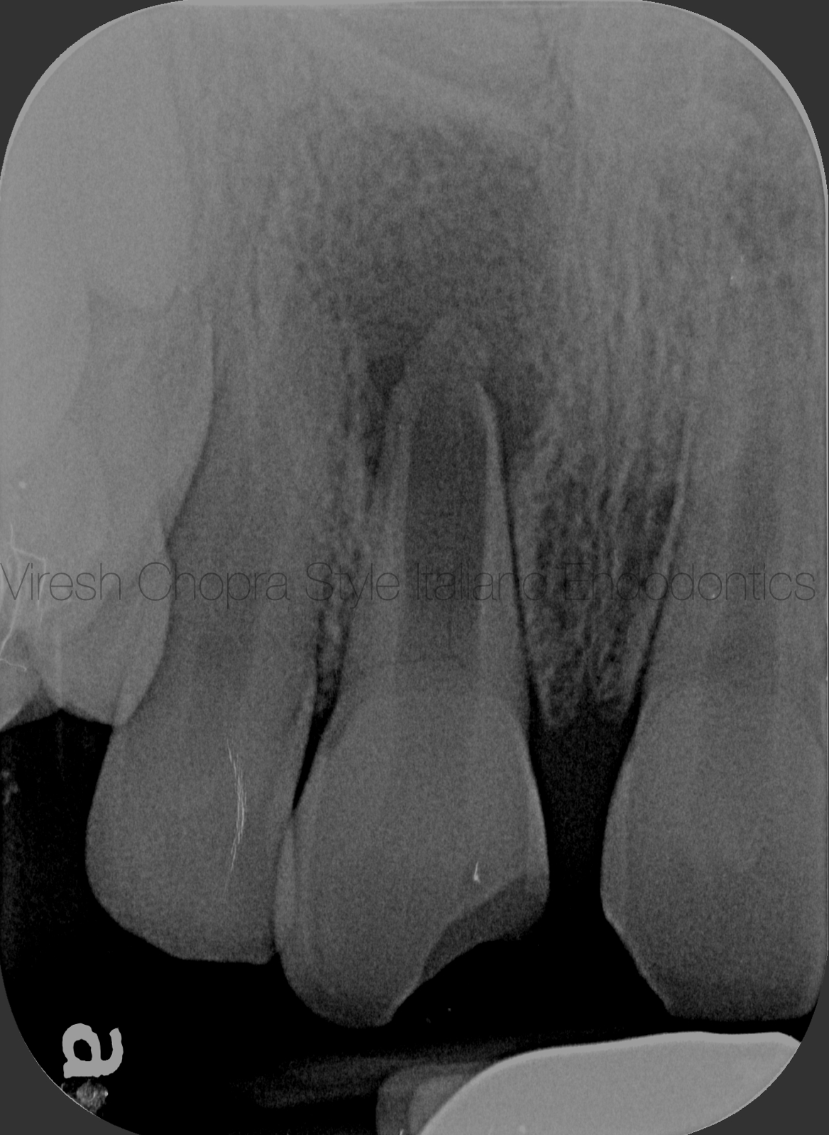
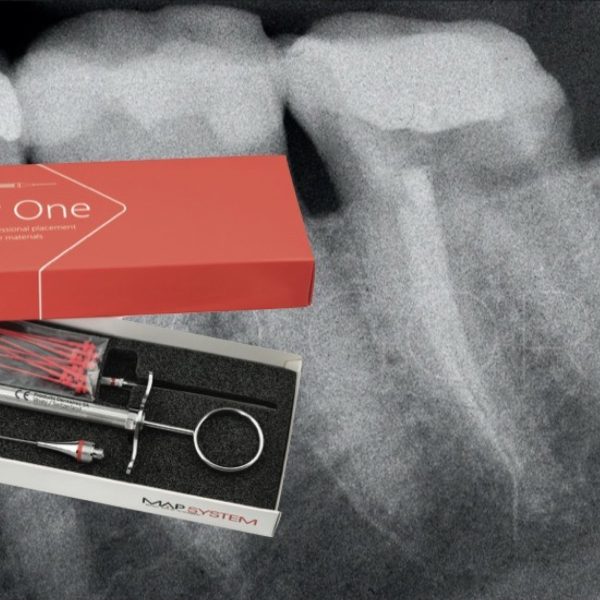
Fig. 2
The patient was referred due to wide open apex. The preoperative radiograph sent was with an endodontic file. Radiograph with the endodontic file revealed wide open apex with thinning of the root canal walls due to extensive resorption. In addition presence of periapical radiolucency was also evident irt tooth 11. Original preoperative radiograph before initiating endodontic treatment was requested.
Original preoperative radiograph revealed wide open apex with periapical radiolucency.
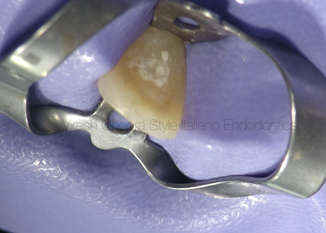
Fig. 3
Treatment plan:
Root canal treatment followed by Apical plug formation and finally obturation with either Gutta percha or Calcium silicate cement.
Step wise procedures in the first visit:
- 1.Buccal infiltration anesthesia was administered, the tooth was isolated with rubber dam isolation and the tooth was seen under the microscope preoperatively.
- 2. The temporary cement was removed . The moment temporary cement was removed there was bleeding on palpation
Step wise procedures in the first visit:
3. The canal was cleaned with profuse irrigation and was minimally shaped with endodontic files.
4. There was profuse continuous bleeding from the canal. Attempts to dry the canal with paper points dipped in homeostatic agent were made.(video 2)
5. After few minutes the bleeding stopped and a good look into the root canal was done under microscope.
Video 2 showing profuse blessing and attempts to stop bleeding with paper points and homeostatic agent.

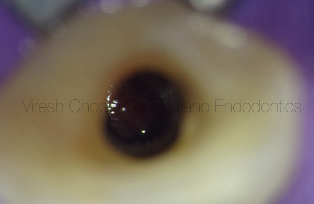
Fig. 4
Once the bleeding stopped, a good look at the root canal was done under microscope. The wide open apex was clearly visible under magnification.
Once the canal was shaped and disinfected with irritants it was decided to obturate the complete canal with Calcium Silicate cement (MTA). The reason for choosing MTA as an obturating material is that there is resorption evident along the length of the root canal wall. Therefore, it would be justified to place a material that can initiate bone formation along the whole surface.
MTA was mixed as per the manufacturers instructions and carried to the root canal with the help of MTA gun. Since the apex was wide and bleeding, attempt was made to carry large pellets of MTA in each turn so as to close the apex soon. (Video 3)
Full length of the root canal was obturated with MTA and verified on the periapical radiograph
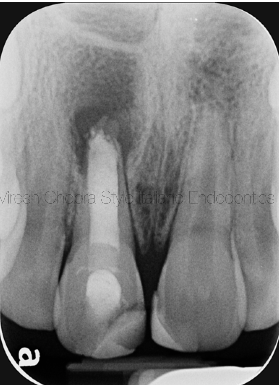
Fig. 5
Complete obturation with MTA
Conclusions
MTA is known to be an effective material to promote obturation of open apices by delivering the MTA right at the apex where it comes in contact with the periapical tissues and some moisture required for its setting. The main advantage of this procedure is the high predictability of apical closure with the reduction of treatment time, number of appointments, and radiographs.
Bibliography
1.Kumar SM, Kumar T, Keshav V, Arora S, Singla A. Open apex solutions: One-step apexification, salvaging necrosed teeth with open apex. Endodontology. 2019 Jul 1;31(2):173.
2.De‐Deus G, Coutinho‐Filho T. The use of white Portland cement as an apical plug in a tooth with a necrotic pulp and wide‐open apex: a case report. International Endodontic Journal. 2007 Aug;40(8):653-60.
3.Costa GM, Soares SM, Marques LS, Gloria JC, Soares JA. Strategy for apexification of wide-open apex associated with extensive periapical lesion in a weakened root. General dentistry. 2013 May 1;61(3):e2-4.