C-Shaped Canal Configuration in Mandibular Second Molar
28/07/2017
Marino Sutedjo
Warning: Undefined variable $post in /var/www/vhosts/styleitaliano-endodontics.org/endodontics.styleitaliano.org/wp-content/plugins/oxygen/component-framework/components/classes/code-block.class.php(133) : eval()'d code on line 2
Warning: Attempt to read property "ID" on null in /var/www/vhosts/styleitaliano-endodontics.org/endodontics.styleitaliano.org/wp-content/plugins/oxygen/component-framework/components/classes/code-block.class.php(133) : eval()'d code on line 2
The C-shaped canal is an anatomical variation that was first reported by Cooke & Cox (1979) and mostly seen in mandibular second molars, although it can also occur in maxillary and other mandibular molars.
The main anatomical feature of C-shaped canals is the presence of a fin or web connecting the individual root canals with the orifice may appear as a single ribbon-shaped opening with a 180° arch, linking the two main canals.
Etiology
Failure of the Hertwig's epithelial root sheath to fuse on the lingual or buccal root surface is the main cause of C-shaped roots, which always contain a C-shaped canal. The C-shaped root may also be formed by coalescence because of deposition of the cementum with time.
Prevalence
This C-shaped variation appears to be genetically determined and may be used in tracing the ethnic origin of the subjects. Studies indicate that the occurrence of C-shaped canals is more frequent in Asian populations than in other races (Yang et al. 1988, Weine 1998, Haddad et al. 1999, Gulabivala et al. 2001, 2002).
East Asian population groups like Chinese (0.6% - 41.27%) and Koreans (31.3% - 45.5%) display a high prevalence of this variant. Among the South Asian countries, Burmese population showed a prevalence of 22.4%, which was much higher than the Indian, Thai or Sri Lankan population.
And when present on one side, a C-shaped canal may be found in the contralateral tooth in over 70% of individuals.
Classification
Melton in 1991 proposed the following classification based on the different configurations of the orifices in C-shaped canal systems.
Class I :
A continuous C-shaped canal, with no separation of the canals.
Class II :
The canal orifices resemble a semicolon, where a C-shaped canal is present buccally or lingually, separated from another distinct canal by a dentine wall.?
Class III :
Two or more separate canals are present, as in a typical lower molar, with three canal orifices.
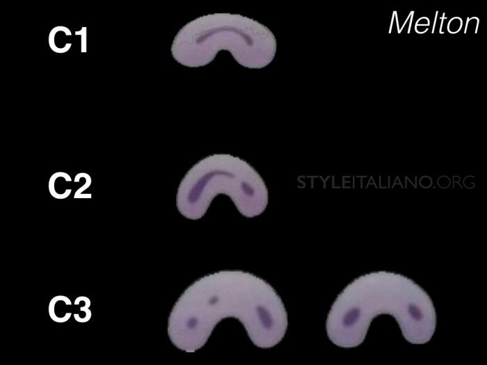
Fig. 1
Melton Classification
In Melton’s classification, there has been no clear description of the difference between categories II and III as well as the clinical significance; therefore in 2009 Zapata described the classification of C-shaped according to their canal configuration which was a modification of Melton’s method that modified by Fan Bing et al. 2004 into the following categories:
I Category (C1):
The shape was an uninterrupted “C” with no separation or division.
II Category (C2):
The canal shape resembled a semicolumn resulting from a discontinuation of the “C” outline, but either angle α or β should be no less than 60°.
III Category (C3):
Two or three separate canals and both angles, α and β were less than 60°.
IV Category (C4):
Only one round or oval canal in that cross- section.
V Category (C5):
No canal lumen could be observed.
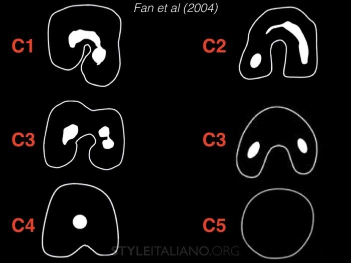
Fig. 2
Fan et al (2004) classification
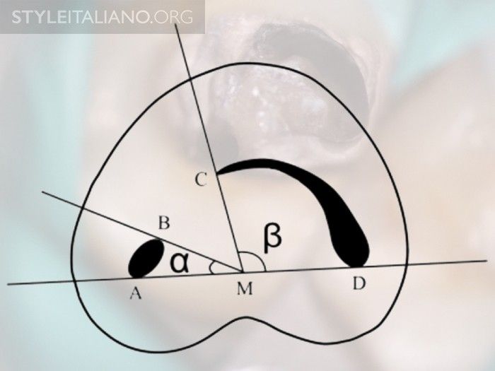
Fig. 3
Measurement of angles for the C2 canal. Angle α and β is more than 60°. A and B: ends of one canal cross-section; C and D: ends of the other canal cross-section; M: middle point of line; AD; α: angle between line AM and line BM; β: angle between line CM and line DM.
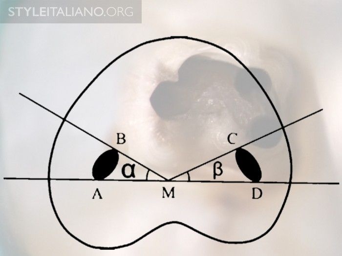
Fig. 4
Measurement of angles for the C3 canal. Both angle α and β are less than 60°. A and B: ends of one canal cross-section; C and D: ends of another canal cross-section;
M: middle point of line AD;
α: angle between line AM and line BM;
β: angle between line CM and line DM.
Diagnosis
The preoperative awareness or diagnosis of a C-shaped canal configuration before treatment can facilitate an effective management. There are two ways that can help us to diagnose or identified C-shape preoperatively.
1. Preoperative radiograph
In all endodontic cases, preoperative radiograph is a must for making a good diagnosis. By taking a preoperative radiograph it will provide us many clues in the identification of any variation in root canal morphology as well as in C-shape canal configuration.
C-shaped root in a mandibular second molar may present radiographically as a single-fused root or as two distinct roots with a communication. When the communication or fin connecting the two roots is very thin, it is not visible on the radiograph and may thus give the appearance of two distinct roots. The radiograph may also reveal a large and deep pulp chamber.
Fan et al (2004) furthermore classified the radiographic appearance of a C-shaped into three different types:
Type I:
Conical or square root with a vague, radiolucent longitudinal line separating the root into distal and mesial parts. There was a mesial and a distal canal that merged into one before exiting at the apical foramen.
Type II:
Conical or square root with a vague, radiolucent longitudinal line separating the root into distal and mesial parts. There was a mesial and a distal canal, and the two canals appeared to continue on their own pathway to the apex.
Type III:
Conical or square root with a vague, radiolucent longitudinal line separating the root into distal and mesial parts. There was a mesial and a distal canal, one canal curved to and superimposed on this radiolucent line when running toward the apex, and the other canal appeared to continue on its own pathway to the apex.
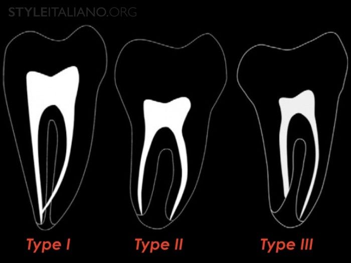
Fig. 5
Radiographic type of C-shaped Canal (Fan et al 2004)
2. Clinical Diagnosis
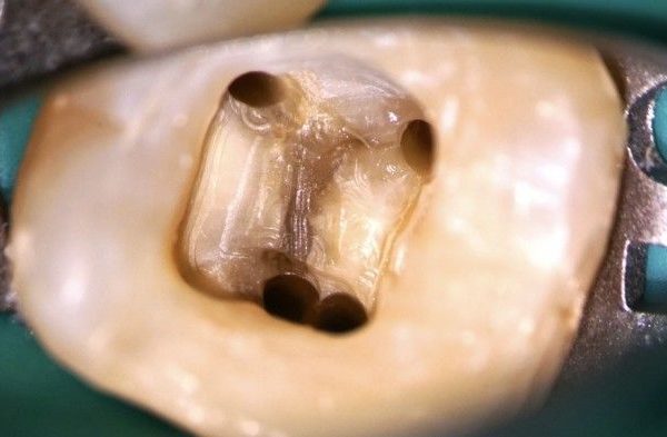
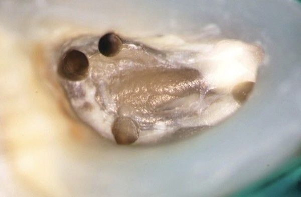
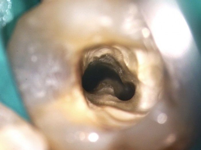
As for clinical diagnosis, following access cavity, the pulp chamber in teeth with C-shaped canals may be large in the occluso-apical dimension with a low bifurcation. Other authors reported that the pulpal floor in C-shaped teeth can vary from peninsula like with a continuous C-shaped orifice to non C-shaped floors as per the classification given (Min et al 2006).
Fan et al 2004 also mentioned that for mandibular second molar to have a C-shaped canal system, it has to exhibit all the following three characteristics:
1. Fused roots.
2. A longitudinal groove on lingual or buccal surface of the root.
3. At least one cross-section of the canal should belong to the C1, C2, C3 or C4 configuration.
A magnification is also important to help us identify this pulp chamber anatomy of C-shaped canals. But, when a C-shaped canal orifice is evident, we must remember that we cannot assume that such a shape continues throughout its length.
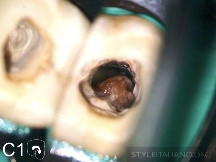
Fig. 6
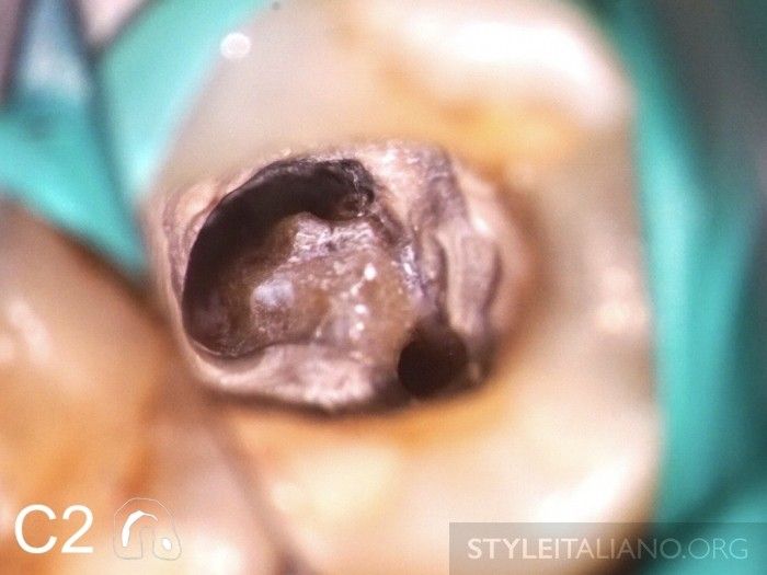
Fig. 7
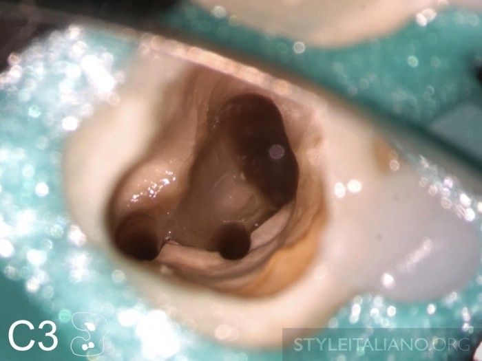
Fig. 8
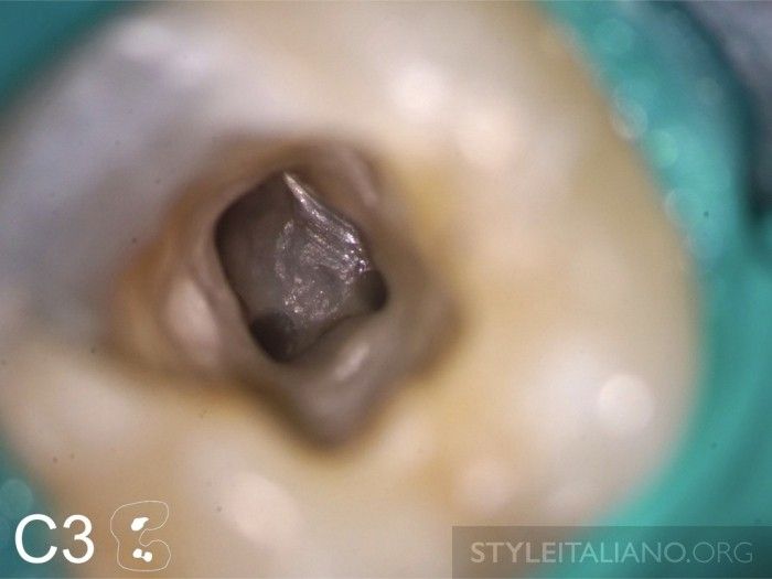
Fig. 9
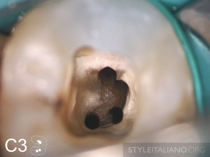
Fig. 10
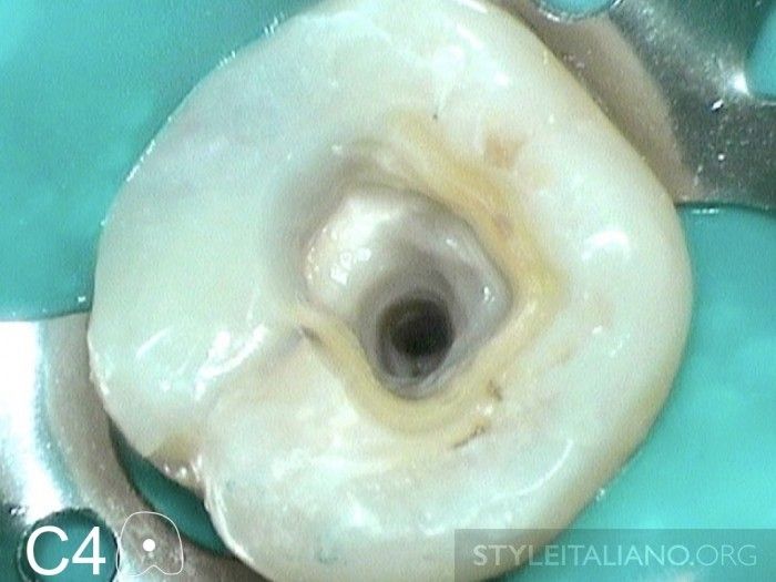
Fig. 11
Management
Basically like any other endodontic treatments, procedures and sequences that of C-shaped canals is also the same. But, since we have more complexity with the anatomical variations such as high percentage of canal irregularities, accessory and lateral canals as well as apical delta that make us more difficult to locate, shape, clean and seal adequately. Therefore we have to put more attention to details for treating this C-shaped canal.
Access Cavity
Access Cavity is very important for teeth with C-shape configuration. We have to remember that the appearance of the pulp chamber and orifices is not the same like regular mandibular molars. This is very important since, in my experience, quite many cases that come in for retreatment of C-shaped canal failed due to under or over access cavity. Sometimes we also need to modify our access for easier identification of orifice location and easier manipulation of our endodontic instruments. Usually when the orifice is continuous C-shape or arch like from mesiobuccal to distal (MB-D), the number of canals can vary from one to three and when the orifice is oval or flat, the number of canals may be one or two. Also when the orifice is round-shaped, there is usually only one canal below the orifice.
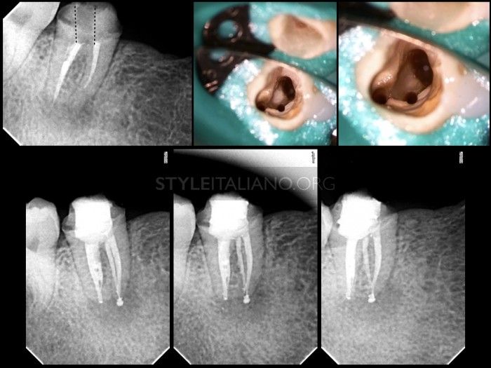
Fig. 12
Retreatment case of second mandibular right molar with C-shaped canal configuration. Due to under access of previous treatment, the operator failed to identify the C-shaped anatomy.
Shaping, Cleaning and Obturation
The complexity of C-shaped canals requires appropriate shaping and cleaning procedure as well as obturation technique. Due to large area of canal space, it is doubtful that our shaping instruments can reach and debride the entire portion of the systems. A large shaping size can also create a high risk of root perforation since these have thinner lingual walls in second mandibular molars C-shaped canal.
In this C-shaped canal, irrigation plays a very important role. Copius irrigation of 5,25% of NaOCl along with sonic or ultrasonic agitation are mandatory. This is needed to ensure maximum disinfection and debridement as well as tissue removal of the large volumetric space in C-shaped canal.
Obturation of C-shaped canals may require technique modifications. Although some canals may be obturated as standard, due to large volumetric extension of the C-shaped canal, the use of thermoplasticized gutta-percha is mandatory to achieve three dimensional obturation.
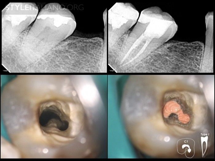
Fig. 13
Case 1
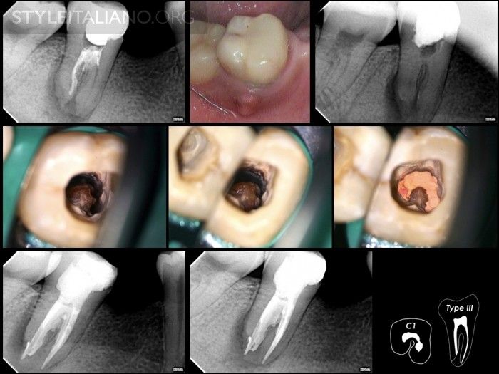
Fig. 14
Case 2
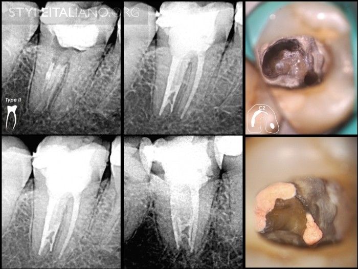
Fig. 15
Case 3
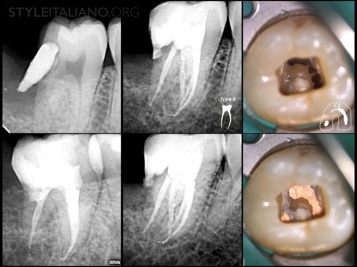
Fig. 16
Case 4
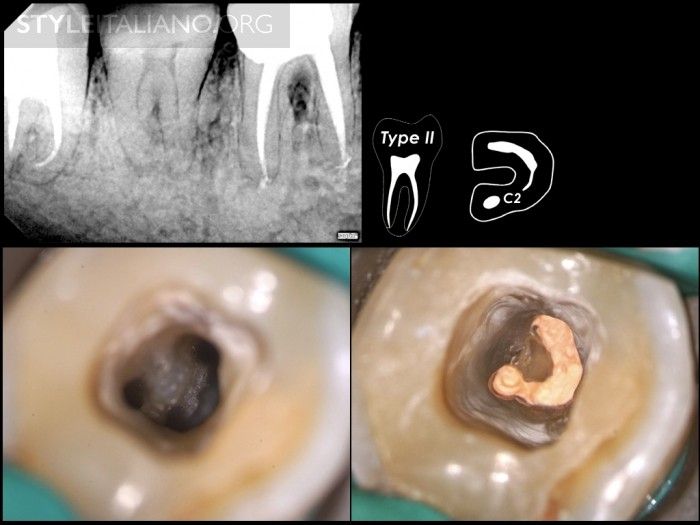
Fig. 17
Case 5a
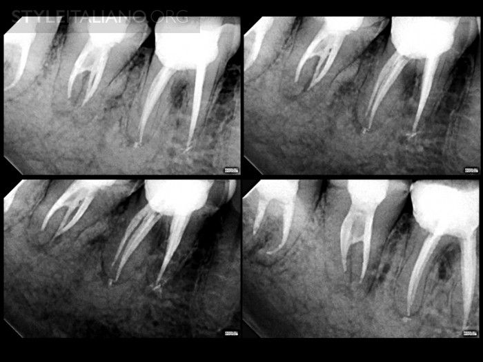
Fig. 18
Case 5b
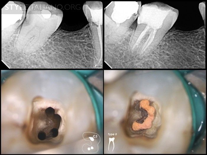
Fig. 19
Case 6
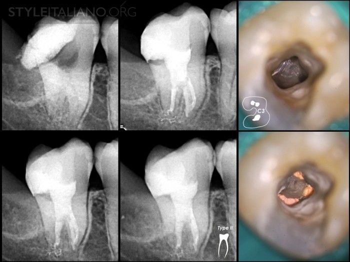
Fig. 20
Case 7
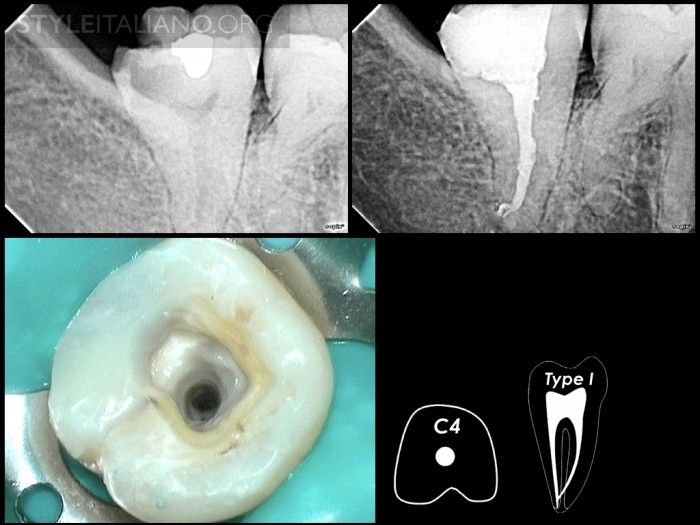
Fig. 21
Case 8
Conclusions
The C-shaped root canal configuration in second mandibular molar has an ethnic predilection. Our understanding in the anatomical presentations of this variation will help us manage this configuration effectively. Diagnosis as well as root canal procedure is important to be carried out adequately to ensure good treatment result.
Bibliography
Cooke HG, Cox FL. C-shaped canal configurations in mandibular molars. J Am Dent Assoc 1979; 99: 836839.
Yang ZP, Yang SF, Lin YL. C-shaped root canals in mandibular second molars in Chinese population. Endod Dent Traumatol 1988; 4: 160163
Manning S. Root canal anatomy of mandibular second molars. Part II C-shaped canals. Int. Endod. J 1990; 23: 40-45
Melton DC, Krell KV, Fuller MW. Histological Anatomical and features of C-shaped canals in mandibular second molars. J Endod 1991; 17: 384-388.
Sabala CL, Benenati FW, Neas BR. Bilateral root or root canal aberra- tions in a dental school patient population. J Endod 1994; 20: 3842.
Haddad GY, Nehme WB, Ounsi HF. Diagnosis, classification, and frequency of C-shaped canals in mandibular ?second molars in the Lebanese population. J Endod 1999; 25: 268-271.
Sidow SJ, West LA, Liewehr FR, Loushine RJ. Root canal morphology of human maxillary and mandibular third molars. J Endod 2000; 26: 675678.
Gulabivala K, Aung TH, Alavi A, Ng YL. Root and canal morphology of Burmese mandibular molars. Int Endod J 2001; 34: 359-370.
Gulabivala K, Opasanon A, Ng YL, Alavi A. Root and canal morphology of Thai mandibular molars. Int Endod J 2002; 35: 5662.
Fan B, Cheung GS, Fan M, Gutmann JL, Bian Z. C-shaped canal system in mandibular second molars: Part I: Anatomical features. J Endod 2004; 30: 899-903.
Min Y, Fan B, Cheung GS, Gutmann JL, Fan M. C-shaped canal system in mandibular second molars Part III: The morphology of the pulp chamber floor. J Endod 2006; 32: 1155-1159.
Fernandes M, Ataide ID, Wagle R. C-shaped root canal configuration: A review of literature. Journal of Cons Dent 2014; 14: 312-319
Raisingani D, Gupta S, Mital P, Khullar P. Anatomic and Diagnostic Challenges of C-Shaped Root Canal System. Int J Clin Pediatr Dent 2014;7(1):35-39.