Bioceramics and adhesion: post space preparation for single cone technique
26/08/2021
Calogero Bugea
Warning: Undefined variable $post in /var/www/vhosts/styleitaliano-endodontics.org/endodontics.styleitaliano.org/wp-content/plugins/oxygen/component-framework/components/classes/code-block.class.php(133) : eval()'d code on line 2
Warning: Attempt to read property "ID" on null in /var/www/vhosts/styleitaliano-endodontics.org/endodontics.styleitaliano.org/wp-content/plugins/oxygen/component-framework/components/classes/code-block.class.php(133) : eval()'d code on line 2
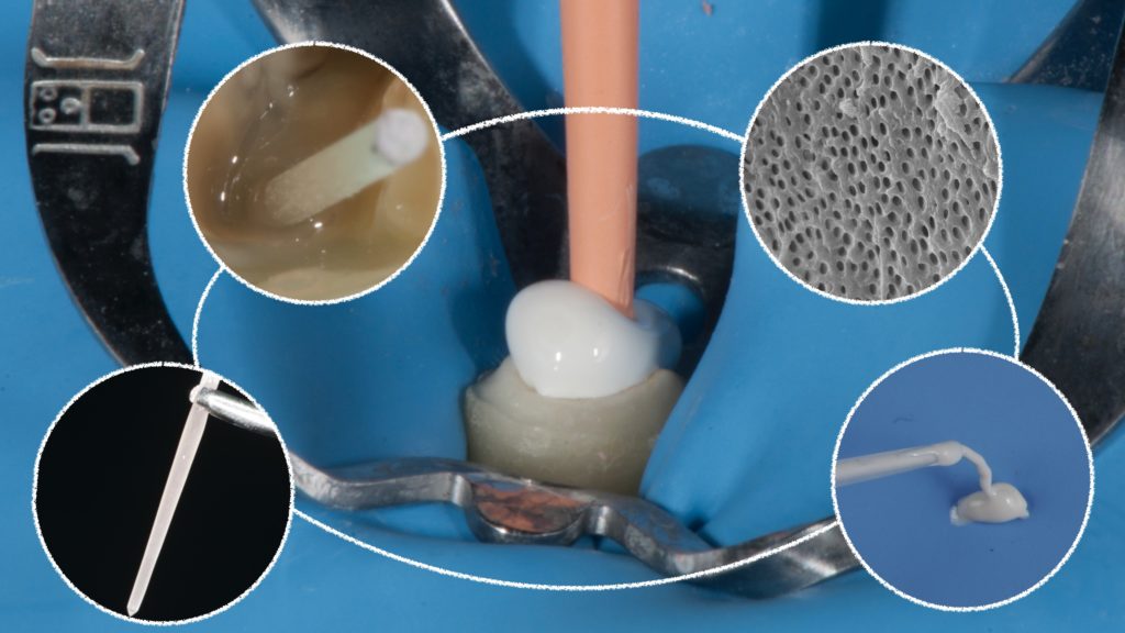
One of the controversial topics about endodontics and restorative dentistry is the interaction between endodontic sealers and adhesion. Regarding fiber post cementation, the cleaning of the endodontic space plays a fundamental role in the adhesion. Bioceramic sealers may interfere with adhesive procedure due to their penetration in the dentinal tubule, alcaline PH and salt precipitation. To increase the adhesion between dentin and composite cement in a canal filled with bioceramic sealer and guttapercha, the literature studies suggests to put a fiber post contextually to the endodontic procedure, instead of delayed post.
In case of single cone technique for obturation, on the other hand, during the preparation of the post space, the operator may alterate the apical sealing with burns.
In order to respect the apical sealing and obtain a clean endodontic space , Dr Ali Nasseh suggests a technique t predispose the canal to receive a post in case of a single cone technique + bioceramic sealer. Another method is to utilize the bioconeless technique that I will describe in another article.
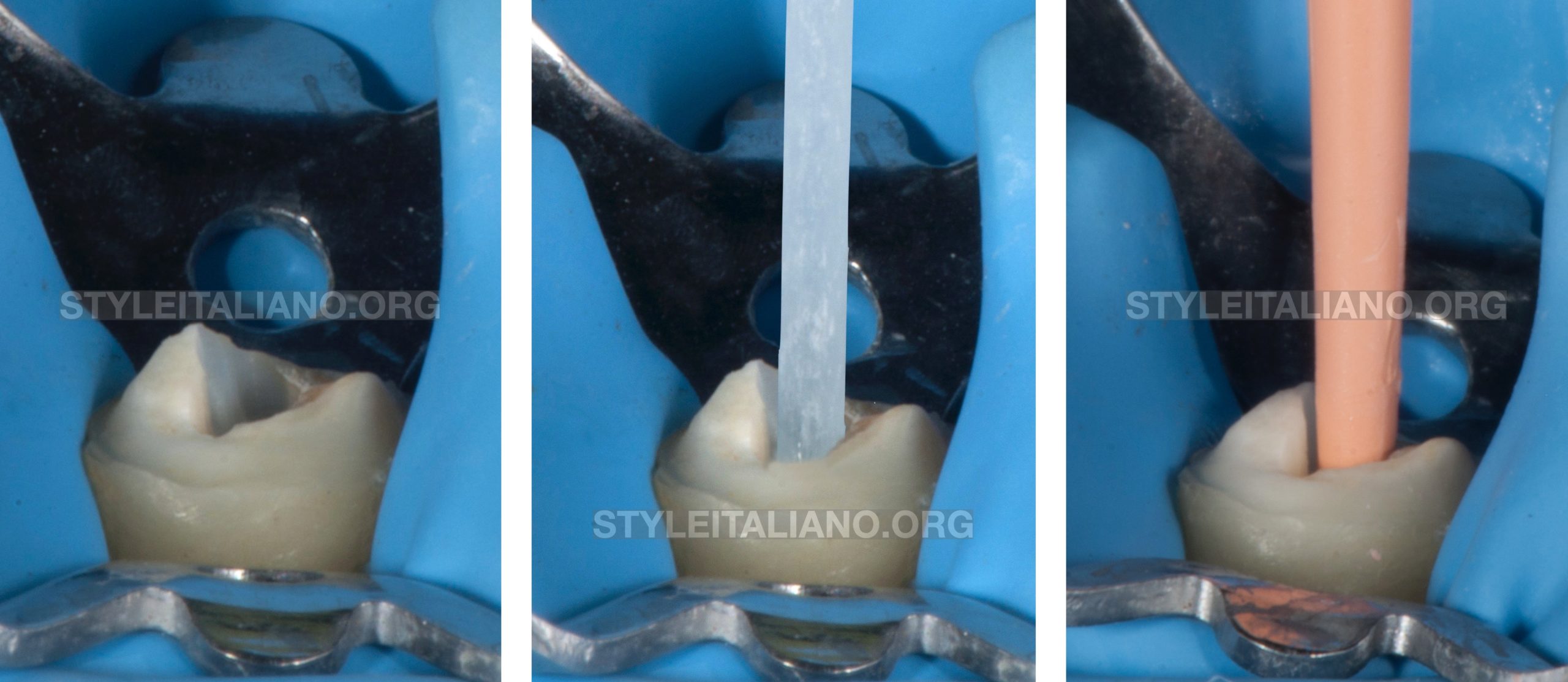
Fig. 1
The ideal post should have the same shape as the endodontic preparation. For this reason I can try different fiber posts and if necessary adapt the post to the canal (not the canal to the post) before the endodontic obturation, to choose the ideal size of the fiber post without canal enlargement
The most important thing is to have an horizontal and vertical ferrule effect instead of a bigger post.
In this case, after the proof of different post this was was the best one.
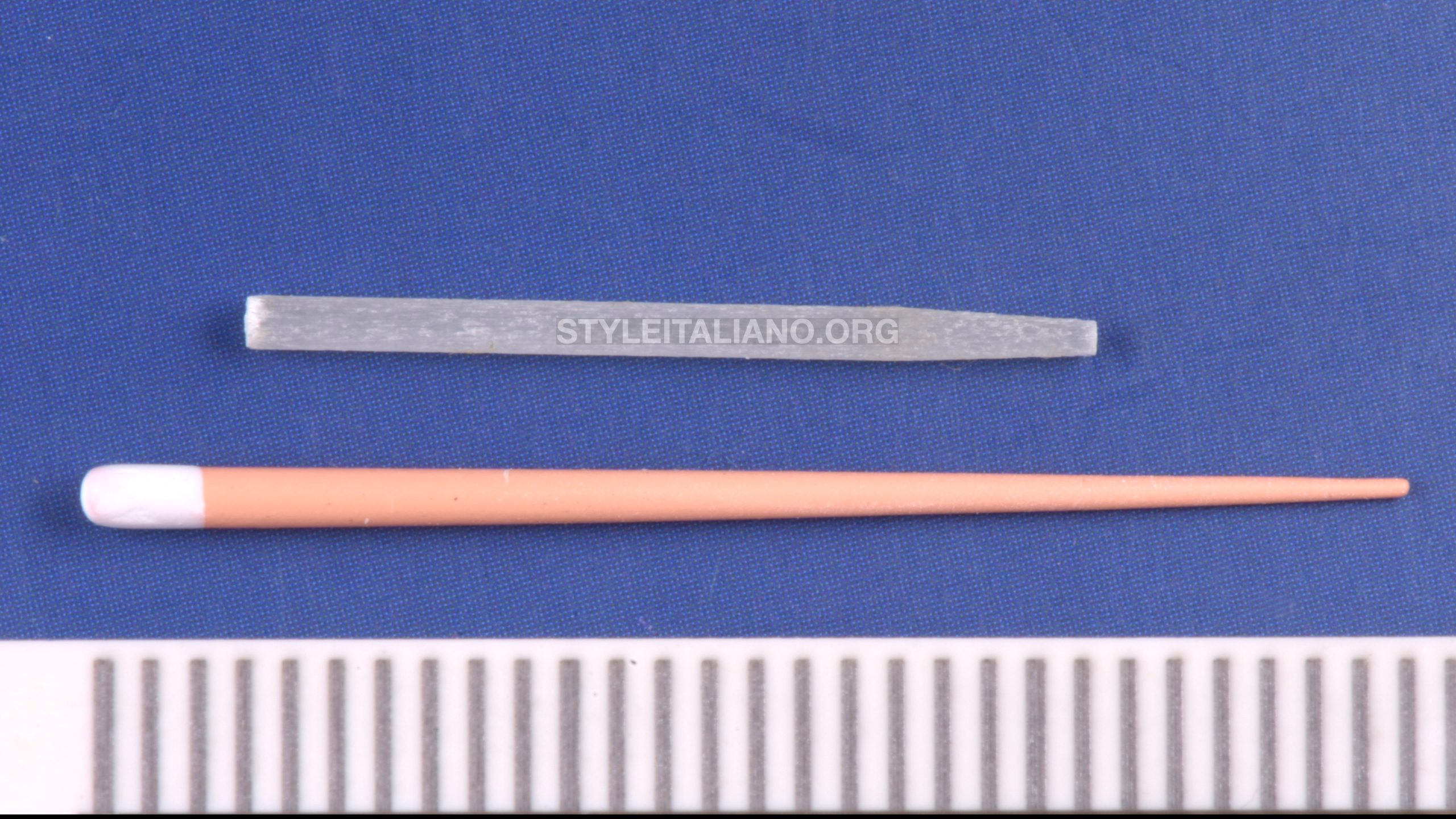
Fig. 2
The second step is to calculate the length of the fiber post inside of the canal and report it on the guttapercha point.
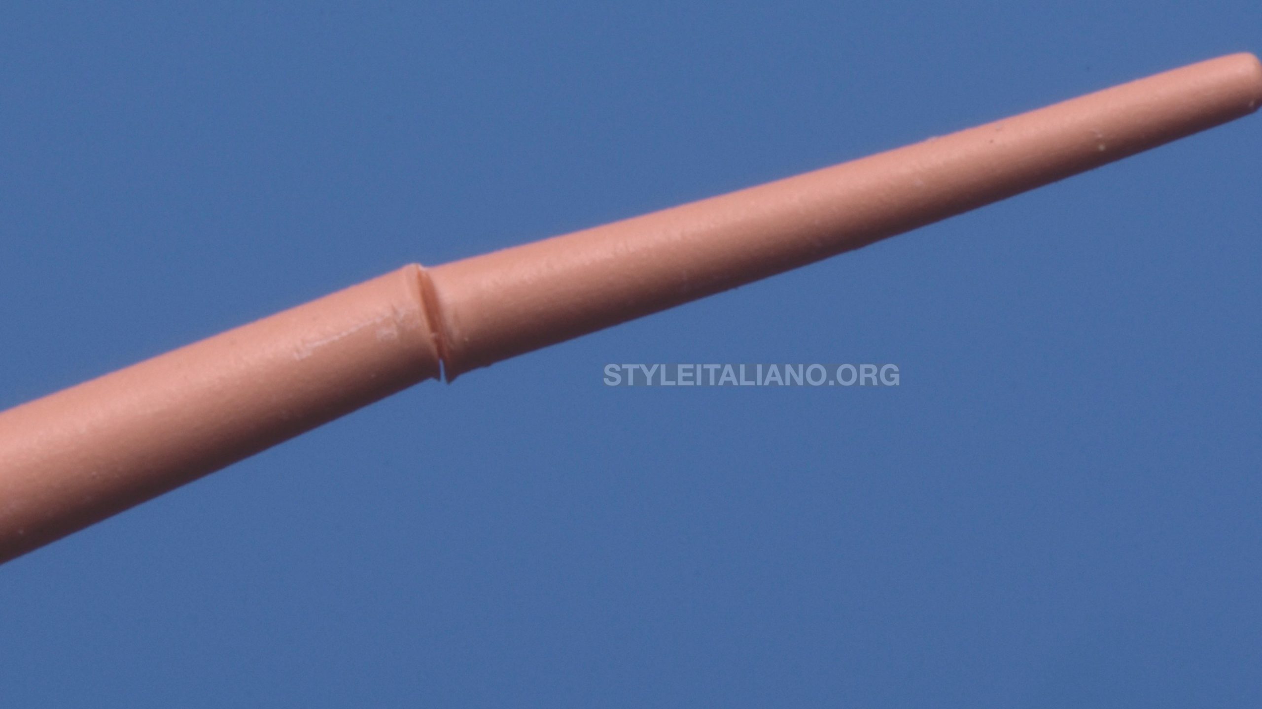
Fig. 3
With a surgical blade the gutta-percha point will be pre-sectioned. This technique was introduced by Dr. A. Castellucci to prevent the guttapercha contamination in case of coronal root perforation. The Cone must be pre-sectioned for 70- 80% of its thickness.
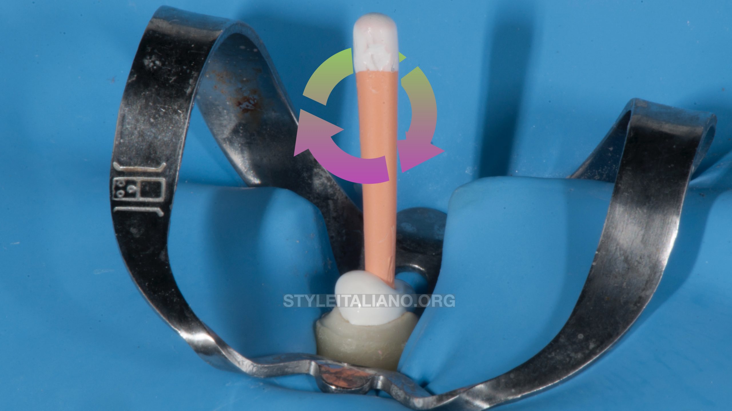
Fig. 4
Until the cone + bioceramic sealer are inserted inside the canal at working length, the endodontist begins to rotate the head of the guttapercha point inside the canal.
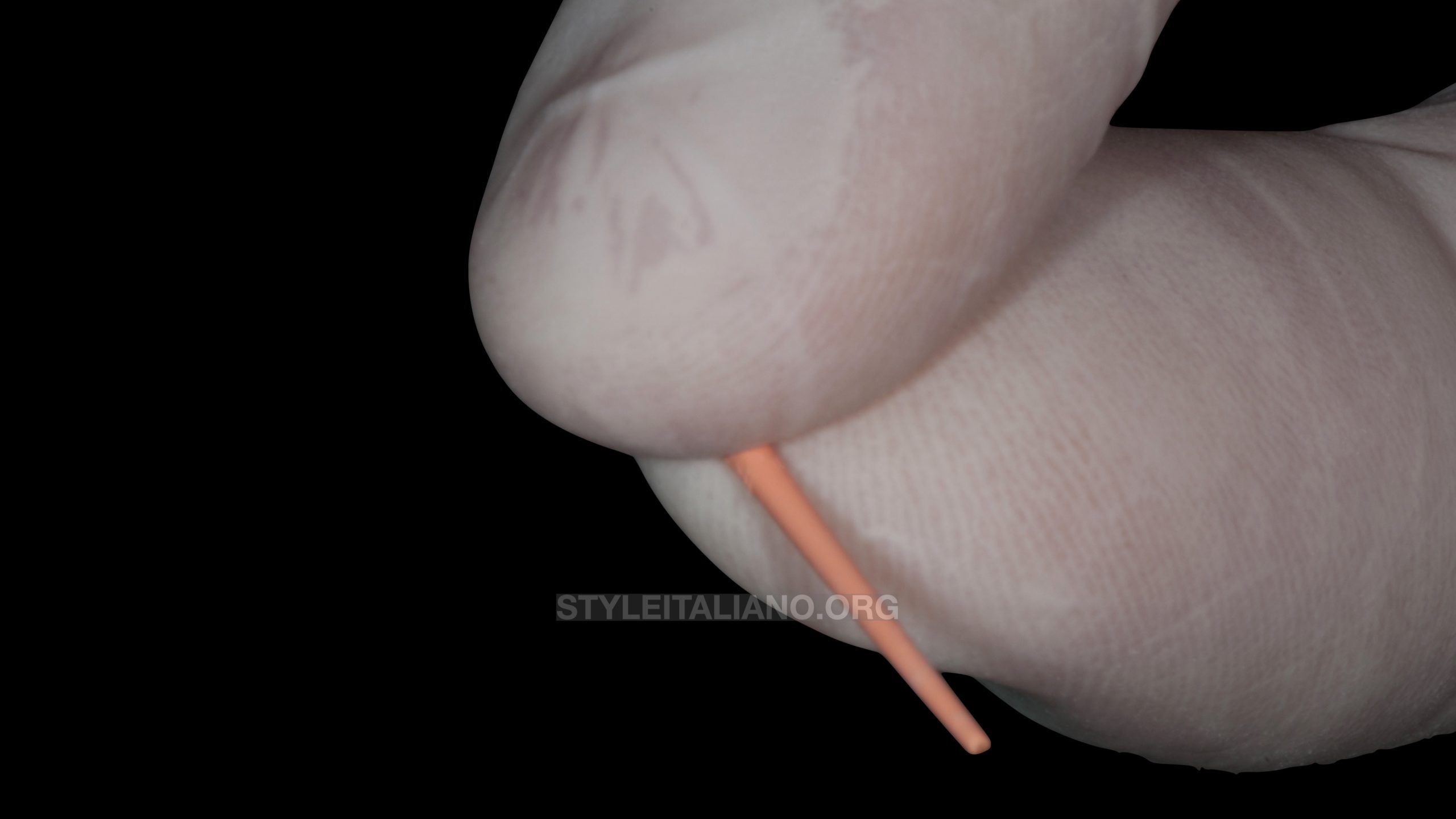
Fig. 5
In this manner the apical part remains inside the canal, instead the post space is free from gutta-percha remnants.
The action of the plugger will pack the gutta-percha in the apical area.
To better understand the procedure, this video shows the cutting movement with the blade and the action of the hand on the gutta percha point
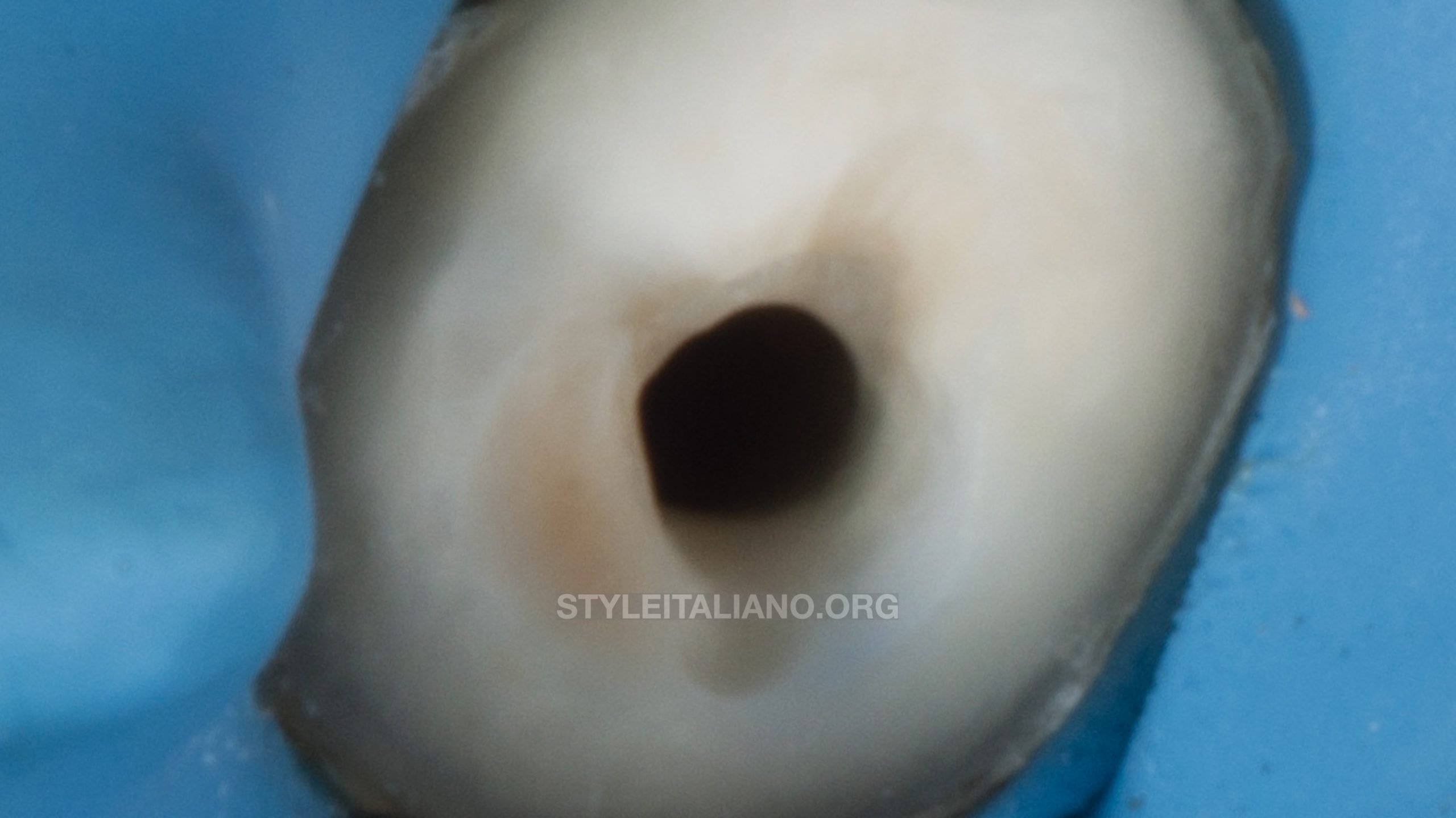
Fig. 6
The post space is gutta-percha free but the canal walls are filled with bioceramic sealer.
The action of an ultrasonic device, under irrigation, may clean the post space from the bioceramic sealer.
The color of the water will change from white to transparent. When the water is transparent it means that the post space is clean.
Literature Studies about ultrasonics are conducted only one week after the root canal obturation. It’s opinion of the author that ultrasonics can play an important role only if they are use immediately after the canal preparation, because the reaction between dentin and bioceramic sealer has not started yet.
For this reason, to maximized the degree of the adhesion, the endodontist has to clean the post space even when the post is not inserted in the same day as the root canal filling.
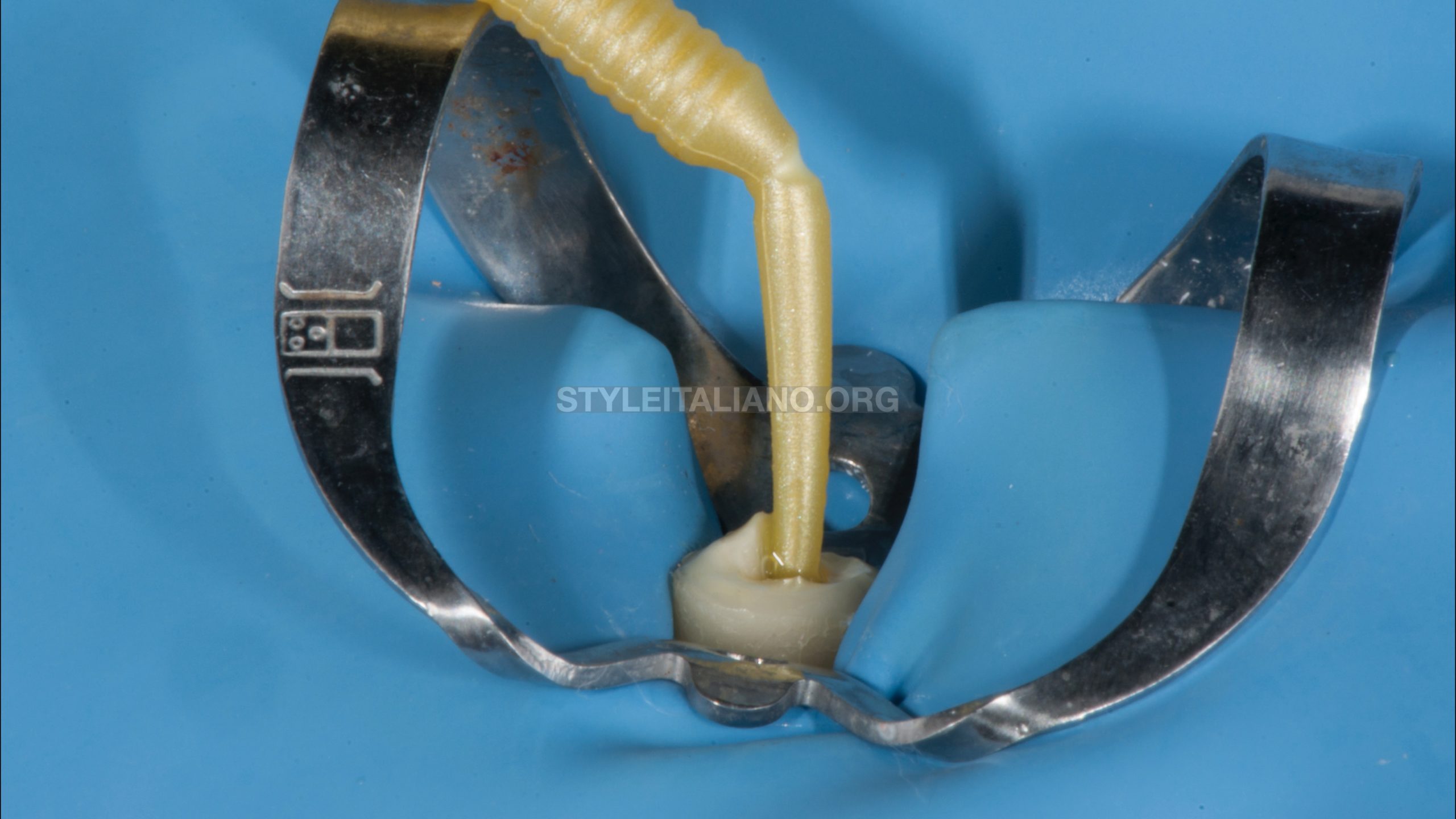
Fig. 7
Start of adhesive procedures
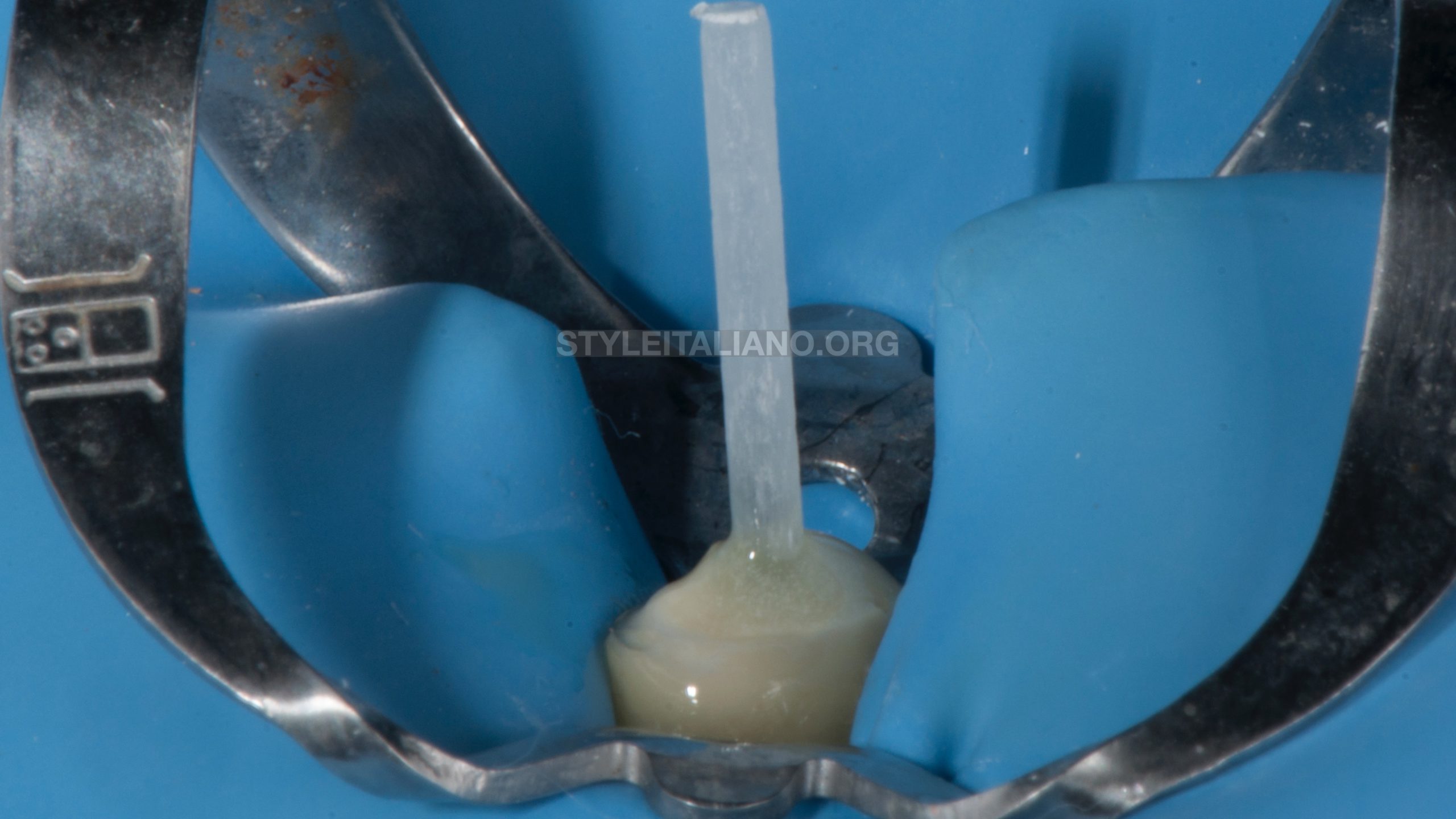
Fig. 8
Fiber post + sealer in situ
Conclusions
The Literature evidences the negative interaction between bioceramic sealers and adhesive procedures. To minimize this negative interaction, the time of the post space preparation, the use of ultrasonics and the respect of the apical sealing plays a fundamental role for the endodontic and prosthetic success.
Bibliography
Effect of different endodontic sealers and time of cementation on push-out bond strength of fiber posts. Clin Oral Investig. 2018 Apr;22(3):1403-1409. doi: 10.1007/s00784-017-2230.
Shokouhinejad N, Gorjestani H, Nasseh AA, Hoseini A, Mohammadi M, Shamshiri AR (2013) Push-out bond strength of gutta-percha with a new bioceramic sealer in the presence or ab- sence of smear layer. Aust Endod J 39:102–106
Effect of ultrasonic cleaning on the bond strength of fiber posts in oval canals filled with a premixed bioceramic root canal sealer. Restor Dent Endod. 2020 Feb 20;45(2):e19.