Vertical Apical Root Fracture
07/06/2020
Riccardo Tonini
Warning: Undefined variable $post in /home/styleendo/htdocs/styleitaliano-endodontics.org/wp-content/plugins/oxygen/component-framework/components/classes/code-block.class.php(133) : eval()'d code on line 2
Warning: Attempt to read property "ID" on null in /home/styleendo/htdocs/styleitaliano-endodontics.org/wp-content/plugins/oxygen/component-framework/components/classes/code-block.class.php(133) : eval()'d code on line 2
Vertical fractures can occur and their diagnosis is not always predictable. Will be presented here the treatment of a 2.6 with a vertical apical fracture in the mesial root.
Vertical root fractures have been described as longitudinally oriented fractures of the root, extending from the canal to the periodontium. It usually happens in endodontically treated teeth and may involve the whole length of the root or only a part of it as in this clinical case of a root canal treated upper first molar.
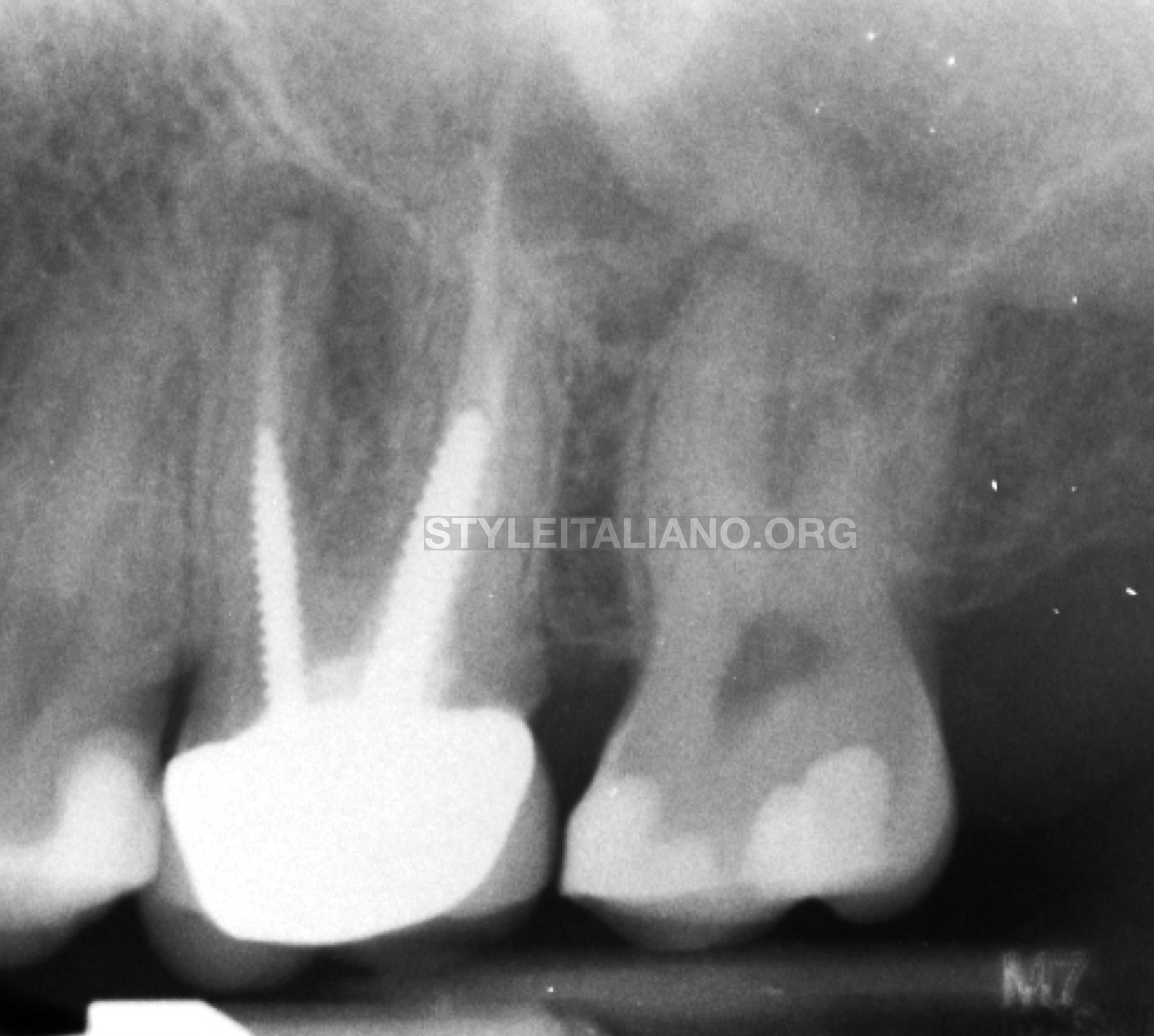
Fig. 1
2.6 upper first molar with an apical lesion on Mesial root.
The patient came with symptoms during biting. From X-ray appeared evident an apical lesion on mesial root and an inappropriate root canal treatment. Considering the presence of screwed metal posts it was supposed the presence of a vertical fracture, but there was no deep probing.
The right and most conservative choice was a surgical approach, in order to investigate better the apical area.
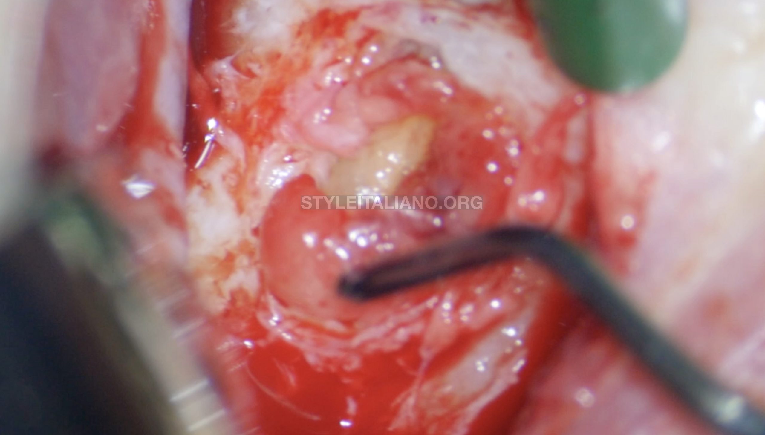
Fig. 2
Surgical access
After soft tissue elevation a huge mesial lesion was present and it was removed with a micro curette.
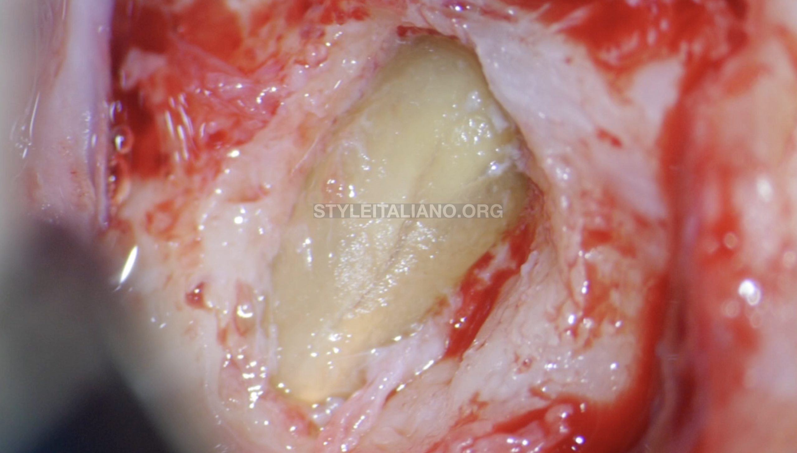
Fig. 3
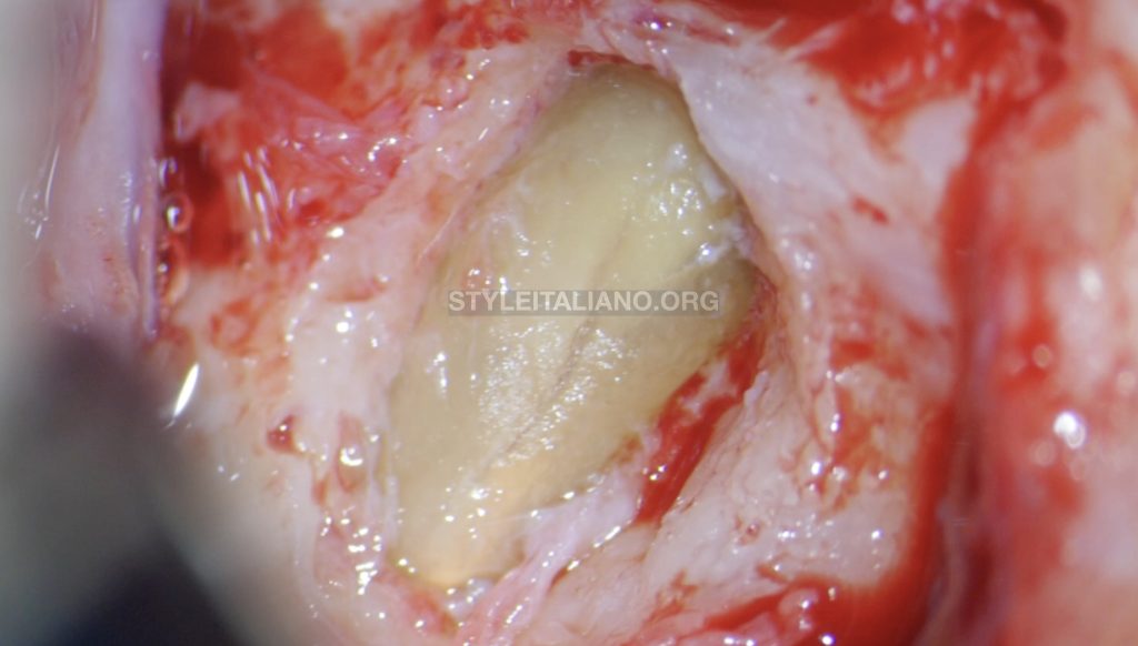
Apical vertical Fracture
An apical vertical fracture appeared with a bone loss at its level
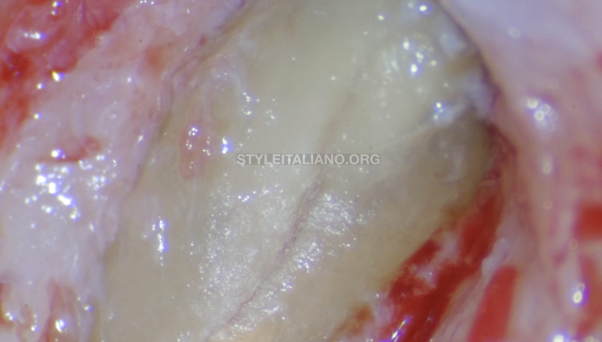
Fig. 4
16x vision of fracture
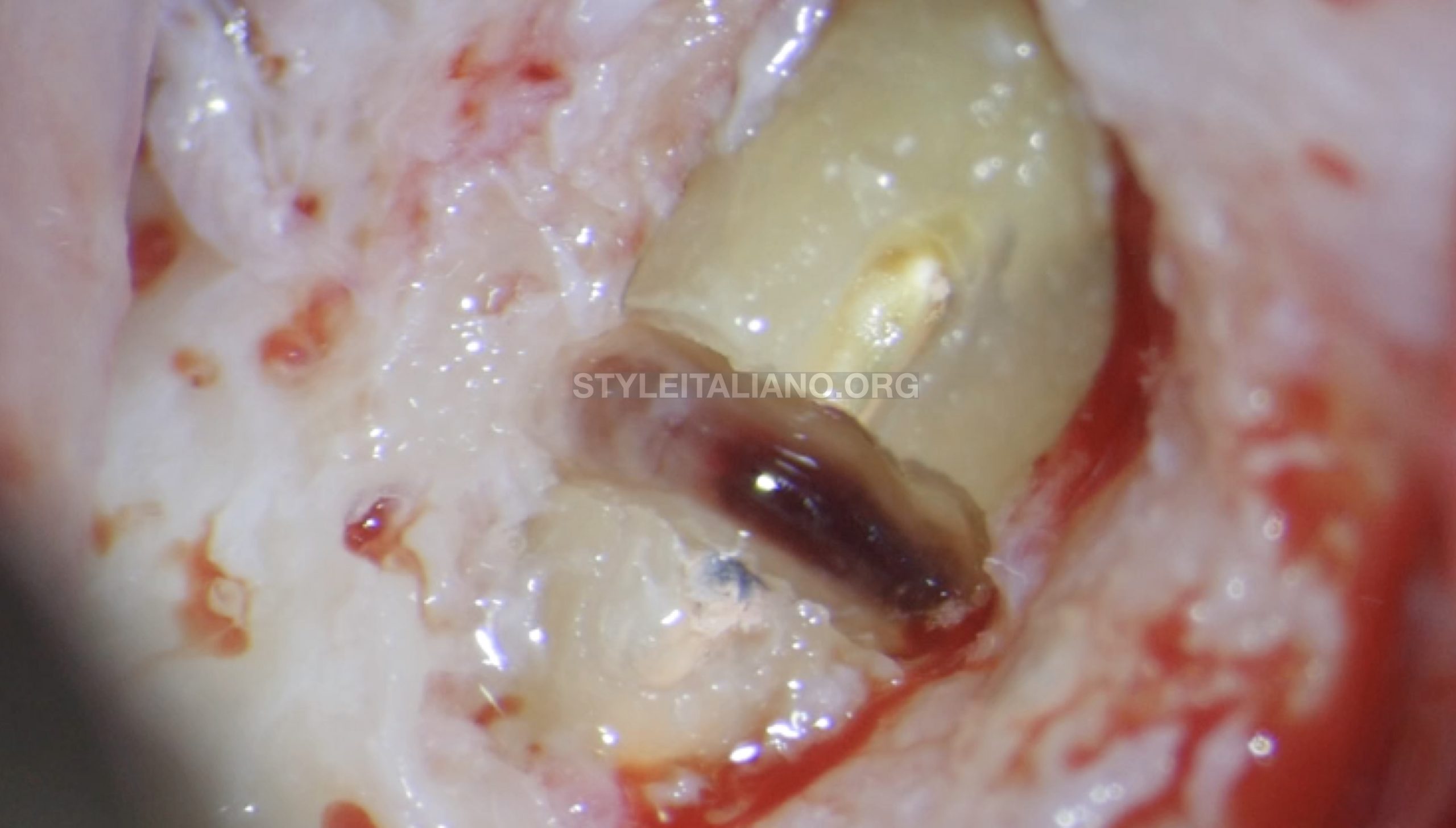
Fig. 5
The vertical fracture was prepare before with a round bur in order to understand its depth. Considering that it involved only the external part of the root , it has been decided to remove surgically the apex leaving the sound part of the root.
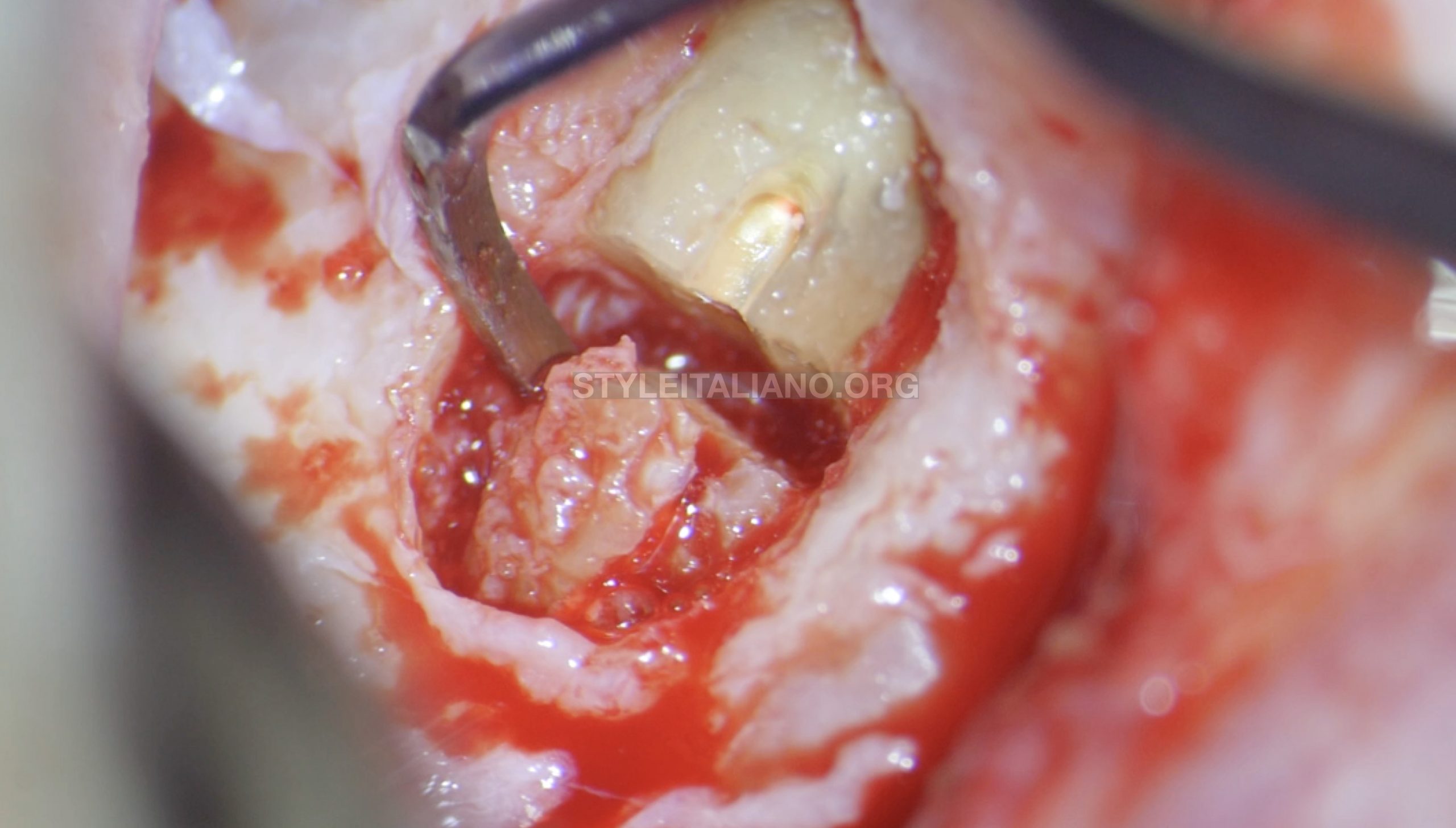
Fig. 6
Apex removal
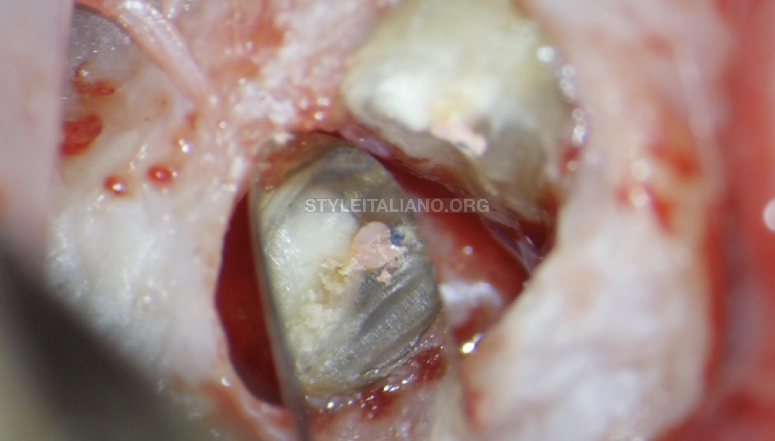
Fig. 7
Micro mirror view
With a micro mirror has been detected that fracture was no more present. Metylen blue in this case is extremely suggested.
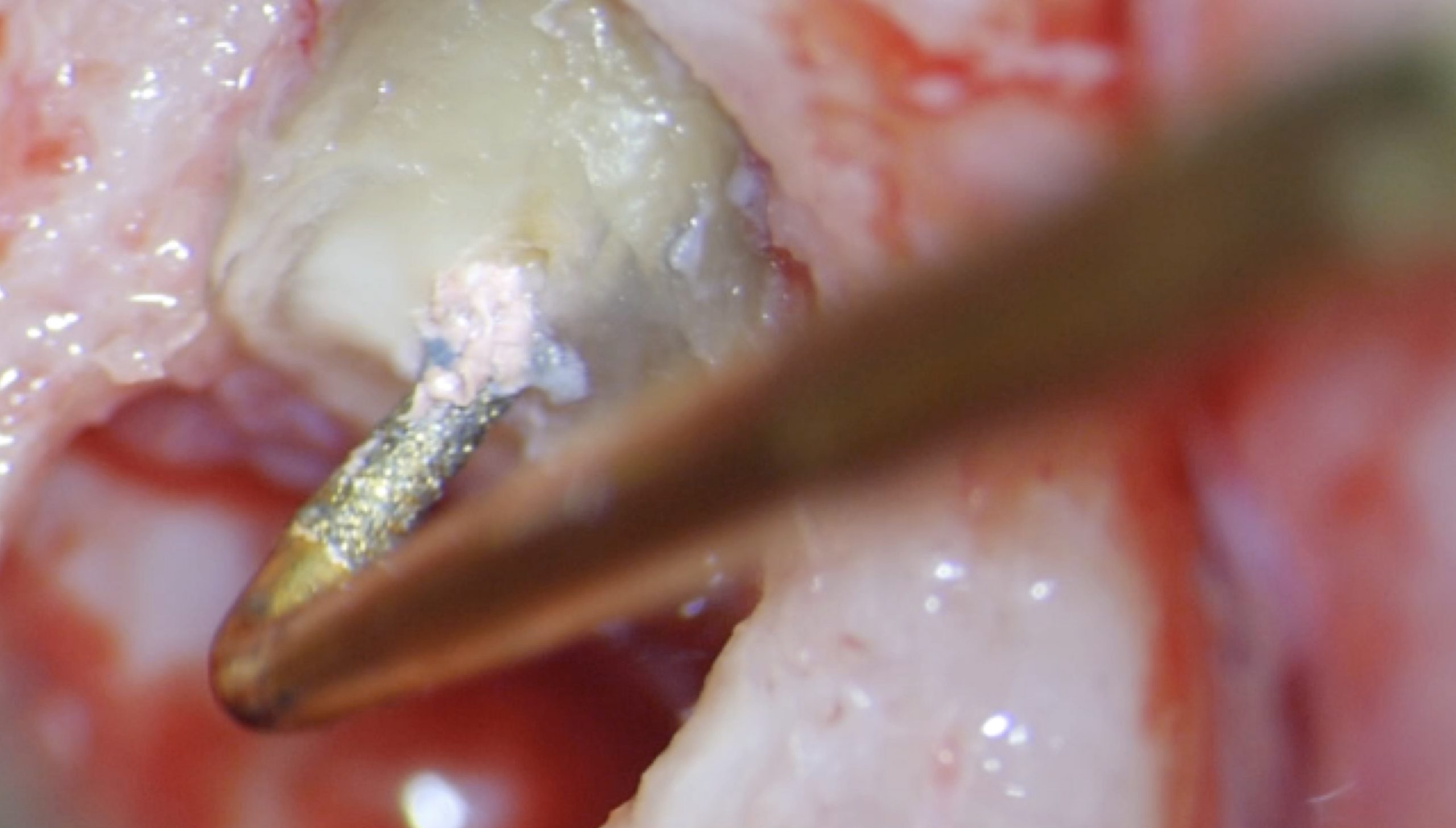
Fig. 8
Retro Prep with a Kid tip
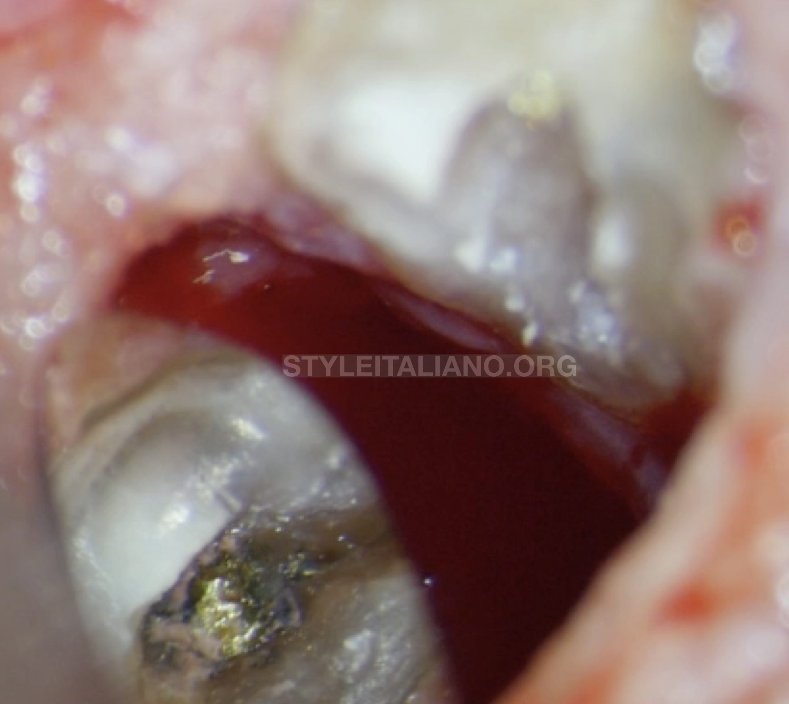
Fig. 9
16x vision through a micro mirror
The retro prep involved also the tip of the screw metal post in order to create more space for retro filling material
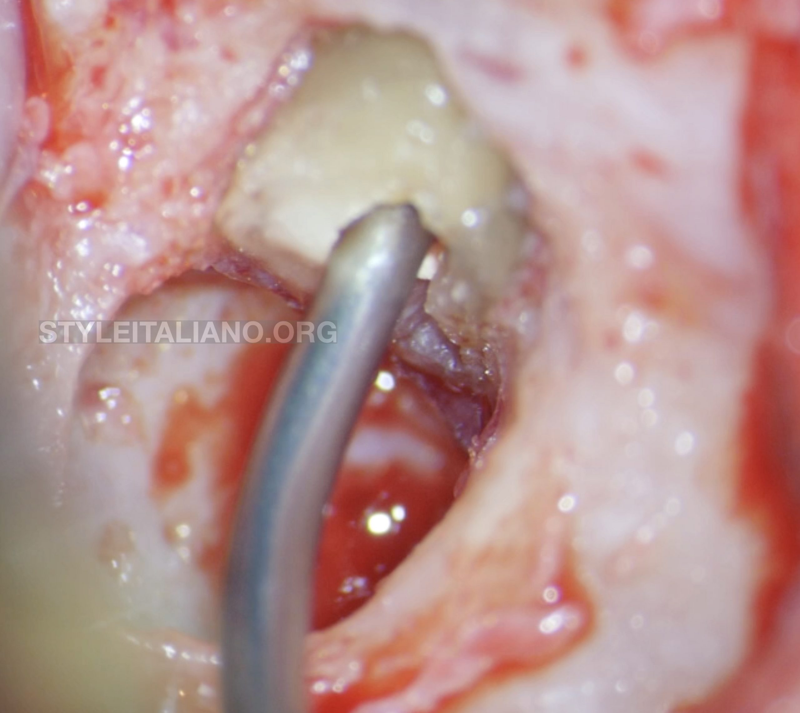
Fig. 10
Retrofilling
Map system with a NiTi tip and PD MTA has been used for managing the retrofilling of the root.
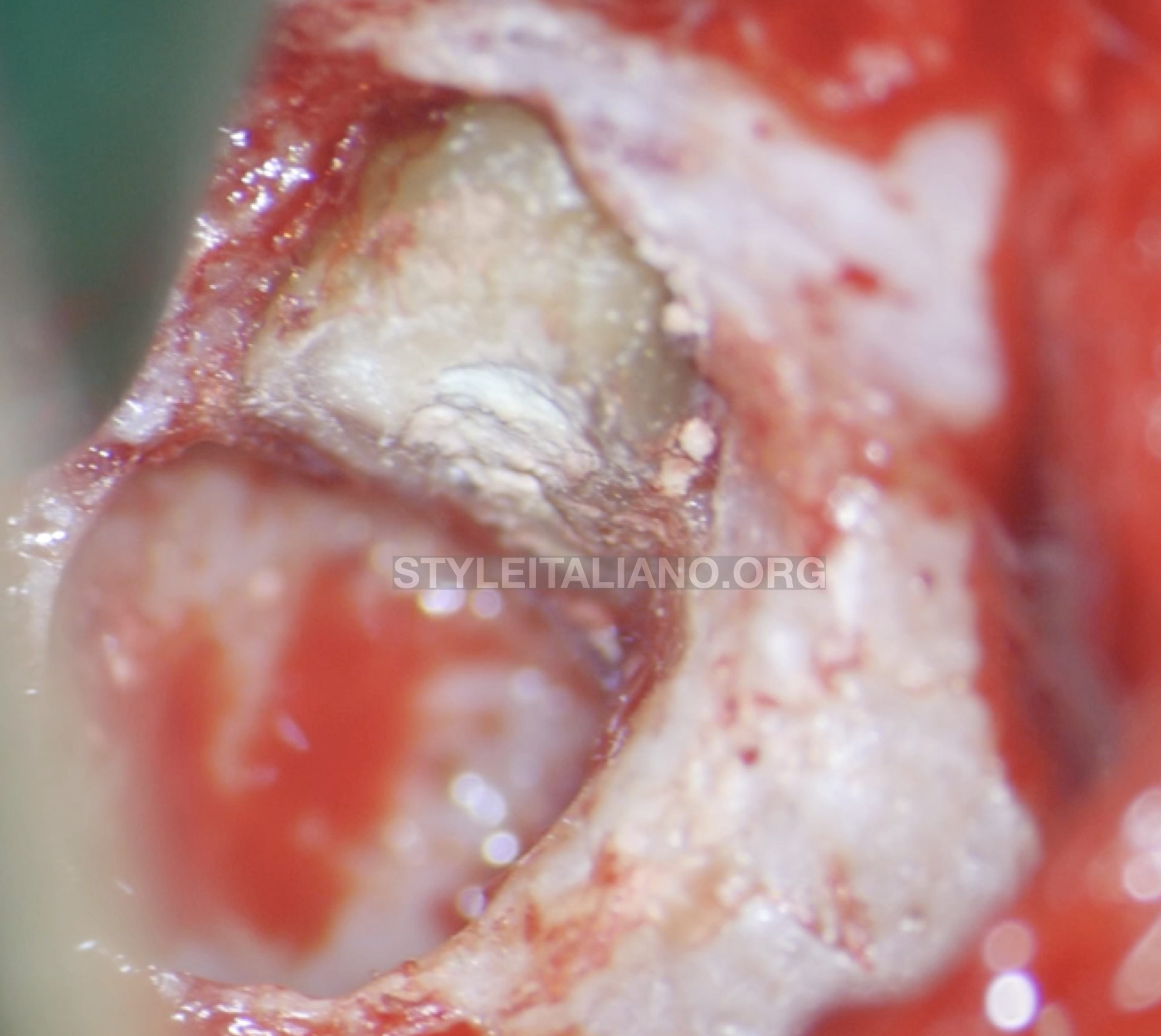
Fig. 11
Surgical area after MTA placement
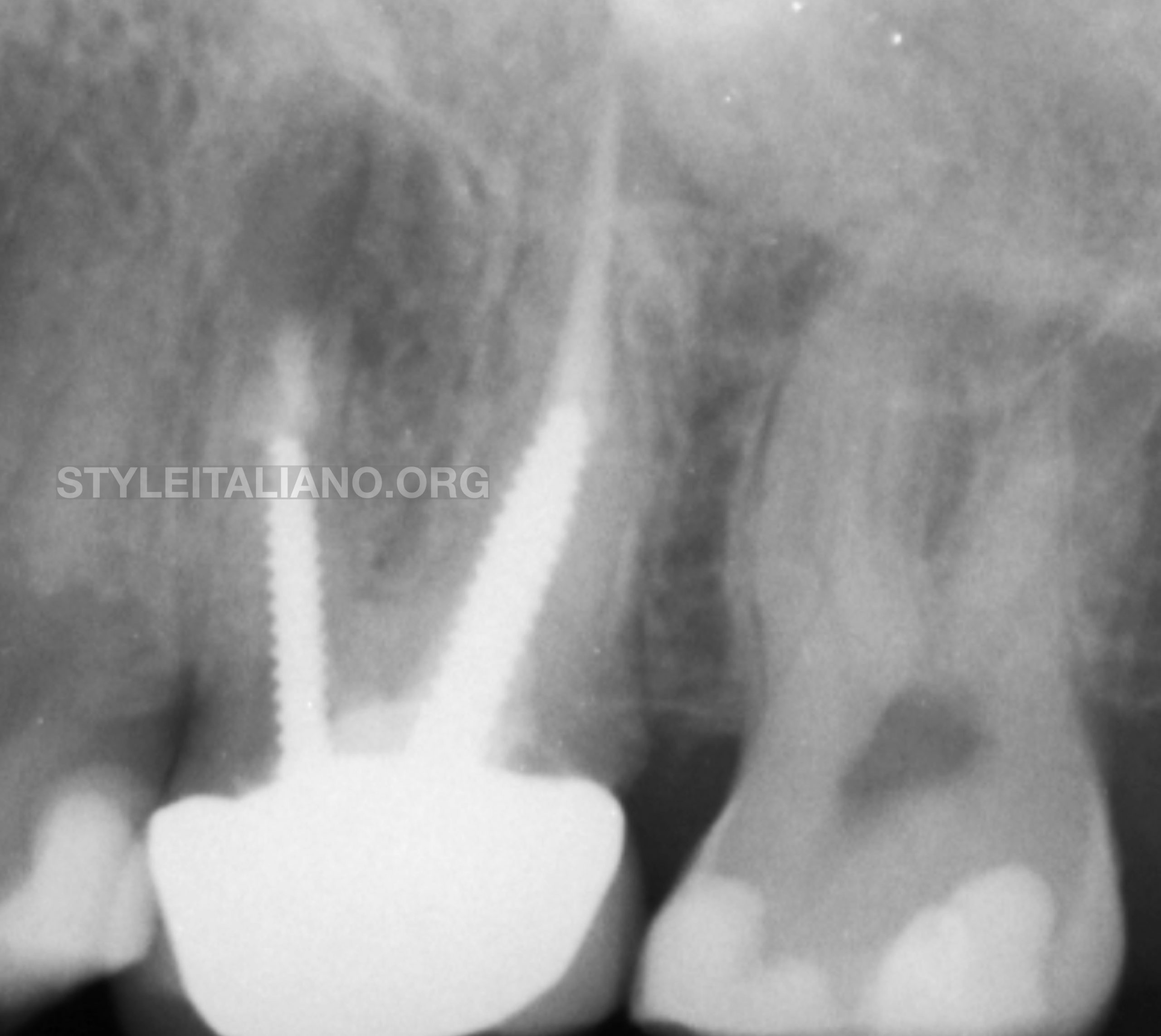
Fig. 12
Post op x Ray
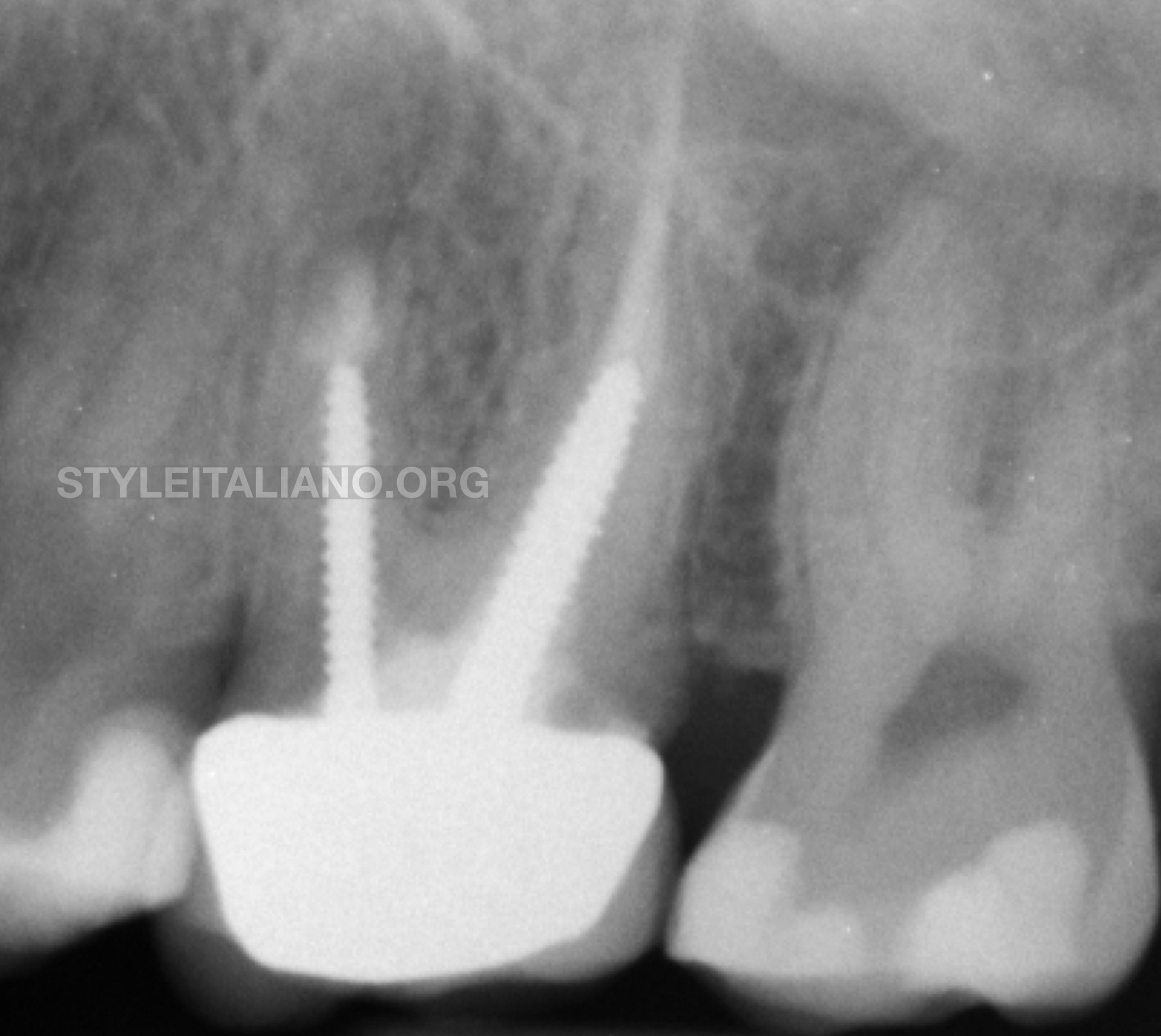
Fig. 13
6 months recall
After 6 months the lesion disappeared and it can be observed a complete healing.
Conclusions
Vertical apical root fracture is one of the most difficult thing to detect, even if a CBCT is used. Like in this case, the surgical approach can be considered as the most conservative ones
Bibliography
Prevalence of vertical root fractures in teeth planned for apical surgery. A retrospective cohort study
M. Maddalone M. Gagliani C. L. Citterio L. Karanxha A. Pellegatta M. Del Fabbro
International Endodontic Journal volume 51 ,issue 9
Apical stress distribution under vertical compaction of gutta‐percha and occlusal loads in canals with varying apical sizes: a three‐dimensional finite element analysis
K. Yuan C. Niu Q. Xie W. Jiang L. Gao R. Ma Z. Huang
International Endodontic Journal 51,233–239, 2018
Resistance to vertical fracture of MTA‐filled roots
Ahmad M. EL‐Ma'aita Alison J. E. Qualtrough David C. Watts
Dental TraumatologyVolume 30, Issue 1

