Upper molar, how to prepare the post space: from A to Z
23/09/2022
Massimo Giovarruscio
Warning: Undefined variable $post in /home/styleendo/htdocs/styleitaliano-endodontics.org/wp-content/plugins/oxygen/component-framework/components/classes/code-block.class.php(133) : eval()'d code on line 2
Warning: Attempt to read property "ID" on null in /home/styleendo/htdocs/styleitaliano-endodontics.org/wp-content/plugins/oxygen/component-framework/components/classes/code-block.class.php(133) : eval()'d code on line 2
Post space preparation is a critical step to achieve clean dentinal surfaces for adhesion of the cementing material. Therefore, complete removal of the root filing materials is essential to enhance the adhesive bond to the dentine and increase post retention. Furthermore, the presence of residual gutta-percha and deficient dentine hybridisation may result in poor sealing of the resin–dentine interface.
Moreover, remnants of endodontic sealers, owing to their chemical compositions, may negatively affect post retention. For example, eugenol inhibits the free radical polymerisation reaction of chemically cured composite resins, which results in a weakened bond when zinc oxide-eugenol sealers are used.
Several techniques have been proposed for removing root canal filing materials, most of them used in root canal retreatment, such as chemical solvents (eucalyptol, xylol, chloroform), heat or hand files to soften and remove the gutta-percha, and rotary instruments (Gates-Glidden, nickel-titanium instruments), which represent the easiest method to clean root canal walls.
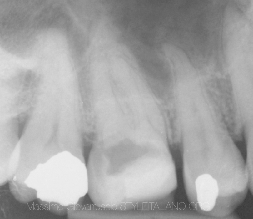
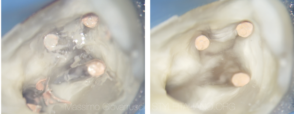
Fig. 1
The upper right molar has been accessed by referral dentist as an emergency appointment. The patient has been referred to our practice to finalize the root canal treatment and the post restoration.
As soon as the root canal treatment has been completed, the post space and the chamber are been cleaned using GP and Sealer solvent. All the procedures have been performed under microscope vision.
The first video shows all the steps to clean and prepare the access cavity and the post space for a proper restoration. Dedicated long neck burs are been used to remove the excess of GP and to achieve a “fresh dentine”for an optimal adhesion. The using of Air Abrasion guarantees a great cleaning of the surface before the adhesive steps.
As soon as the post space is cleaned, we have to choose the right post for the canal. We usually prefer to do not enlarge the canal to fit the post but we prefer to find the right post that fits passively into the canal. Usually the best canal is the Palatal root for upper molar and Distal root for lower molar.
The glass fibre post should be treated to achieve the best adhesion with cement or composite or bulk material. The Itena Clinical Silane has been applied on the post and an equal amount of Bond and Activator has been applied too. The cavity access and the canal space are been etched and prepared with Primer and Bonding (Itena Clinical - Bond A and Bond B + Activator). Light curing is requested for at least 40 seconds.
The last video is showing the final step to restore the tooth using a glass fibre post and dual composite (Itena Clinical - Dentocore). It is important to restore the tooth with incremental steps to avoid composite shrinking as much as possible. A soon as we completed the restoration and the shaping of the tooth, we want to cover the glass post head with composite to avoid a fluid or saliva infiltration. This is an additional step, but is very important to do not leave the glass post head in contact with saliva due to the potential bacteria infiltration.
The restored tooth is now ready for final crown preparation. The final shape can be used to prepare a temporary crown, saving time for the patient.
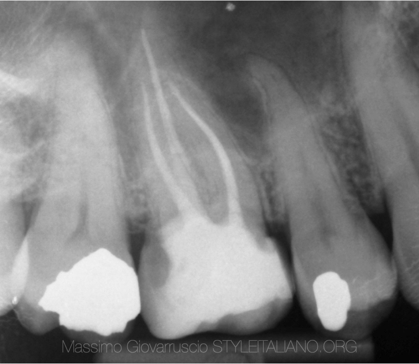
Fig. 2
Post operative X-Ray
Conclusions
The weak point of the whole tooth-posts-restoration system may be identified in the adhesion between the substratum and the resinous cements. Many in vivo and in vitro studies have showed how the fiber post reconstruction failure is dependent on the bonding-dentin interface. To achieve the best adhesion the optimal interface preparation is related to the bonding agents. The bonding mechanism exploits the dentinal tubules penetration by the resin and the collagen fiber exposition.
The latest adhesive material, as Itena Clinical Products, are been used on this case and show a great result in terms of application of the material and great clinical result.
Bibliography
1. Bhuva B, Giovarruscio M, Rahim N, Bitter K, Mannocci F. “The restoration of root filled teeth: a review of the clinical literature” Int Endod J. 2021 Apr;54(4):509-535. doi: 10.1111/iej.13438. Epub 2021 Jan 5. PMID:33128279.
2. Ivanovic Coniglio, Elisa Magni, Cecilia Goracci, Ivana Radovic, Carlos Augusto Carvalho, Simone Grandini, Marco Ferrari “Post space cleaning using a new nickel titanium endodontic drill combined with different cleaning regimens” J Endod 2008 Jan;34(1):83-6.
3. Giuseppe Lo Giudice, Angelo Lizio,Roberto Lo Giudice, Antonio Centofanti,1Giuseppina Rizzo, Michele Runci, Angela Alibrandi, and Marco Cicciù. “The Effect of Different Cleaning Protocols on Post Space: A SEM Study”. Open Access Vol 2016, Article ID 1907124
4. Dino Re, Davide Augusti, Gabiele Augusti, Francesca Cerutti, Antonio Cerutti.“Cleanliness of dentinal walls following post space preparation using magnification”. ENDO (Lond Engl) 2010;4(3):207-214