Three roots in a first maxillary premolar: A case report
17/02/2025
Fellow
Warning: Undefined variable $post in /home/styleendo/htdocs/styleitaliano-endodontics.org/wp-content/plugins/oxygen/component-framework/components/classes/code-block.class.php(133) : eval()'d code on line 2
Warning: Attempt to read property "ID" on null in /home/styleendo/htdocs/styleitaliano-endodontics.org/wp-content/plugins/oxygen/component-framework/components/classes/code-block.class.php(133) : eval()'d code on line 2
Missed anatomy is the main factor of endodontic failure according to the literature. So, we need to have sound knowledge of anatomic variations in the tooth we are going to treat, in order to devise a proper treatment plan. In this article, I will address the anatomical variation of three roots in a maxillary first premolar. According to the literature, prevalence of three roots, 2 buccal and one palatal, in these teeth is 5%. In order this anatomy, ideally, we would use a CBCT from the patient. Even if it is old, we would have signs that there are three roots. Nowadays, most of the patients may have such exam so we should ask. Then, assessing the pre-op periapical xray might reveal the three roots under examination. Finally, intraoperatively, we can assess by examining the chamber floor with magnification.
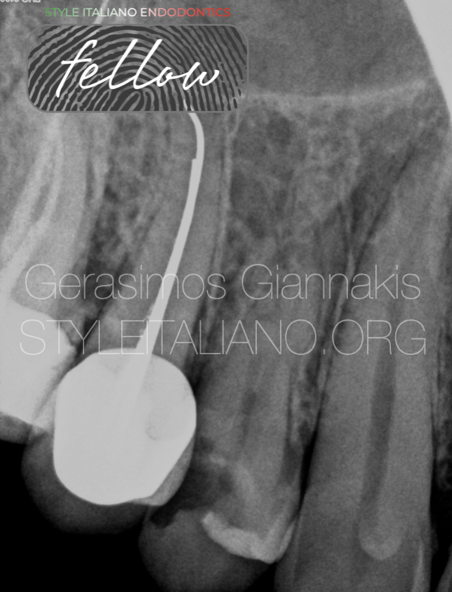
Fig. 1
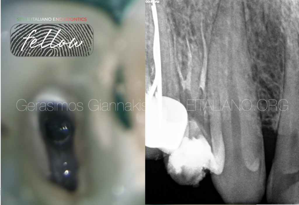
A patient with clear medical history presented to the clinic reporting pain in the upper jaw. After radiographical and clinical examination, we diagnosed irreversible pulpitis in tooth #14. As we can see in the xray, after close examination we may see the lamina dura of three canals.
Fig. 2
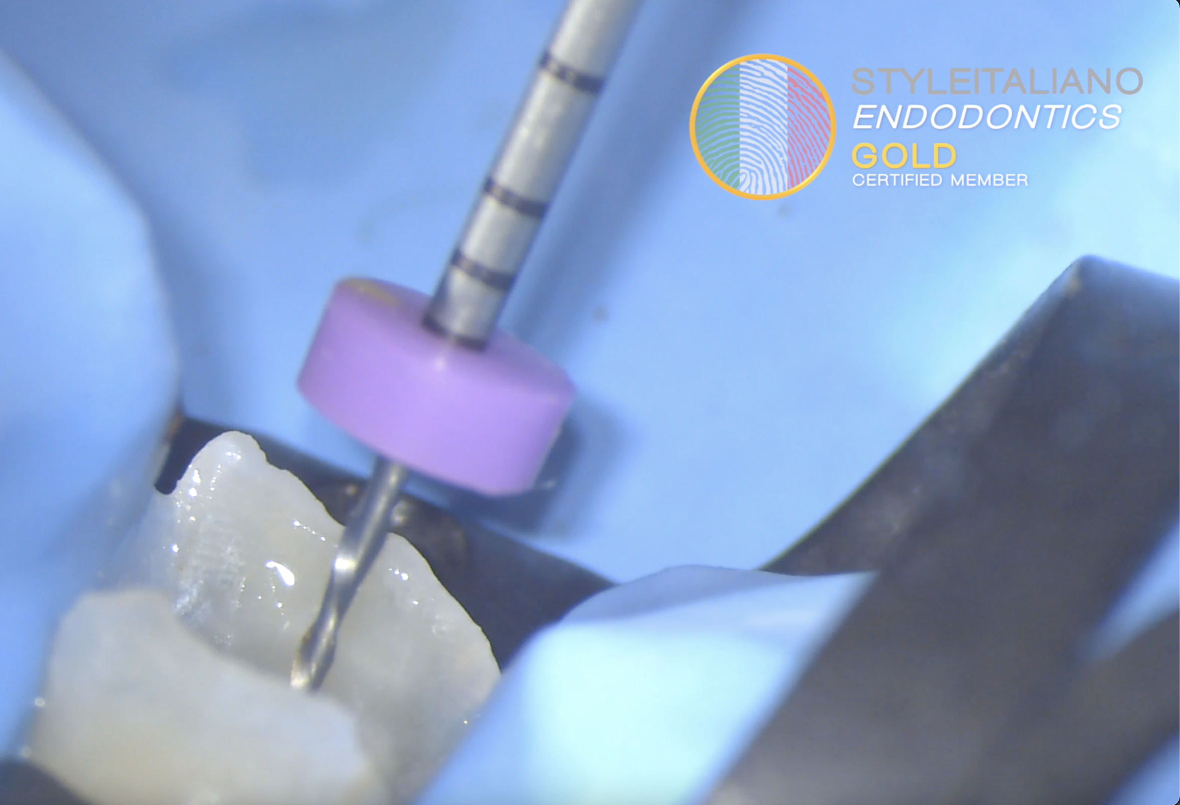
After anesthesia and proper isolation, we performed the access cavity. Pre-endo build up is essential in such cases. Following the pulp chamber floor anatomy and having in mind the possibility of three canals we identified this anatomy.
A clinical tip in absence of magnification is to check the orientation of the file in the buccal canal. If it is not in the center of the tooth, but goes in a mesial or distal direction this is a sign that there is another canal.
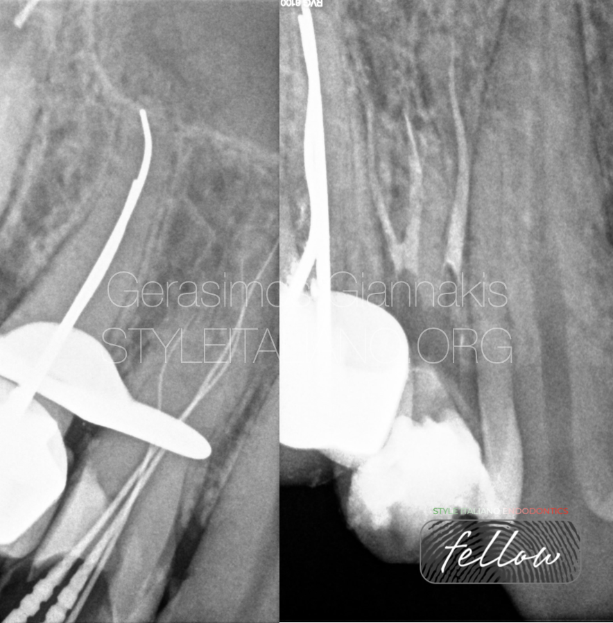
Fig. 3
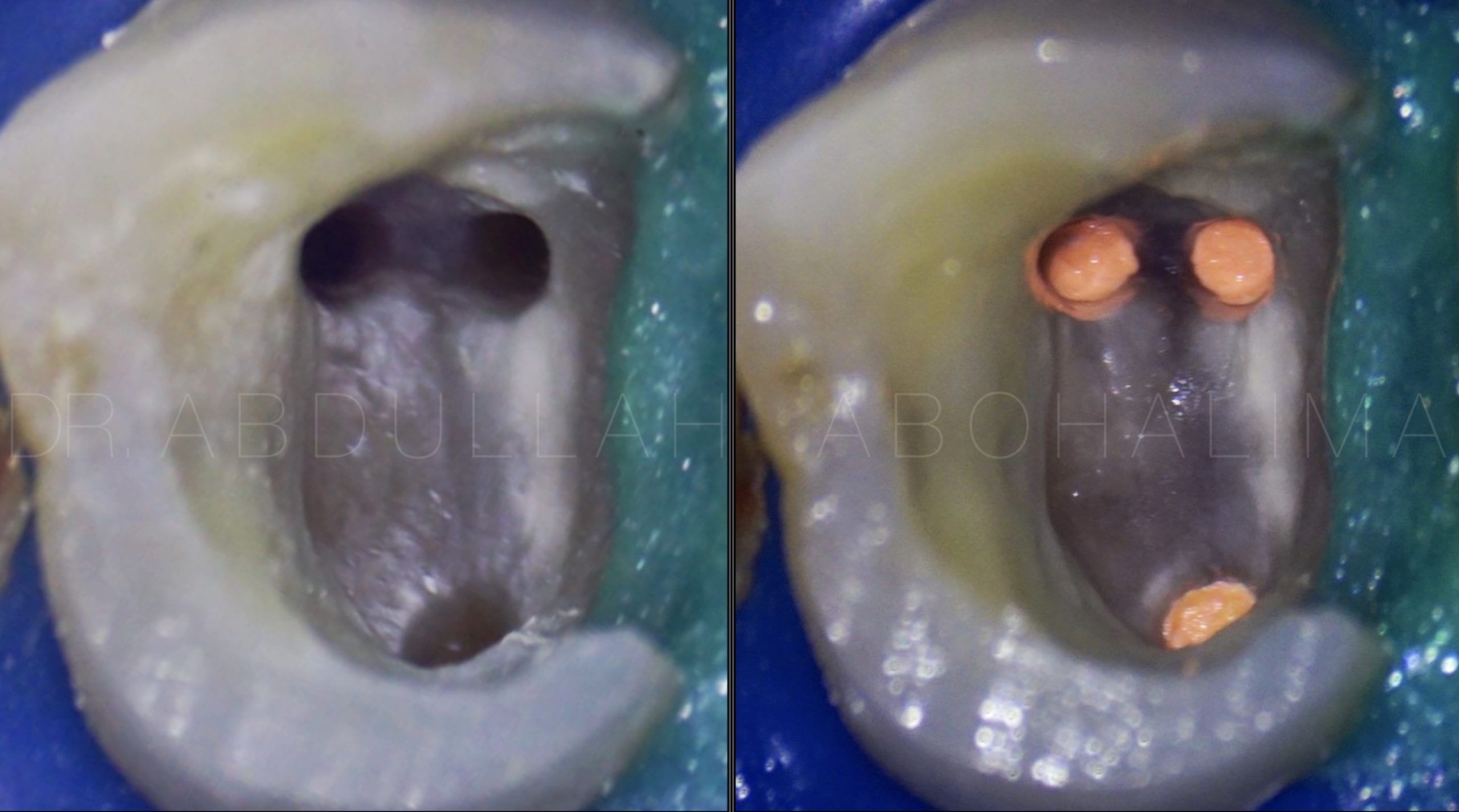
Identifying the extra canal is one thing, now we have to prepare the anatomy. The deeper the split of the canal, the harder it is to prepare. Magnification is vital to such cases. We need to be able to identify in which canal we introduce the file and not do it blindly. In this case, the canal split was in a midrooot location. So, I did not have to do a lot of coronal preflaring in expense to dentine. The final preparation for both buccal canals was 20.05 and palatal 25.04.
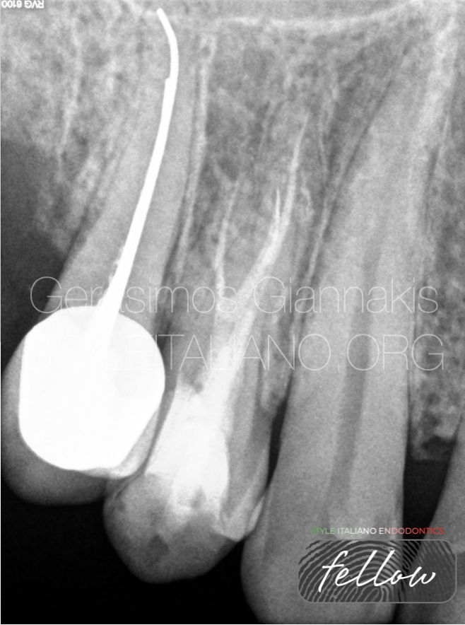
Fig. 4
In the final xrays, we can appreciate the anatomy. This is the 5 months follow-up of the case. The tooth is asymptomatic and functioning well. Now it is planned for cuspal coverage restoration.

Fig. 5
About the author:
Gerasimos Giannakis
Gerasimos Giannakis received the Doctor of Dental Surgery degree from the University of Athens in 2018.
He has special interest in the field of endodontics, restorative dentistry and microsurgical techniques.
Since then has completed
STYLE ITALIANO Daily menu 40th edition , Milan 2020
TOOTH WEAR MASTER COURSE, by Didier Dietschi 2021
EXPERT COURSE IN MASTERING SOFT TISSUE SURGERY,
In cooperation with the University of Cologne 2022
MICROSCOPE SIMULATION COURSE ON CANAL BLOCKAGE MANAGEMENT by Chaniotis, Sousa Dias, 2020
He is working as an endodontics associate in a clinic, responsible also for the restoration of endodontically treated teeth.
Working exclusively under the microscope and rubber dam, has the opportunity to document the cases.
In the Style Italiano Congress in Greece 2023, was part of the bootcamp and became fellow of the Style Italiano Endodontics community.
Conclusions
The split location defines the difficulty level of the treatment. However, without magnification and knowledge of the anatomy it is possible that we could miss the canal. Xray evaluation, CBCT or periapical, is very important to properly address the anatomy of such teeth.
Bibliography
Root canal morphology of the maxillary first premolar
F J Vertucci, A Gegauff
Root and Root Canal Morphology of Maxillary First Premolars: A Literature Review and Clinical Considerations
Ahmad IA, Alenezi MA.
Evaluation of the Root Canal Anatomy of Maxillary and Mandibular Premolars in a Selected German Population Using Cone-beam Computed Tomographic Data
Bürklein S, Heck R, Schäfer E.