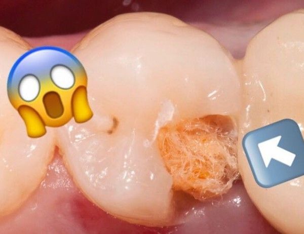
The removal of filling material during retreatments
30/06/2020
Marc Kaloustian
Warning: Undefined variable $post in /home/styleendo/htdocs/styleitaliano-endodontics.org/wp-content/plugins/oxygen/component-framework/components/classes/code-block.class.php(133) : eval()'d code on line 2
Warning: Attempt to read property "ID" on null in /home/styleendo/htdocs/styleitaliano-endodontics.org/wp-content/plugins/oxygen/component-framework/components/classes/code-block.class.php(133) : eval()'d code on line 2
The prognosis of initial endodontic treatment is mostly favorable . Nevertheless, failure can occur due to the persistence of bacteria following initial treatment or due to inadequate disinfection, shaping and obturation of the root canal system. The possibility of a secondary infection is also not to be excluded, especially when a defective coronary reconstruction leads to percolation.
In such cases non surgical retreatment is required.
The success of the retreatment will depend amongst other criteria on the complete removal of all filling material debris, such as gutta-percha or sealer. In fact, the old filling material can contain necrotic tissues and bacteria often assembled in biofilm. These bacteria and their toxins can diffuse into the peri-radicular tissues and cause symptomatic or asymptomatic apical periodontitis.
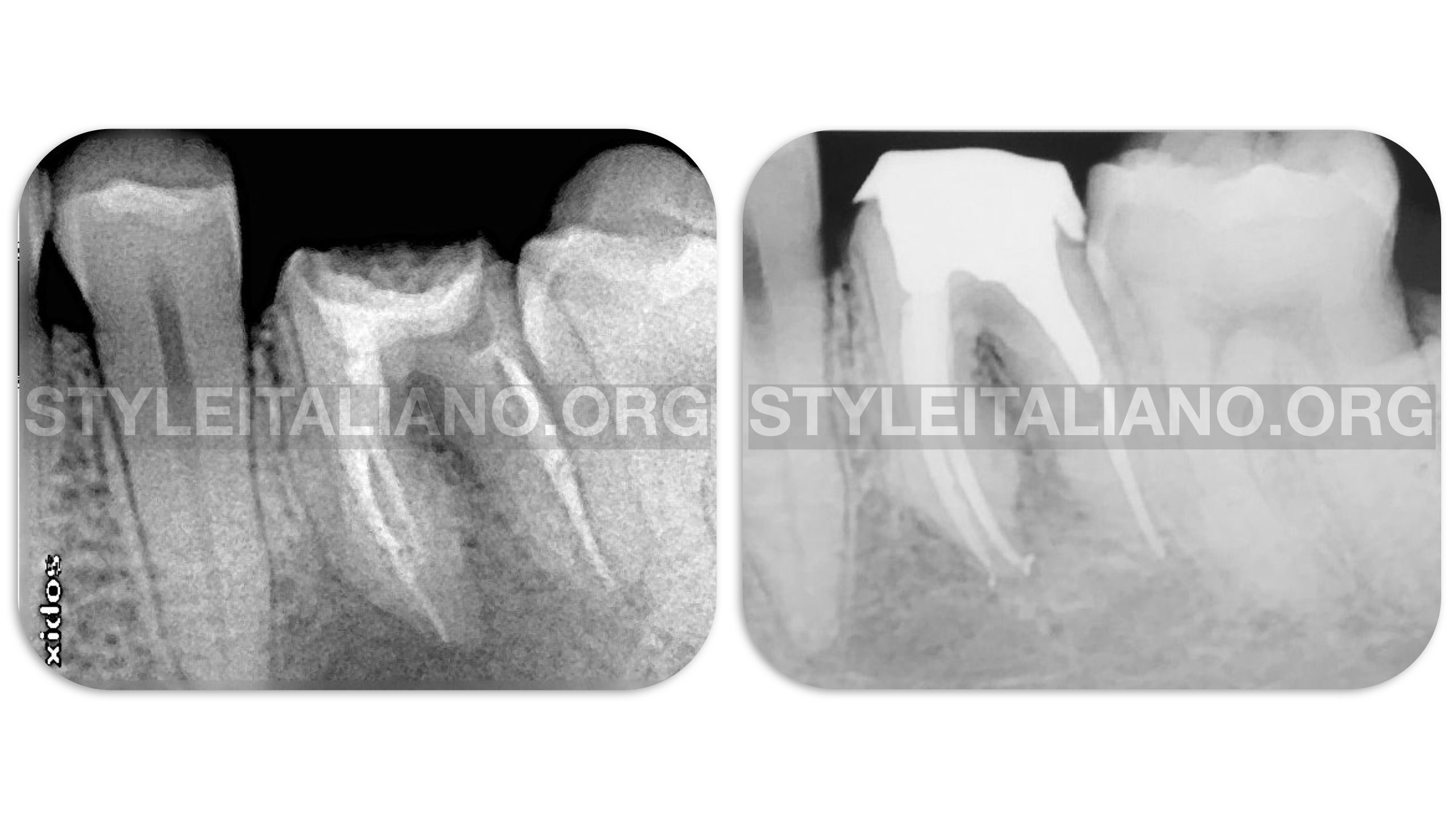
Fig. 1
A lower mandibular molar shows a large peri-apical periodontitis after first root canal therapy with poor obturation. After retreatment, removal of all the filling material, cleaning, shaping and filling, a post and core and a temporary crown was placed on the tooth by the referring dentist. A 6 months Follow up shows healing.
Traditionally obturation material was removed with hand files, but since many years rotary and reciprocating files are used in retreatment cases. Some exclusive retreatment systems exists, but many clinicians also use shaping system to remove old filling material.
Instruments combining good cutting efficiency, flexibility, resistance to fracture and a safe tip are more likely to remove old filling material without harming the original anatomy of the root canal system.
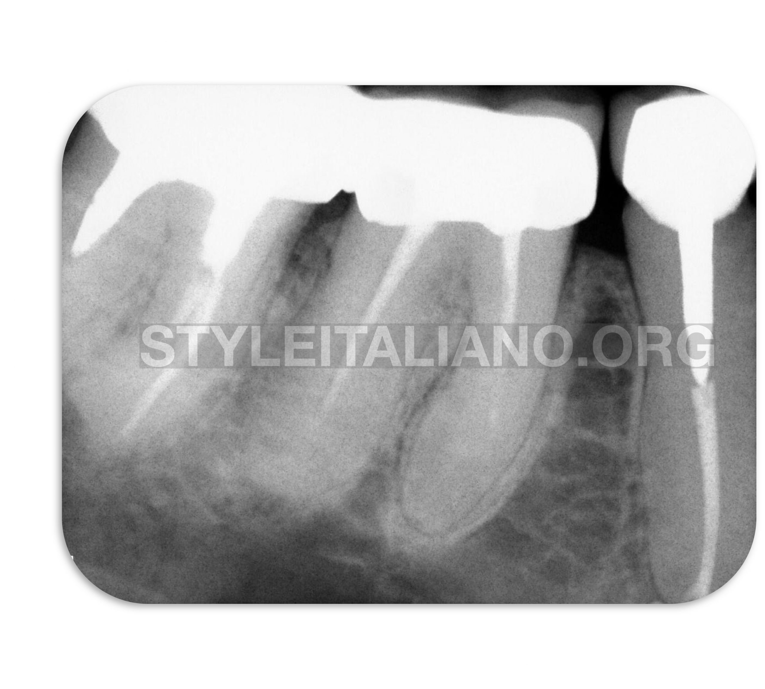
Fig. 2
The case of a lower right first mandibular molar with short obturations in all canals and a ledge in the mesial root.
The obturation material is removed without the use of any solvent with the a rotary instrument and activation with Ultra X (Eighteeth, Changzhou, China)
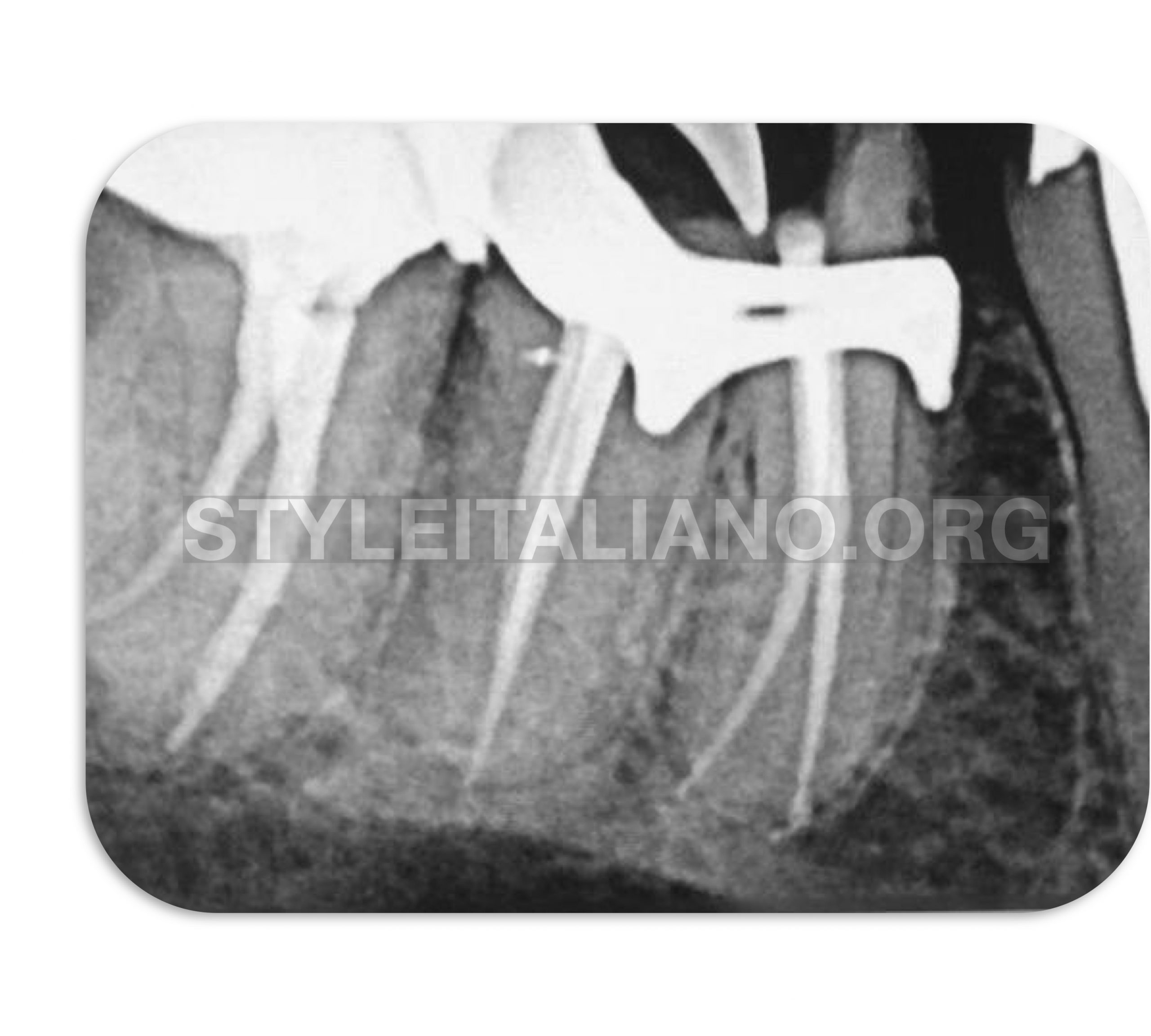
Fig. 3
The final X ray after shaping, cleaning and obturation in one session.
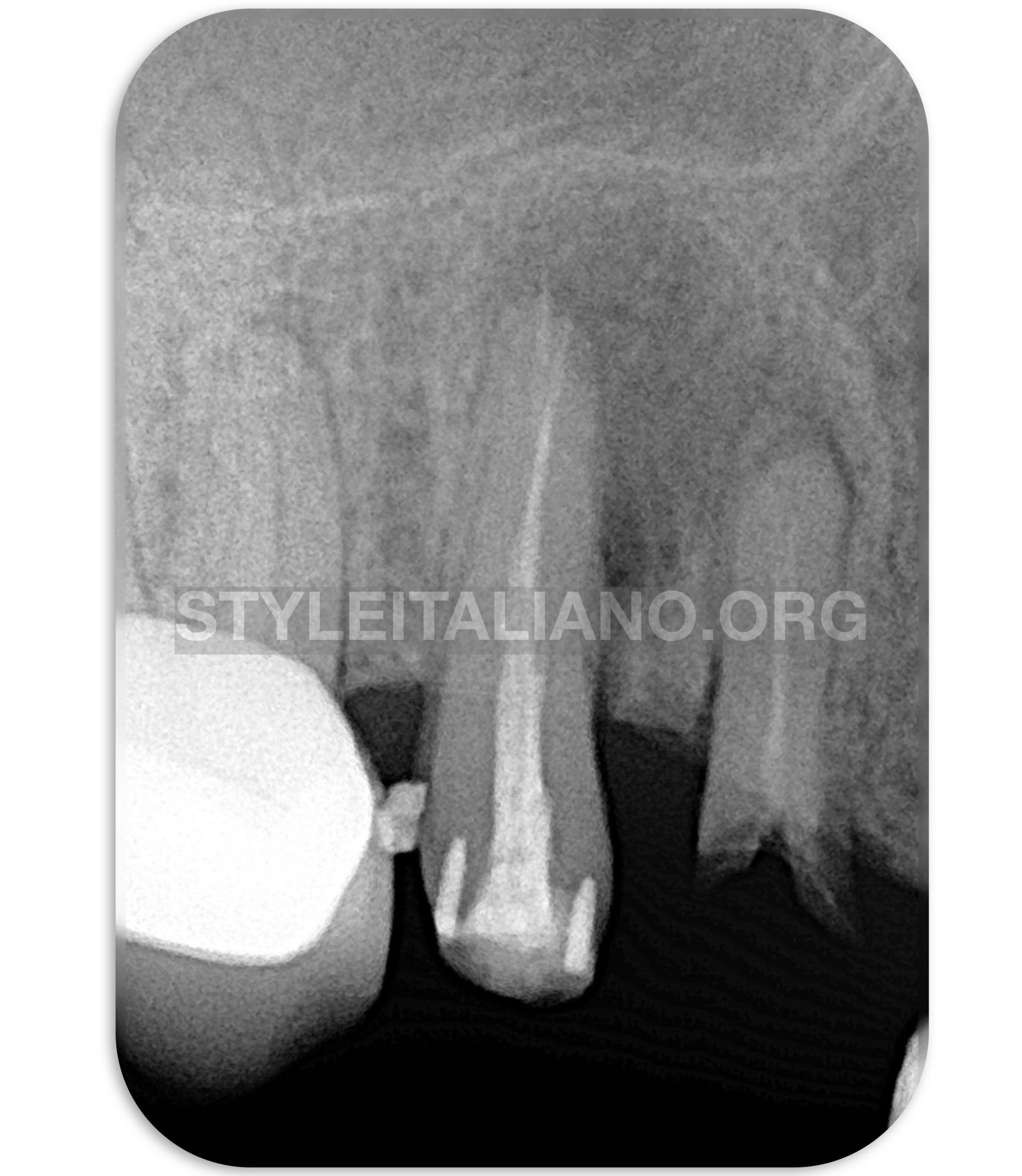
Fig. 4
Exclusive retreatment instruments are designed specifically for the removal of filling material. This case was managed with these instruments.
Initial periapical Xray showing a large periapical periodontitis. Clinically, the tooth was asymptomatic and no probing was noticed.
During the the retreatment procedure gutta-Percha was removed with an exclusive retreatment instrument.
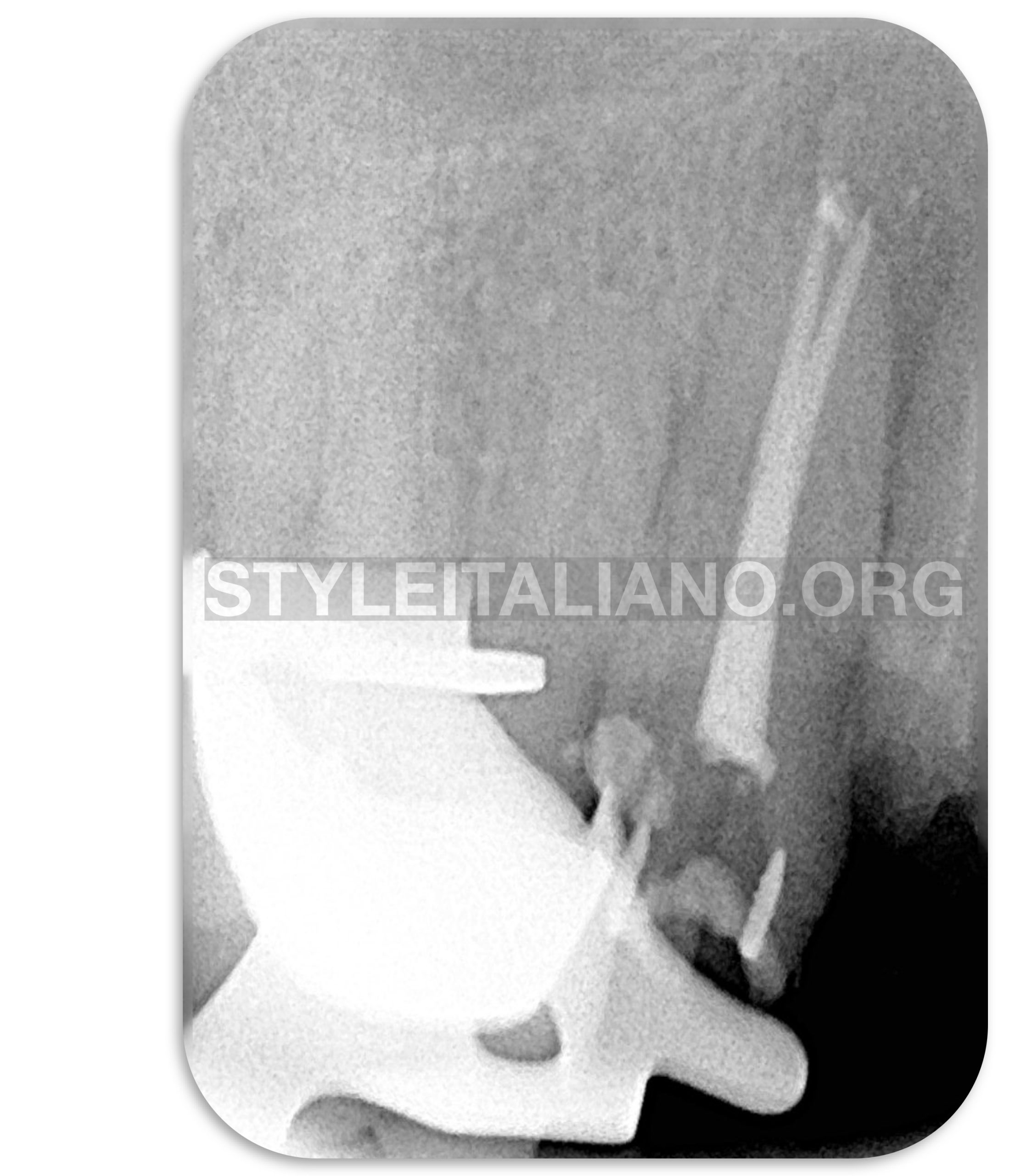
Fig. 5
Final Peri-apical X ray after one session retreatment.
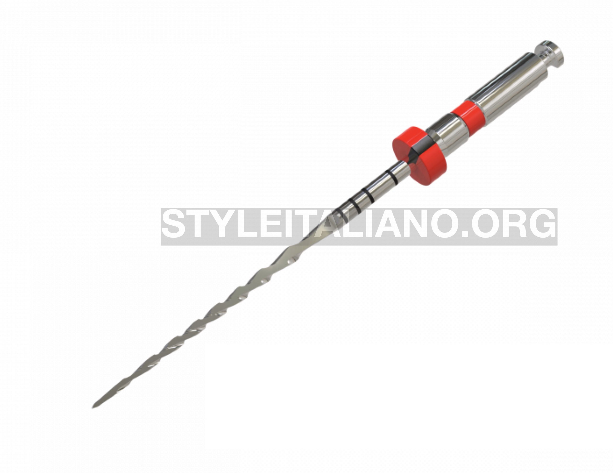
Fig. 6
Fanta F-one
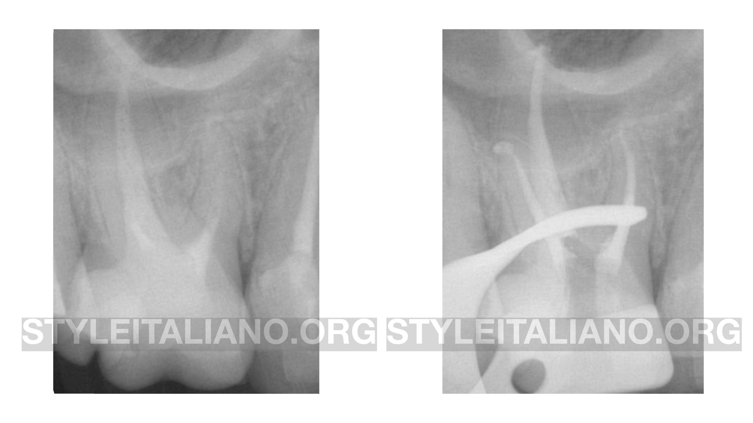
Fig. 7
Clinical case retreatment of an upper molar by Dr. Calogero Bugea
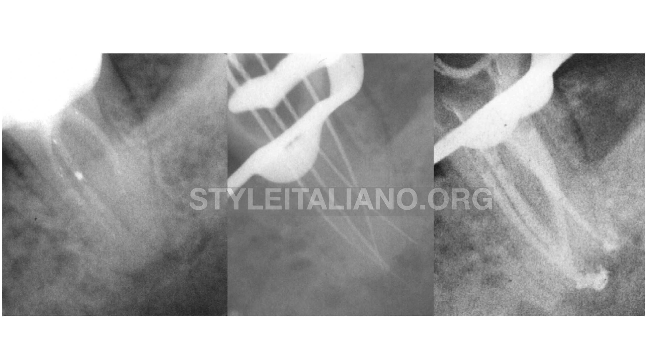
Fig. 8
The complete elimination of the material can be assessed clinically, by observing the canals under high magnification or radiographically by taking a peri-apical X ray confirming the working length determined with an apex locator
Nevertheless, in many cases, the remaining material can’t be detected neither with the microscope nor with the radiograph, because it can be hidden by anatomical obstacles such as curvatures or anastomosis. According to many studies, the removal of filling material from the apical third is very challenging.
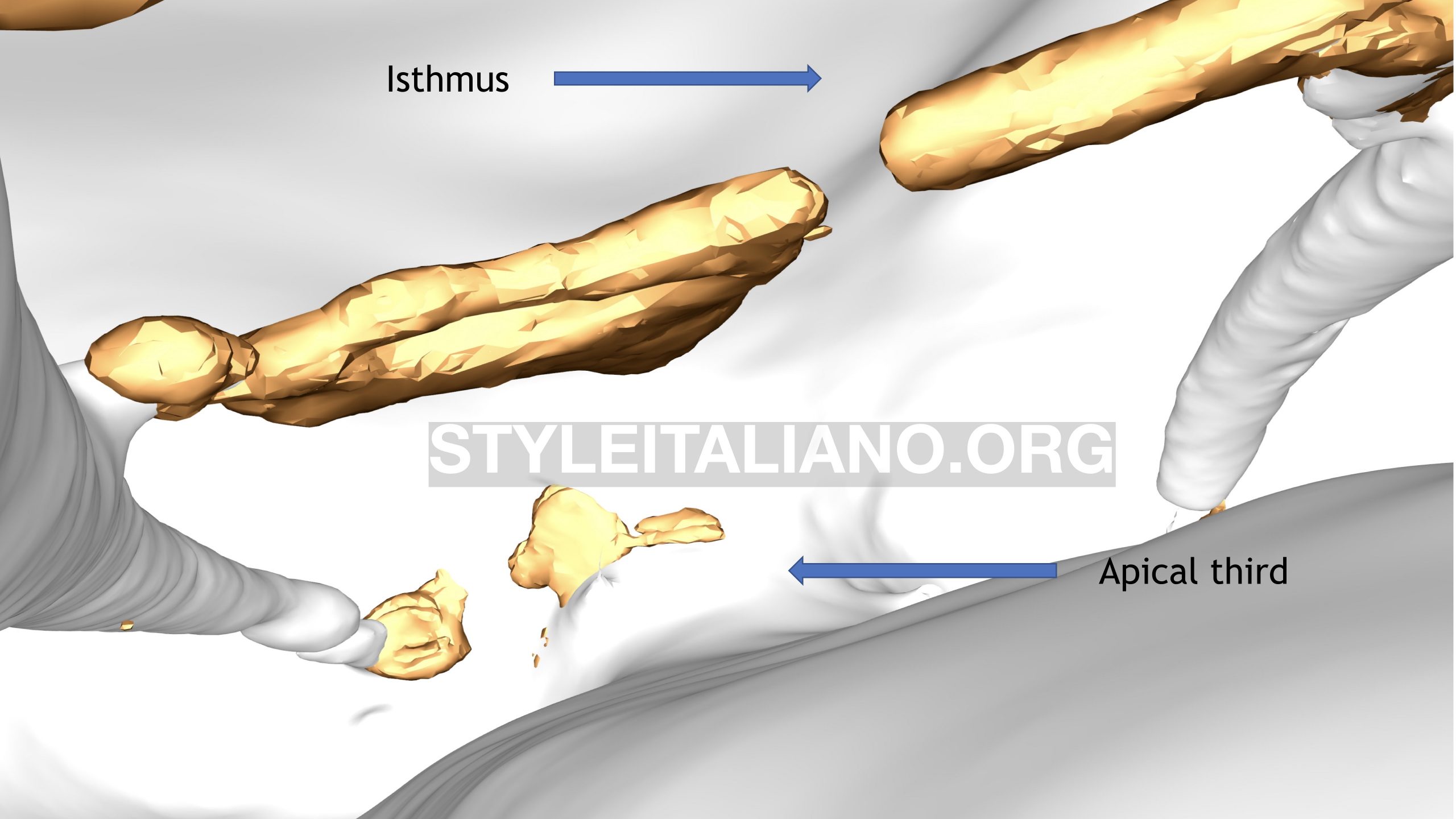
Fig. 9
A 20 microns micro CT image reconstructed from an extracted mesial root of mandibular molar after material removal, shows material remnants in the isthmus area and in the apical part of the canals.
This 3D animation after micro CT 3D reconstruction showing the presence of material remnants after retreatment on an extracted tooth.
Until now, almost all publications confirmed the impossibility to completely eliminate all the filling material during retreatment. The usage of a supplementary approach such as ultrasonic activation (UAI) or sonic activation (SI) seems to enhance the removal of remaining material.
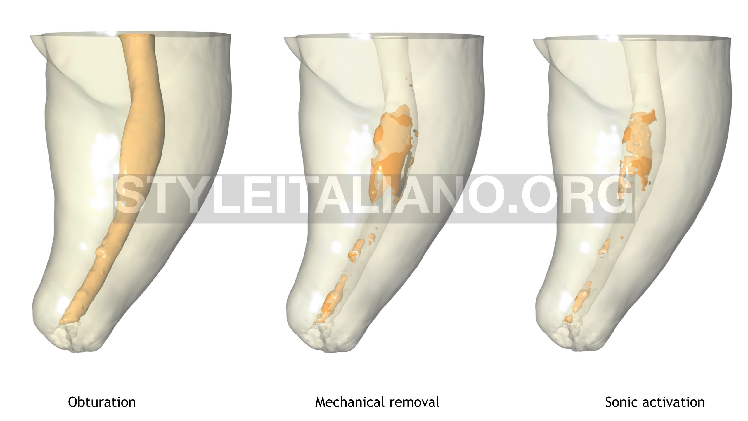
Fig. 10
Micro CT images confirming the effectiveness of the UAI EndoUltra (Vista Dental Products, Racine, WI,USA) in removing more remnants.
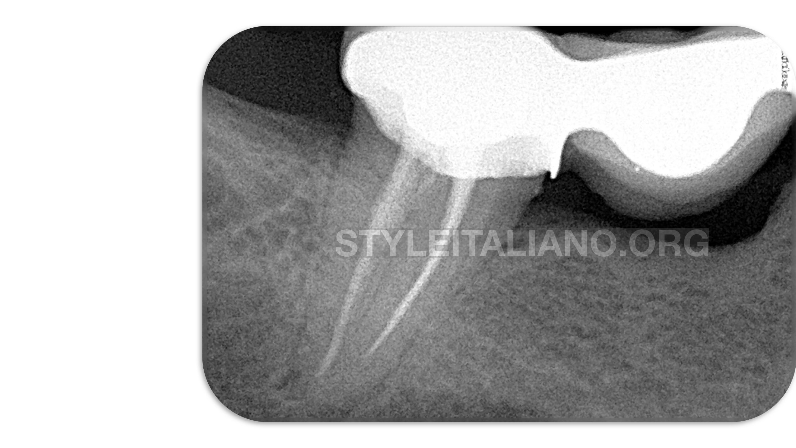
Fig. 11
A case of a mandibular symptomatic molar retreated through the bridge.
Removal of carrier based obturation with Reciproc Blue (VDW) from a C shaped canal.
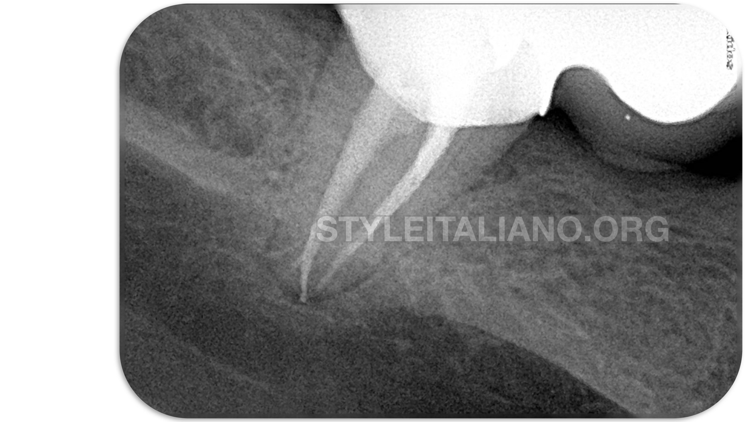
Fig. 12
Final X ray after filling material removal, shaping, cleaning and 3D filling.
Conclusions
No clinical protocol has yet shown the possibility of eliminating the filling material entirely. However the usage of an instrument combining a high cutting efficiency, a non-active point, a high resistance to cyclic fatigue and a flexibility necessary to respect the canal anatomy optimizes the elimination of filling material in difficult anatomical areas.
Supplementary approach methods must also be used with to achieve a rapid, simple and effective filling material removal protocol.
Bibliography
- Castro RF de, Melo J do SS, Dias LC de L, Silva EJNL, Brandão JM da S. Evaluation of the efficacy of filling material removal and re-filling after different retreatment procedures. Braz Oral Res. 2018;32.
- Crozeta BM, Silva-Sousa YTC, Leoni GB, Mazzi-Chaves JF, Fantinato T, Baratto-Filho F, et al. Micro-Computed Tomography Study of Filling Material Removal from Oval-shaped Canals by Using Rotary, Reciprocating, and Adaptive Motion Systems. J Endod. 2016 May;42(5):793–7.
- Dammaschke T, Steven D, Kaup M, Ott KHR. Long-term survival of root-canal-treated teeth: a retrospective study over 10 years. J Endod. 2003 Oct;29(10):638–43.
- De-Deus G, Belladonna FG, Zuolo AS, Cavalcante DM, Carvalhal JCA, Simões-Carvalho M, et al. XP-endo Finisher R instrument optimizes the removal of root filling remnants in oval-shaped canals. Int Endod J. 2019 Jun;52(6):899–907.
- De-Deus G, Belladonna FG, Zuolo AS, Simões-Carvalho M, Santos CB, Oliveira DS, et al. Effectiveness of Reciproc Blue in removing canal filling material and regaining apical patency. Int Endod J. 2019 Feb;52(2):250–7.
- Inan U, Aydin C. Comparison of cyclic fatigue resistance of three different rotary nickel-titanium instruments designed for retreatment. J Endod. 2012 Jan;38(1):108–11.
- Kaloustian MK, Nehme W, El Hachem C, Zogheib C, Ghosn N, Mallet JP, Diemer F, Naaman A. Evaluation of two shaping systems and two sonic irrigation devices in removing root canal filling material from distal roots of mandibular molars assessed by micro CT. Int Endod J. 2019 Nov;52(11):1635-1644.
- Kang M, In Jung H, Song M, Kim SY, Kim H-C, Kim E. Outcome of nonsurgical retreatment and endodontic microsurgery: a meta-analysis. Clin Oral Investig. 2015 Apr;19(3):569–82.
- Nevares G, de Albuquerque DS, Freire LG, Romeiro K, Fogel HM, Dos Santos M, et al. Efficacy of ProTaper NEXT Compared with Reciproc in Removing Obturation Material from Severely Curved Root Canals: A Micro-Computed Tomography Study. J Endod. 2016 May;42(5):803–8.
- Nguyen TA, Kim Y, Kim E, Shin S-J, Kim S. Comparison of the Efficacy of Different Techniques for the Removal of Root Canal Filling Material in Artificial Teeth: A Micro-Computed Tomography Study. J Clin Med. 2019 Jul 7;8(7).
- Rôças IN, Jung I-Y, Lee C-Y, Siqueira JF. Polymerase chain reaction identification of microorganisms in previously root-filled teeth in a South Korean population. J Endod. 2004 Jul;30(7):504–8.
- Schirrmeister JF, Meyer KM, Hermanns P, Altenburger MJ, Wrbas K-T. Effectiveness of hand and rotary instrumentation for removing a new synthetic polymer-based root canal obturation material (Epiphany) during retreatment. Int Endod J. 2006 Feb;39(2):150–6.
- Silveira SB, Alves FRF, Marceliano-Alves MF, Sousa JCN, Vieira VTL, Siqueira JF, et al. Removal of Root Canal Fillings in Curved Canals Using Either Mani GPR or HyFlex NT Followed by Passive Ultrasonic Irrigation. J Endod. 2018;44(2):299-303.