Surgical Extrusion of upper lateral incisor with oblique fracture
02/10/2025
Israr Saeed
Warning: Undefined variable $post in /home/styleendo/htdocs/styleitaliano-endodontics.org/wp-content/plugins/oxygen/component-framework/components/classes/code-block.class.php(133) : eval()'d code on line 2
Warning: Attempt to read property "ID" on null in /home/styleendo/htdocs/styleitaliano-endodontics.org/wp-content/plugins/oxygen/component-framework/components/classes/code-block.class.php(133) : eval()'d code on line 2
A 20-year-old male patient reported to our clinic with a history of trauma involving the maxillary right central and lateral incisors. Clinical examination revealed an oblique crown fracture of the right lateral incisor, with the fracture line extending subgingivally. Considering the extent and position of the fracture, surgical extrusion of the affected tooth was planned as the treatment of choice.
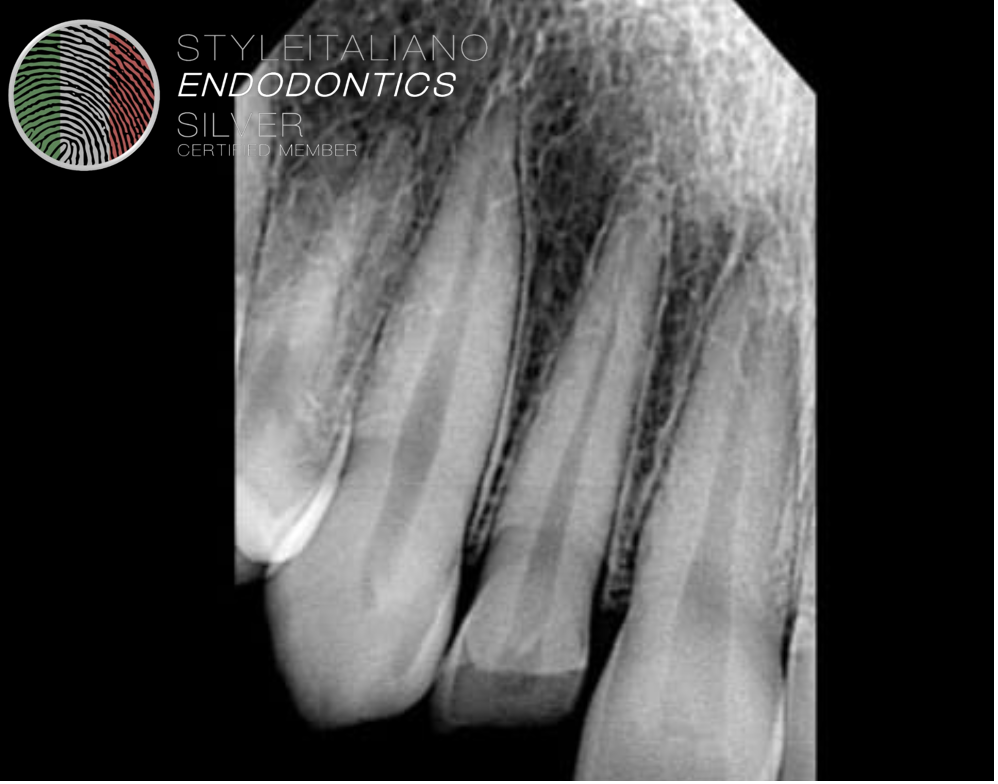
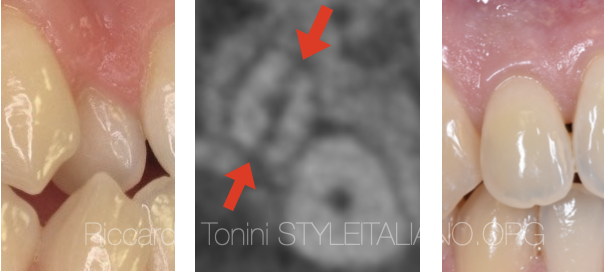
Fig. 1
Pre op radiograph showing fractured right lateral incisor.
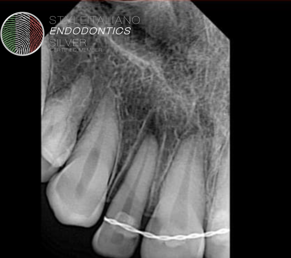
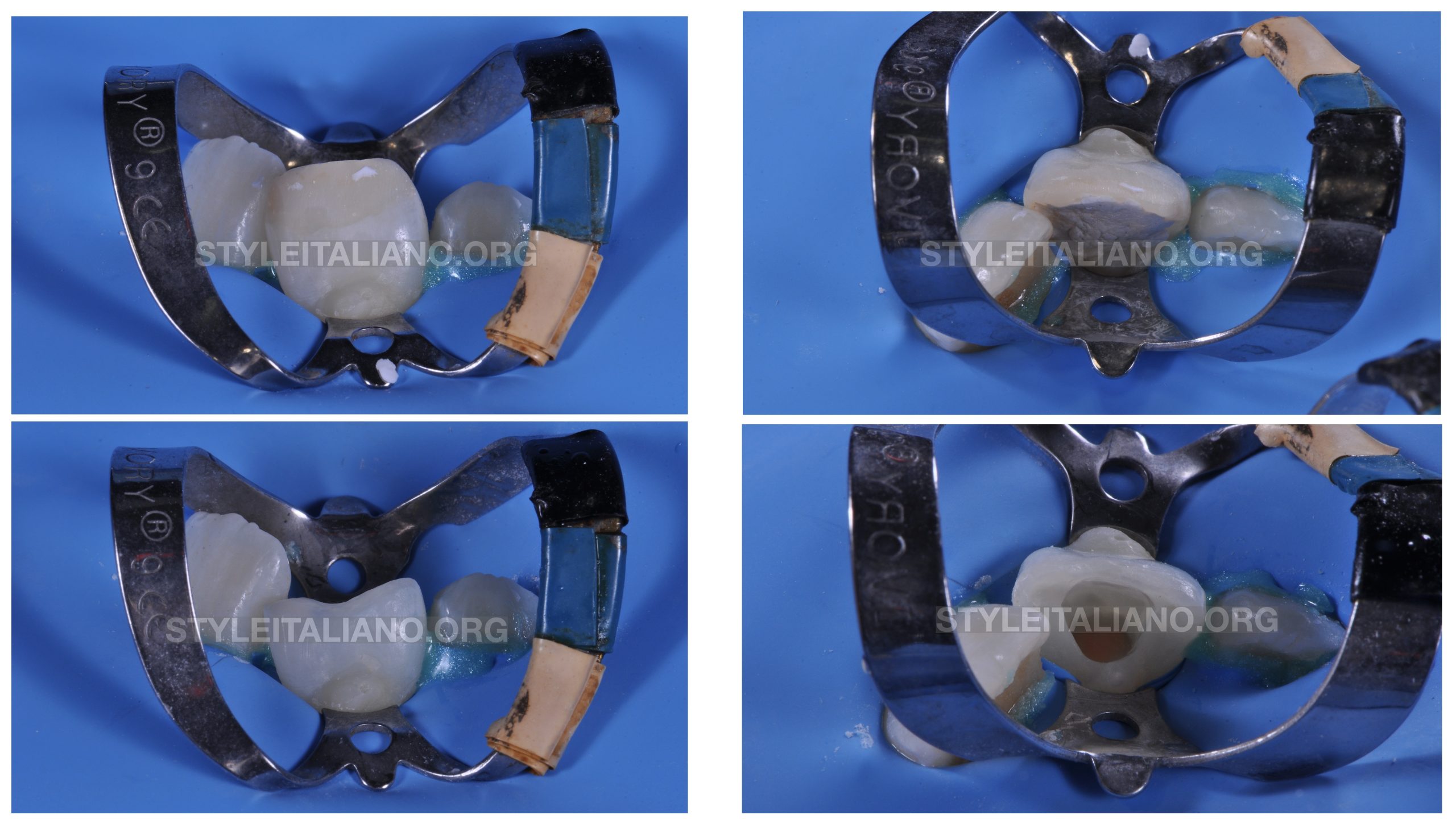
Fig. 2
Tooth was carefully extruded with the help of luxators and forceps about 4mm inside the socket. And splinted with the help of wire.

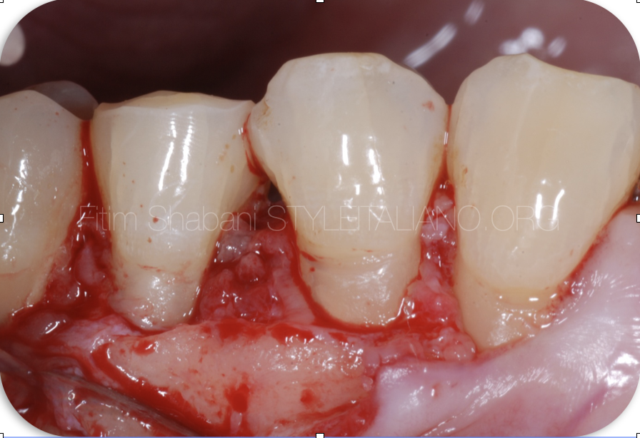
Fig. 3
Splint was removed after two weeks.. Patient was advised to come for endodontic treatment after two weeks.
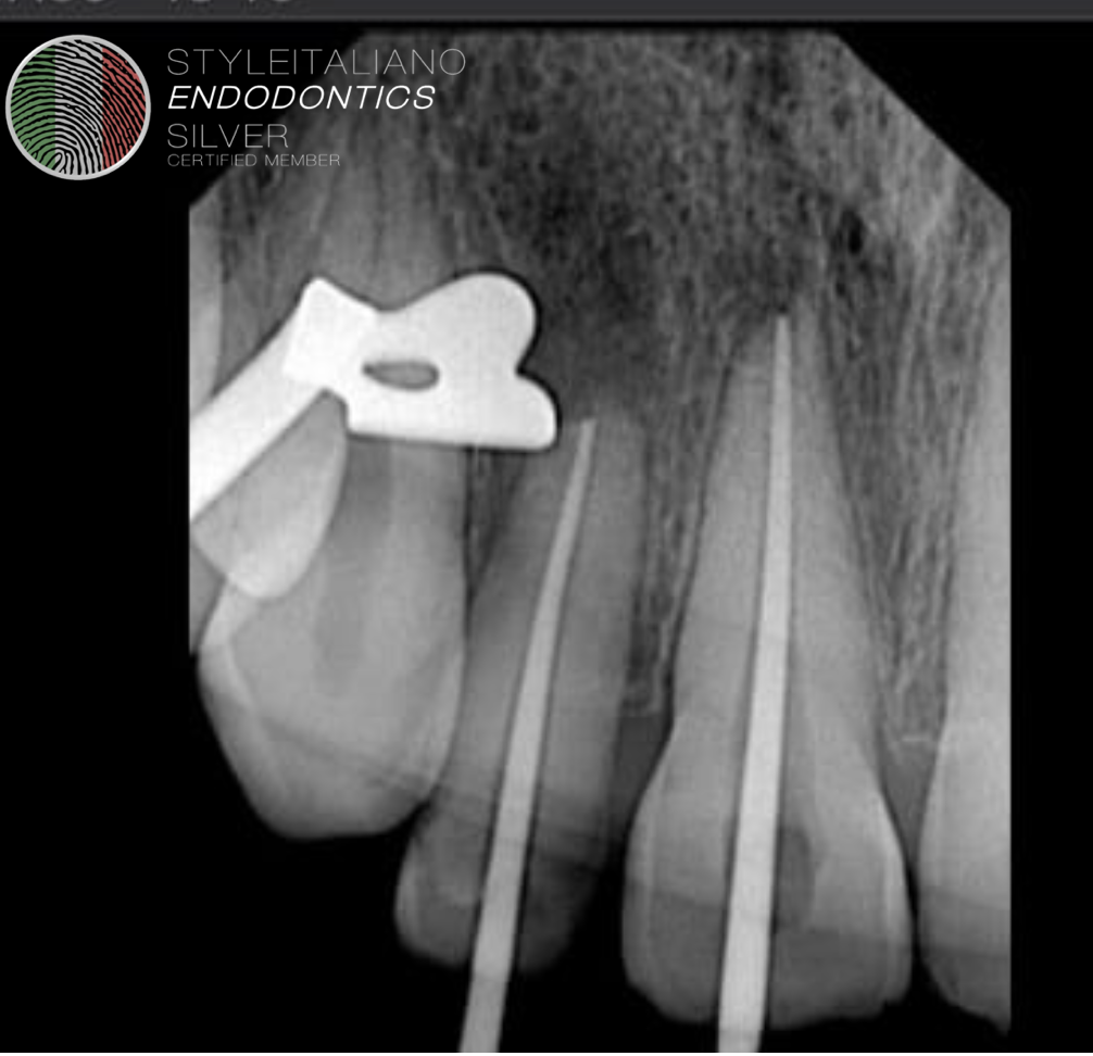
Fig. 4
Patient was not able to report on time. And root resorption was seen after 2 months time.
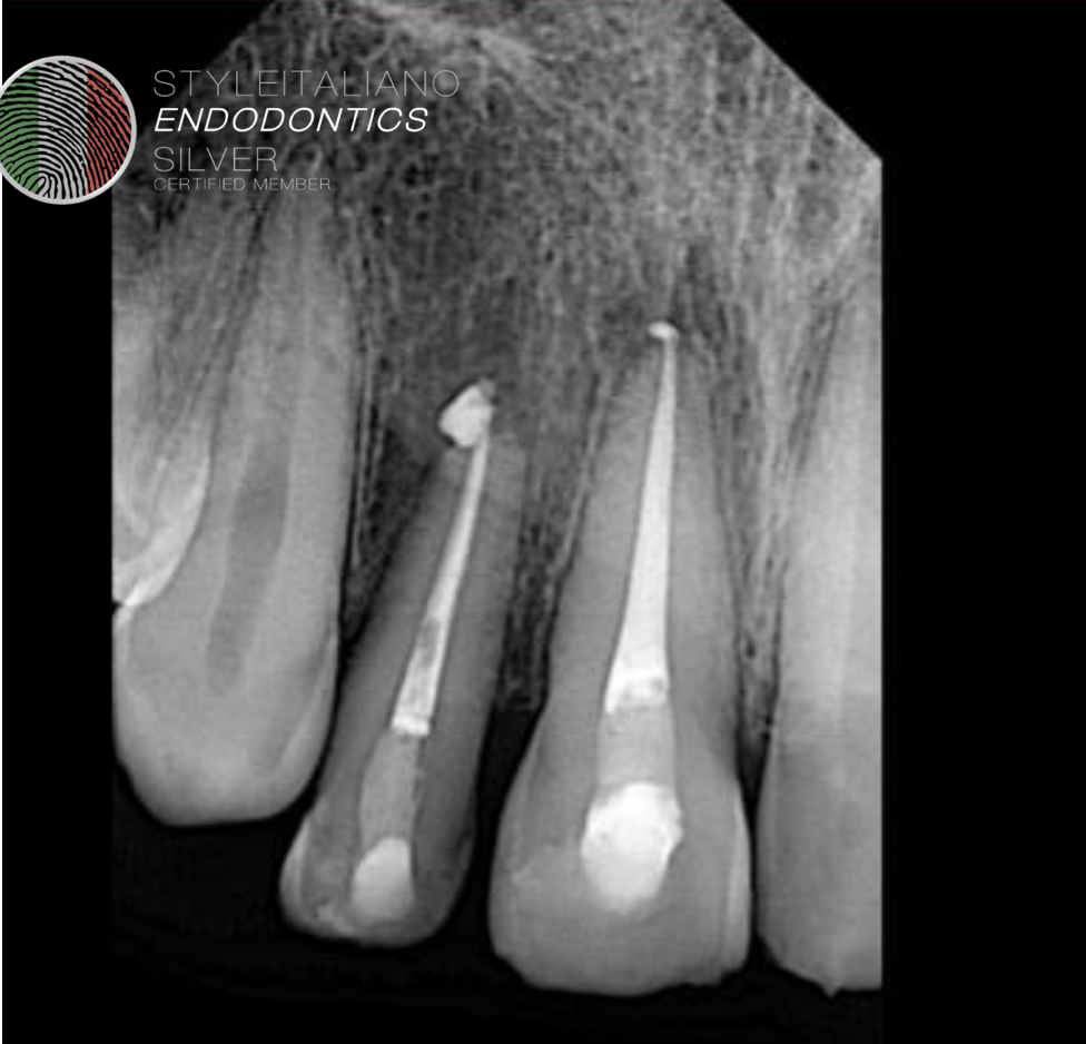
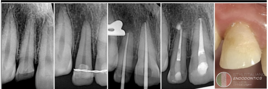
Fig. 5
Post operative radiograph.
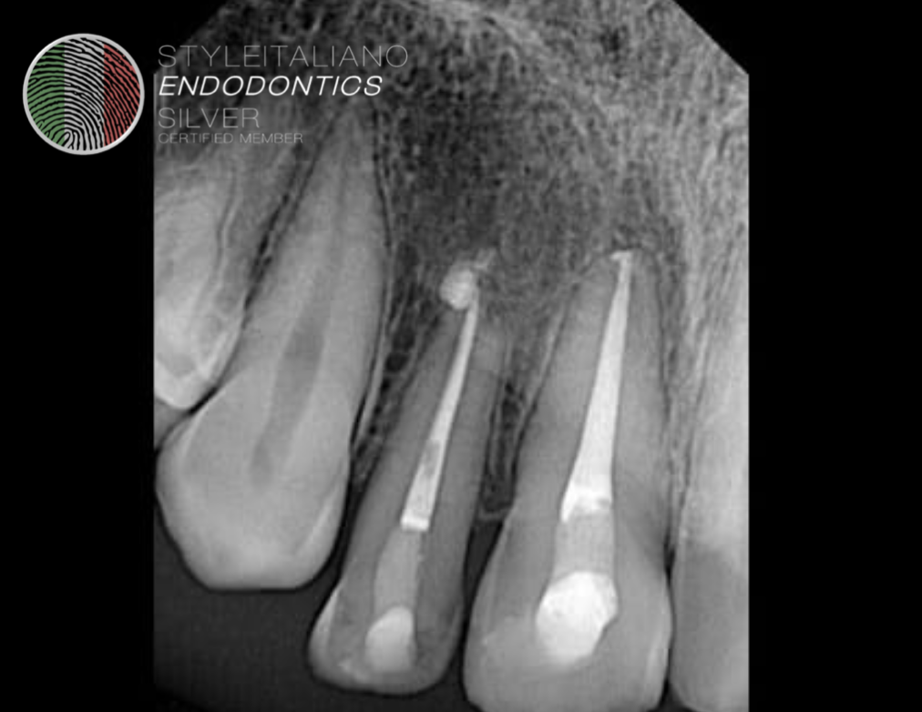
Fig. 6
Follow up after one year.
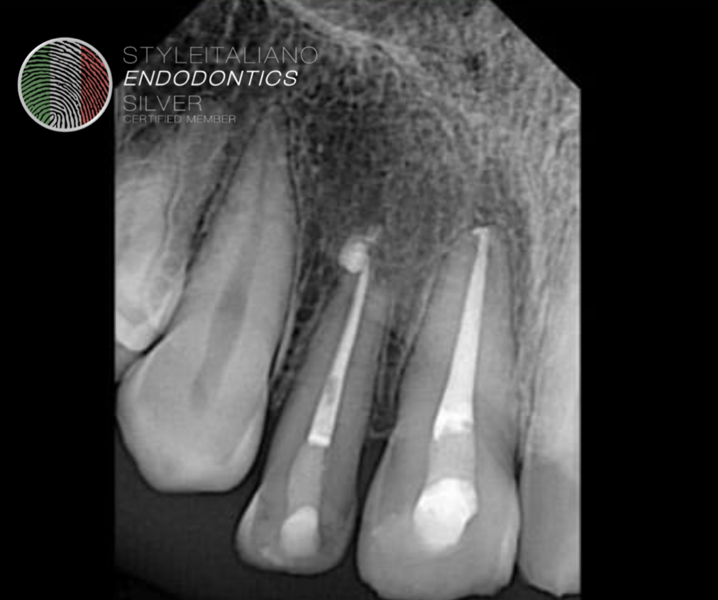
Fig. 7
Follow up after Two years.
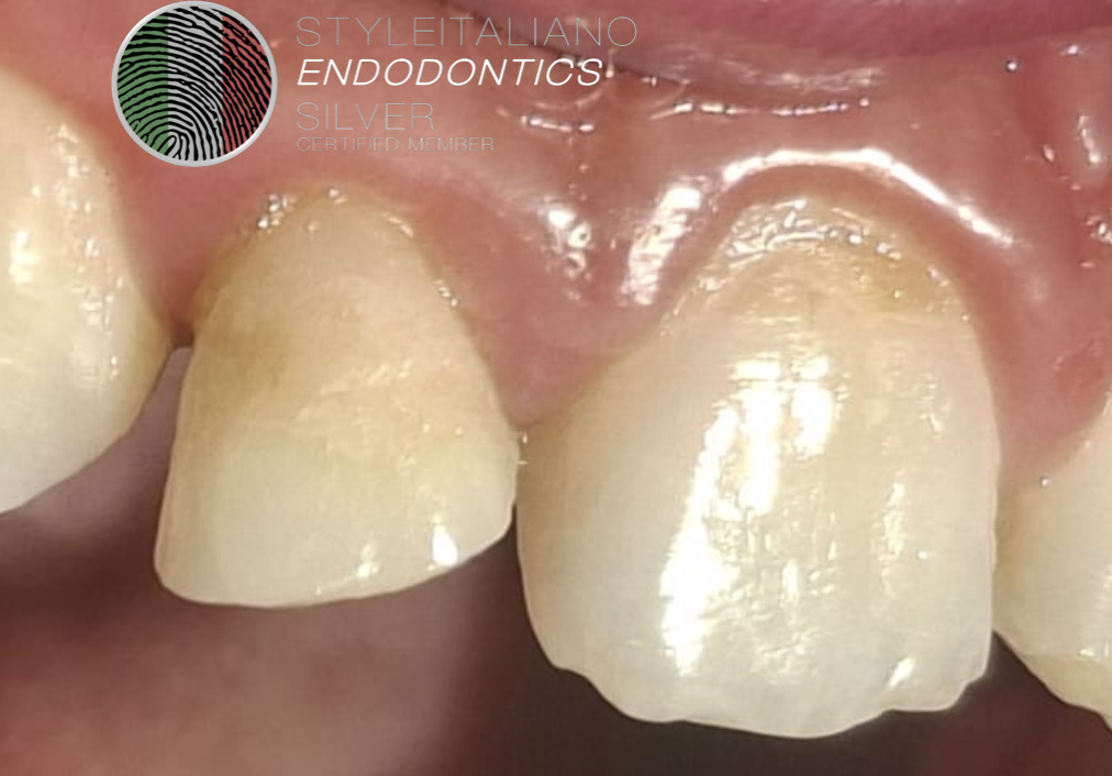
Fig. 8
Clinical image after two years. Buccal view.
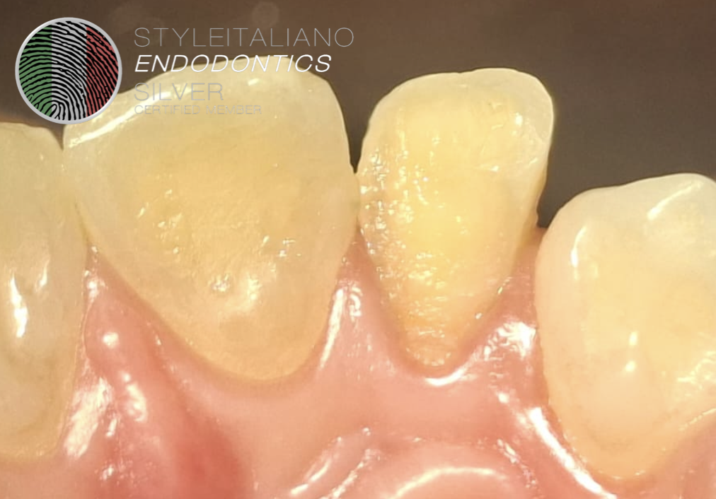
Fig. 9
Clinical image after two years. Palatal view..
Conclusions
In conclusion, teeth presenting with subgingival oblique fractures can be effectively managed through surgical extrusion. However, meticulous care is essential during the procedure, as improper handling may result in irreversible damage to both the tooth structure and the supporting periodontal tissues.
Bibliography
Estrela C, Corrêa CG, Malvezzi AL, et al. Clinical applications and outcomes of the surgical tooth extrusion technique: A bibliometric analysis from 1982 to 2023. J Prosthet Dent. 2024;131(2):187-95.
Tsukiboshi M, Yamauchi N, Tsukiboshi Y. Periodontal and periapical outcomes of surgical extrusion: A prospective clinical volumetric study. Dent Traumatol. 2021;37(6):813-21.
Caliskan MK, Turkun M, Gökay N. Adverse events of surgical extrusion in treatment for crown-root and cervical root fractures: A systematic review of case series/reports. Dent Traumatol. 2013;29(6):403-9.
Krastl G, Filippi A, Zitzmann NU, Walter C, Weiger R. Surgical extrusion of crown-root fractured teeth—a retrospective study. Dent Traumatol. 2009;25(5):508-13.
Singh M, Malhotra A, Rastogi P. Treatment of complicated crown root fracture by means of surgical extrusion: A case report. Univ J Dent Sci. 2022;8(2):107-11.
Lee SJ, Jung JH, Choi YJ, Choi YS. Esthetic restoration of subgingival crown-root fractured maxillary anterior tooth using surgical extrusion. Restor Dent Endod. 2014;39(3):215-9.
Deepthi A, Ramesh A, Thomas MS. Periodontal status of anterior teeth following clinical crown lengthening by minimally traumatic controlled surgical extrusion. J Clin Exp Dent. 2018;10(9):e857-63
Gao Z, Zhang X, Wang X, Zhao Y. Combined treatment of surgical extrusion and crown lengthening procedure for severe crown-root fracture of a growing patient: A case report. BMC Oral Health. 2024;24:1187.