Step by step management of necrotic immature tooth
08/08/2020
Mohamad Zaafrany
Warning: Undefined variable $post in /home/styleendo/htdocs/styleitaliano-endodontics.org/wp-content/plugins/oxygen/component-framework/components/classes/code-block.class.php(133) : eval()'d code on line 2
Warning: Attempt to read property "ID" on null in /home/styleendo/htdocs/styleitaliano-endodontics.org/wp-content/plugins/oxygen/component-framework/components/classes/code-block.class.php(133) : eval()'d code on line 2
Obturation and filling to the root canal system is considered the last step of pulp space treatment.
Providing a 3D filling to root canal space and adapting intracanal filling material to cleaned surfaces is required for long term success of the treatment to prevent reinfection.
Using MTA as an artificial barrier and apical filling material is considered a treatment option in cases of immature root with necrotic pulp due to the superior sealing ability of the material along with the chemical and physical properties of the material
3 main signs or root maturation
Root length
Root thickening
Apical closure
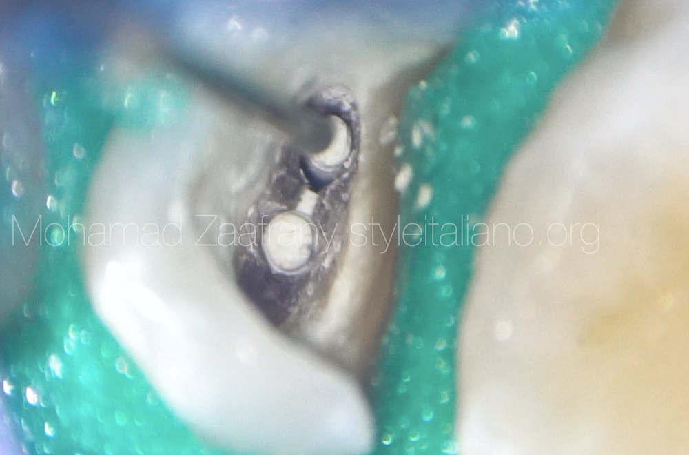
Representing today a case of 11 years old patient with necrotic pulp of tooth no. 25 with immature root expressed by insufficient apical thickening of the root and large apical foramen, gauged by size 80 K file.
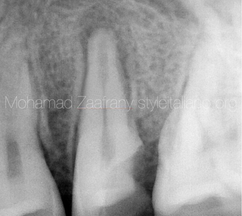
Fig. 1
Preoperative radiograph showing widening of the pulp space in the apical portion of the root as a sign of incomplete maturation.
Radiographically it is the typical appearance of internal resorption but, according to patient's age, the root was still in maturation period. That’s why it was diagnosed as incomplete root thickening.
Chronic periapical abscess diagnosed associated with presence of sinus tract.
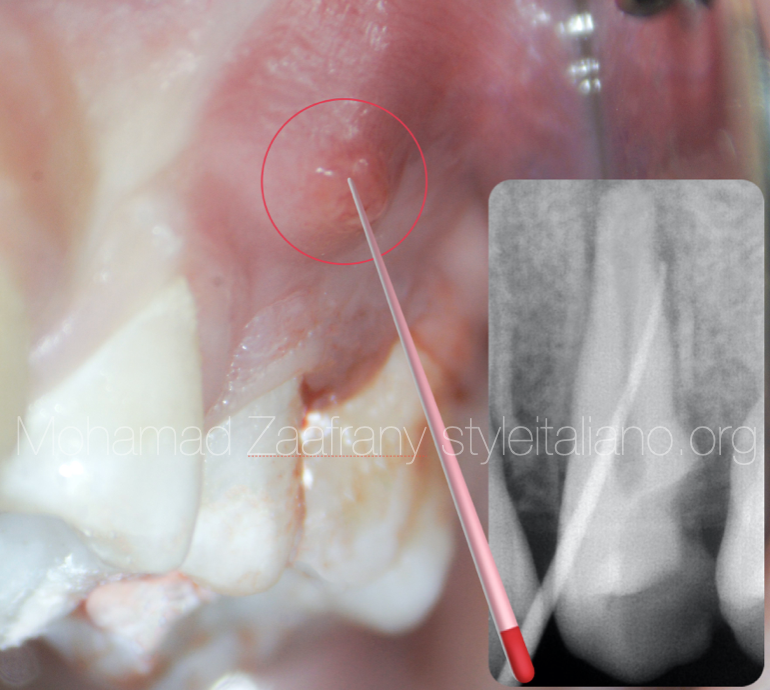
Fig. 2
Sinus tract opening present on the mesial aspect of the tooth
Tracing done using size 25 4% GP cone
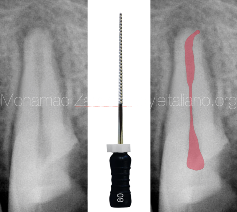
Fig. 3
After access opening, the apical foramen was gauged with a size 80 K file.
As long as the purpose of shaping procedures is to allow sufficient irrigation and chemical disinfection to reach more deep apically, minimal mechanical instrumentation was done and more attention was given to chemical disinfection through the use of copious irrigation, giving sufficient flow of irrigant solutions apically for debridement and smear layer detachment.
Activation was done using ultrasonic technique
Insertion of non setting calcium hydroxide paste into the canal.
The tip should be free inside the canal and not locked to avoid over extrusion of the material beyond the apex.
The purpose of using Calcium Hydroxide as intracanal medication in this case is, besides its antimicrobial activity, the function to raise the PH so the environment would be more suitable to receive the root filling material “MTA”
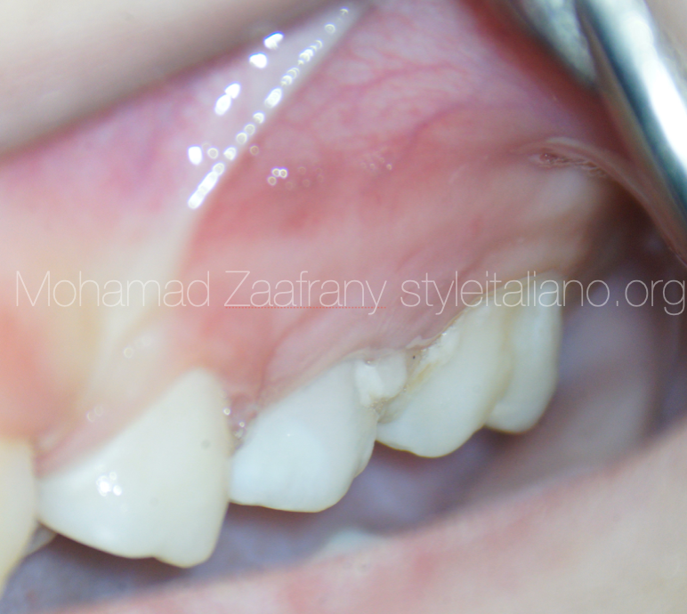
Fig. 4
One week after Calcium Hydroxide dressing the sinus tract opening disappeared and healthy tissue could be noticed.
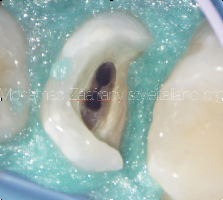
Fig. 5
The picture shows the tooth immediately after washing away calcium hydroxide and drying the canals.
Now canals are ready to be filled
Application of MTA to the irregular apical portion of the root
Fast setting MTA was used delivered to the canal using MAP system. No apical barrier was used because of good control over application of MTA as the size of the foramen was not too large.
MTA provides good sealing to the apical portion of the canal specially with the extension of the isthmus deep in the canal.
After 10 to 15 minutes and hardening of MTA, a little amount of sealer applied to the canal walls to allow better flow and adaptation of warm gutta-percha to the middle and coronal portion of the canal.
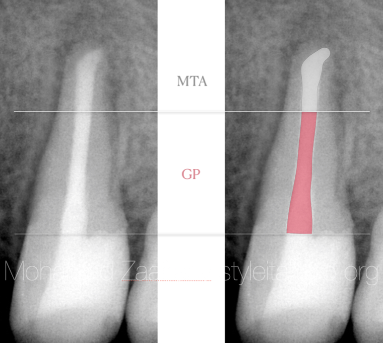
Fig. 6
The radiographic image shows the obturation and filling of the whole canal followed by immediate coronal restoration.
Conclusions
Root maturation and apical closure occurs up to 3 years after tooth eruption.
Pulpal disease or infection during this period might be a reason for pulp necrosis and subsequently periapical lesion that’s why during this period much attention to patient oral hygiene also to treatment of carious lesion to maintain the pulp vitality during the maturation period.
Managing immature roots providing 3D sealing to the root canal system might represent a challenge to the operator.
Using biocompatible filling materials like MTA or Bioceramics that provide superior sealing properties over conventional Gutta percha in this kind of cases is considered a solution for more predictable results.
Fast set MTA should be considered in single setting session after placement.
Clinical judgement is considered a factor on selection of the obturation material that would provide sealing and filling to the apical portion of the canal specially with irregularities.
Bibliography
MTA: A clinical review Peter Z Tawil, DMD, MS, FRCD, Dip. ABE, Derek J. Duggan, BDentSc, MS, Dip. ABE, and Johnah C. Galicia, DMD, MS, PhD. Compend Contin Educ Dent. 2015 Apr; 36(4): 247–264.
Antimicrobial effect of calcium hydroxide as an intracanal medicament in root canal treatment: a literature review - Part I. In vitro studies Dohyun Kim and Euiseong Kim, Restor Dent Endod. 2014 Nov; 39(4): 241–252.
Sealing properties of Mineral Trioxide Aggregate orthograde apical plugs and root fillings in an In vitro apexification model. JOE 2007 Mar;33(3):272-5. doi: 10.1016/j.joen.2006.11.002.

