Smart ledge crossing with FANTA files
26/01/2023
Massimo Giovarruscio
Warning: Undefined variable $post in /var/www/vhosts/styleitaliano-endodontics.org/endodontics.styleitaliano.org/wp-content/plugins/oxygen/component-framework/components/classes/code-block.class.php(133) : eval()'d code on line 2
Warning: Attempt to read property "ID" on null in /var/www/vhosts/styleitaliano-endodontics.org/endodontics.styleitaliano.org/wp-content/plugins/oxygen/component-framework/components/classes/code-block.class.php(133) : eval()'d code on line 2
During preparation ledges are inadvertently created and make further instrumentation more difficult as file tips snag in the ledge rather than progressing smoothly to the working length.
Stiff instruments or drills will tend to straighten in the curved root canal. Ledges are most likely on the outer curvature either at the primary curvature or apically.
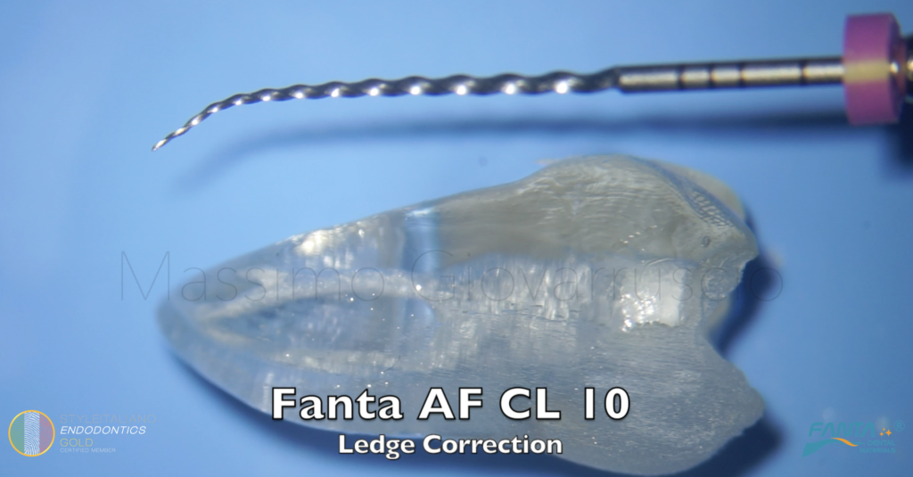
The aim of this article is to share a contemporary new clinical approach to cross a ledge using a SMART pre-bent file used with reciprocation movement.
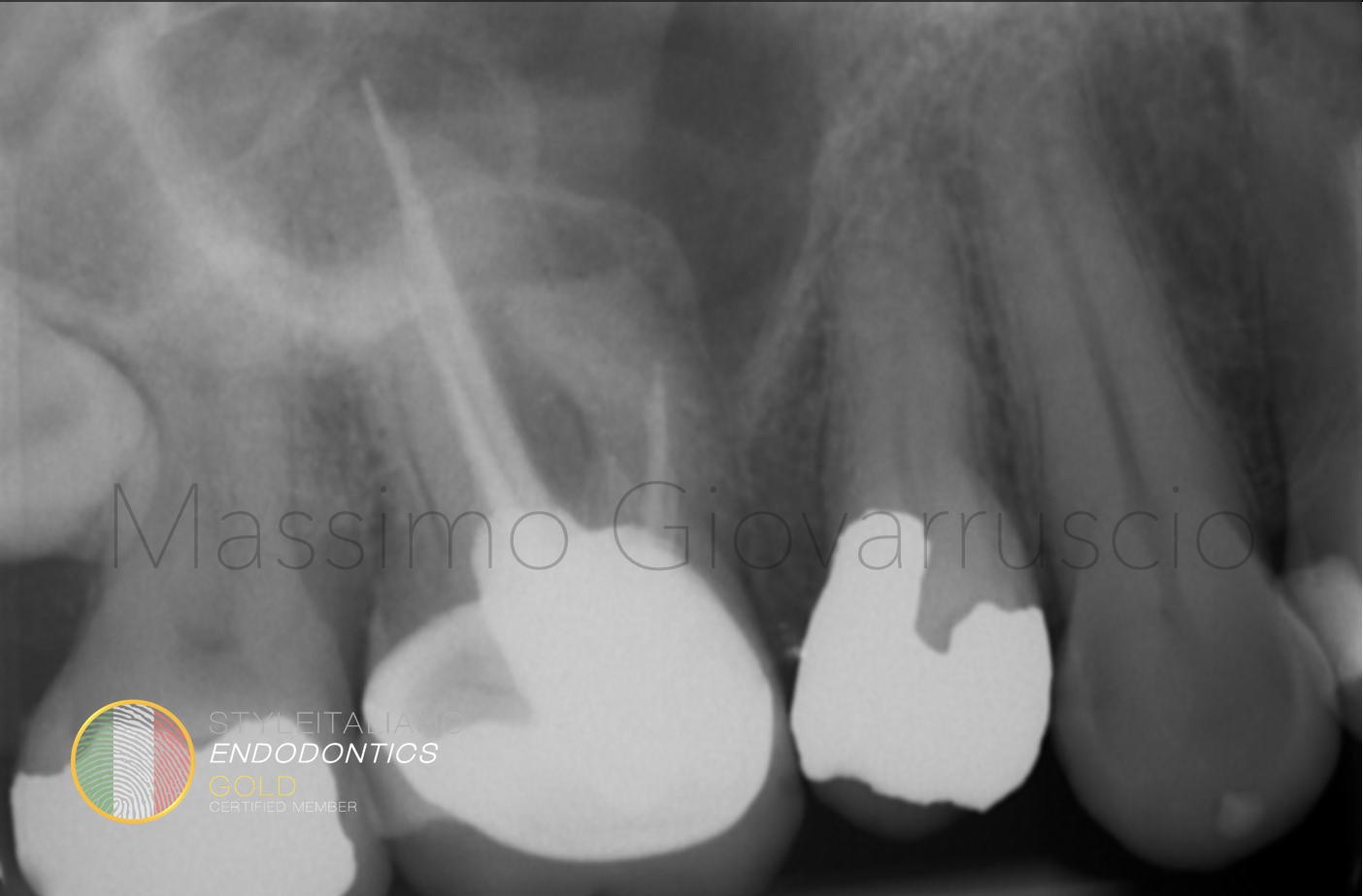
Fig. 1
A patient comes to my attention referring pain to the upper right molar, previously treated by a colleague.
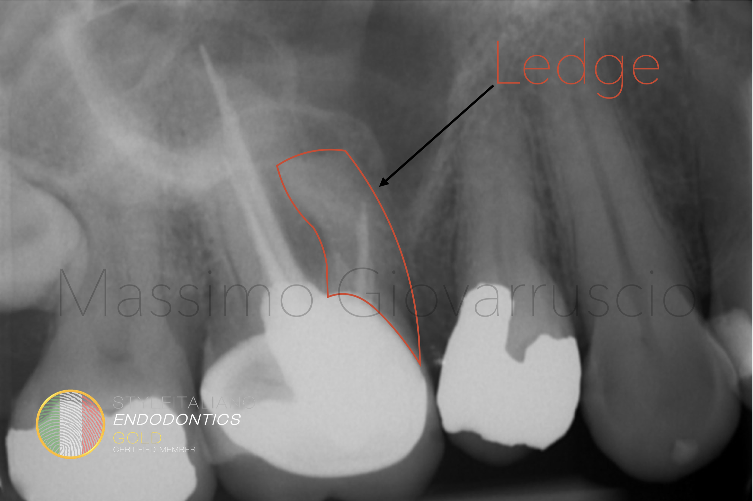
Fig. 2
The presence of a straight-line preparation in the curved mesial root suggests the presence of a ledge that made it impossible to reach the working length.
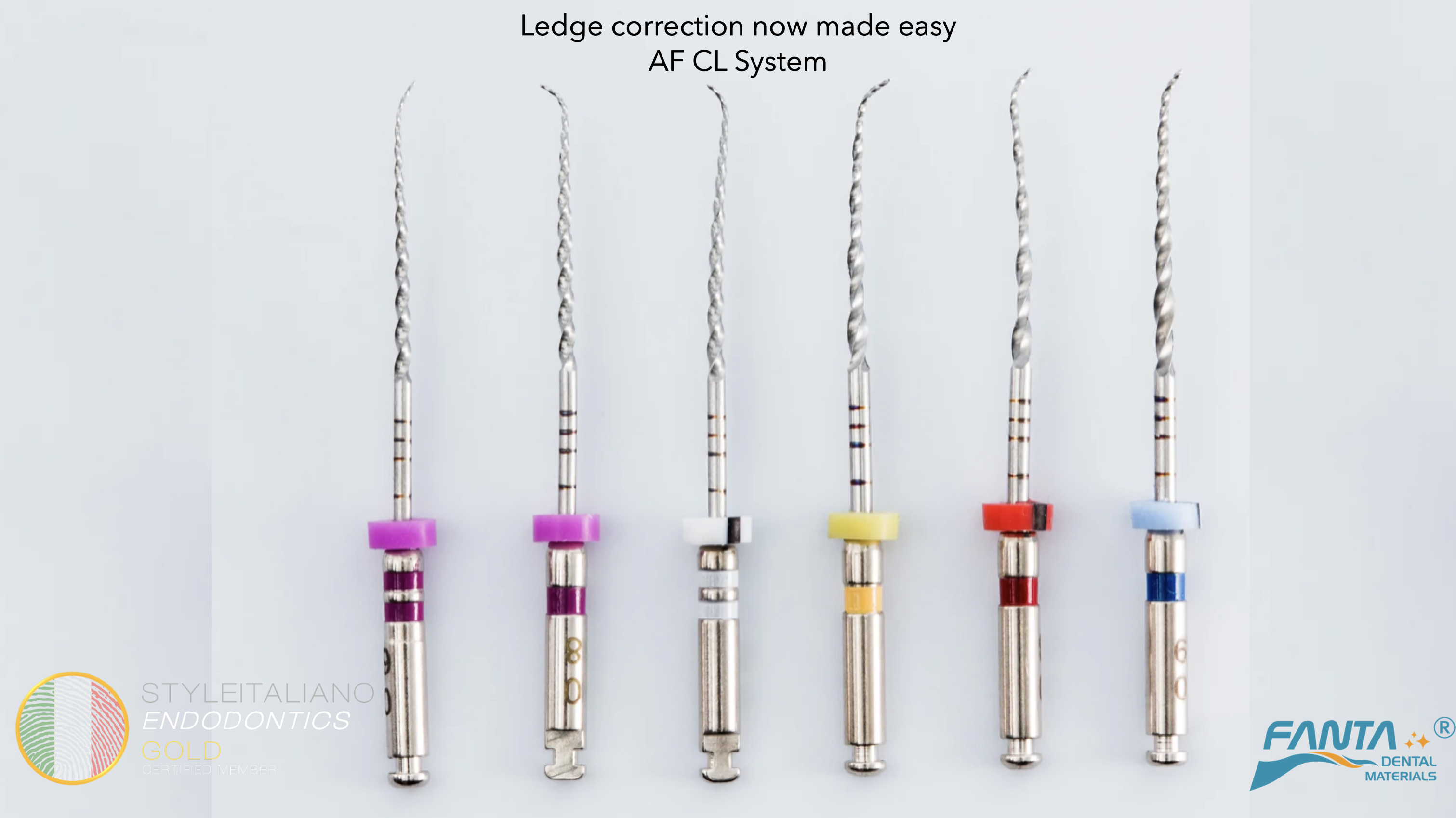
Fig. 3
I decided to cross the ledge with the AF CL System by FANTA
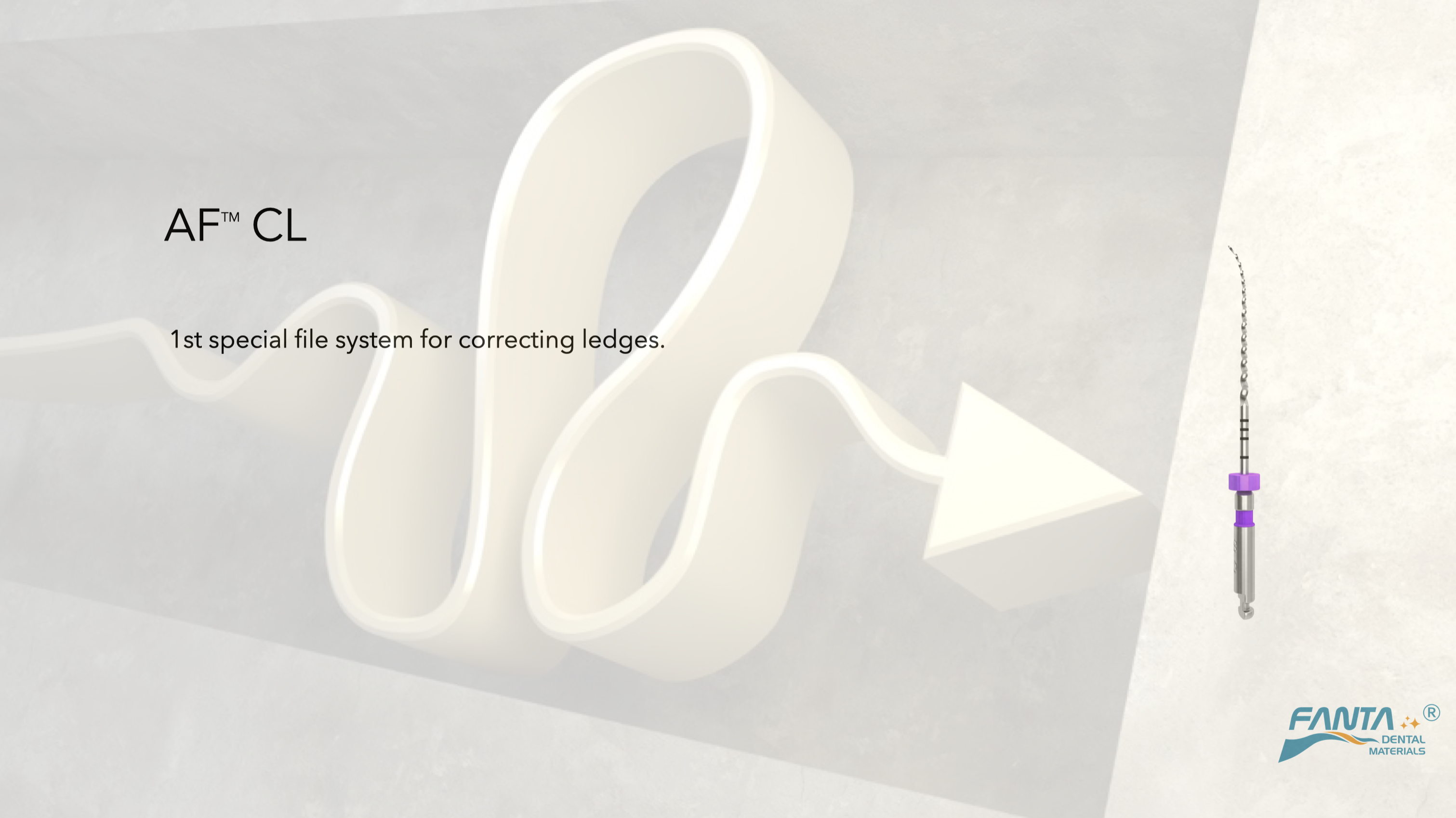
Fig. 4
The AF CL is the first file system specifically designed to cross ledges
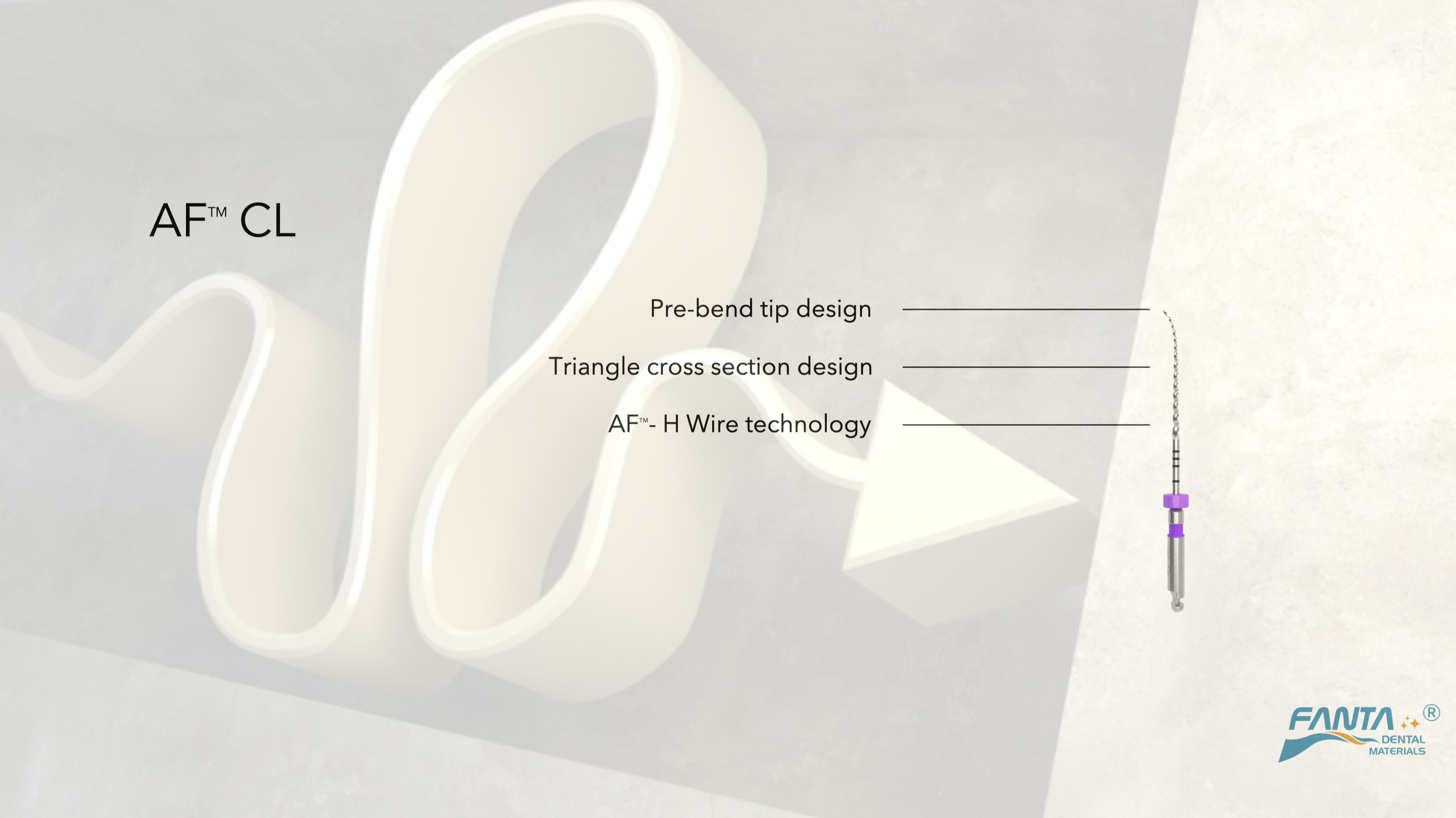
Fig. 5
They are flexible, with a pre-bent tip and a triangular cross section
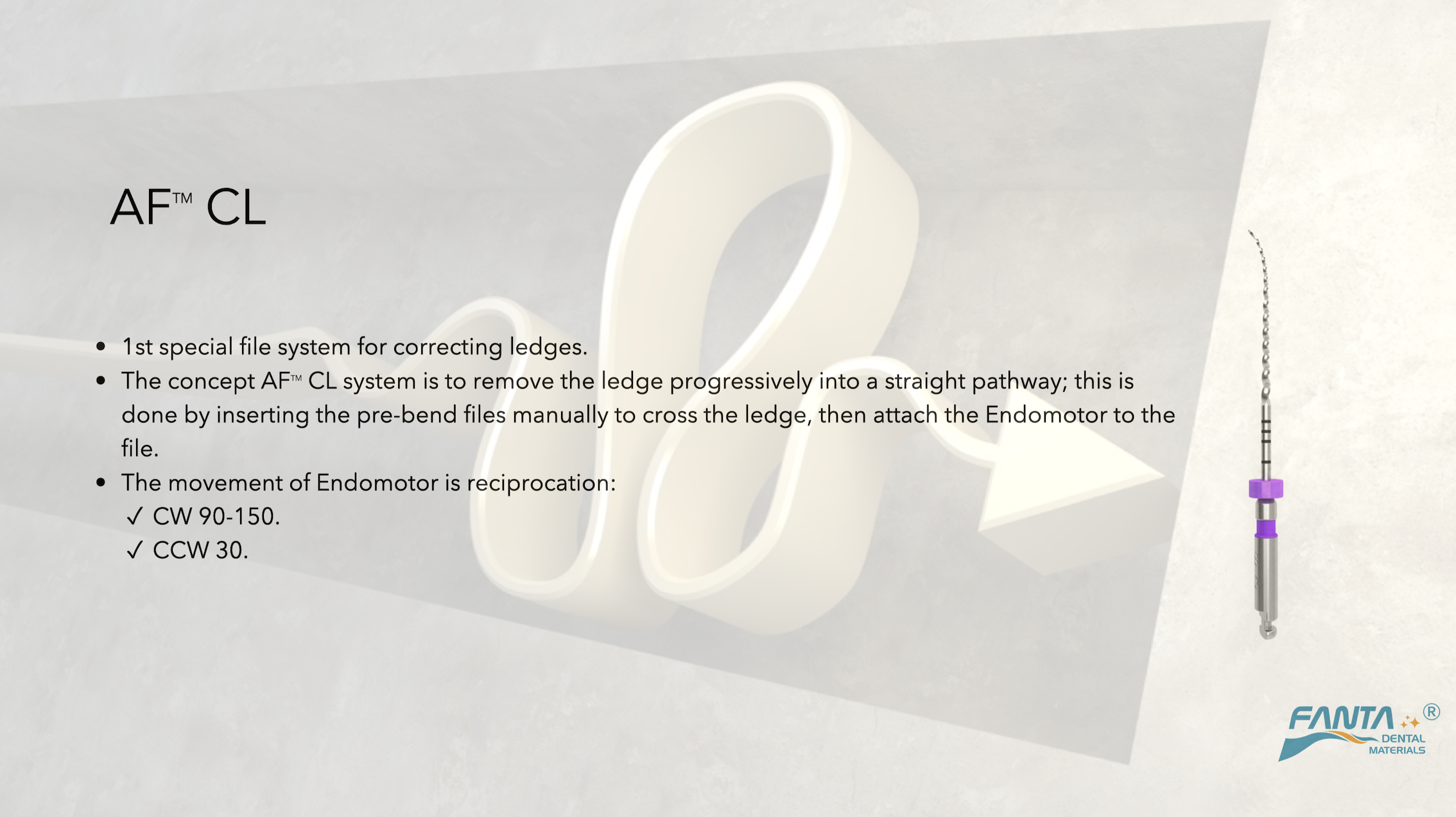
Fig. 6
The first file is inserted manually and crosses the ledge, then the endo motor is connected and the file starts moving in reciprocation motion
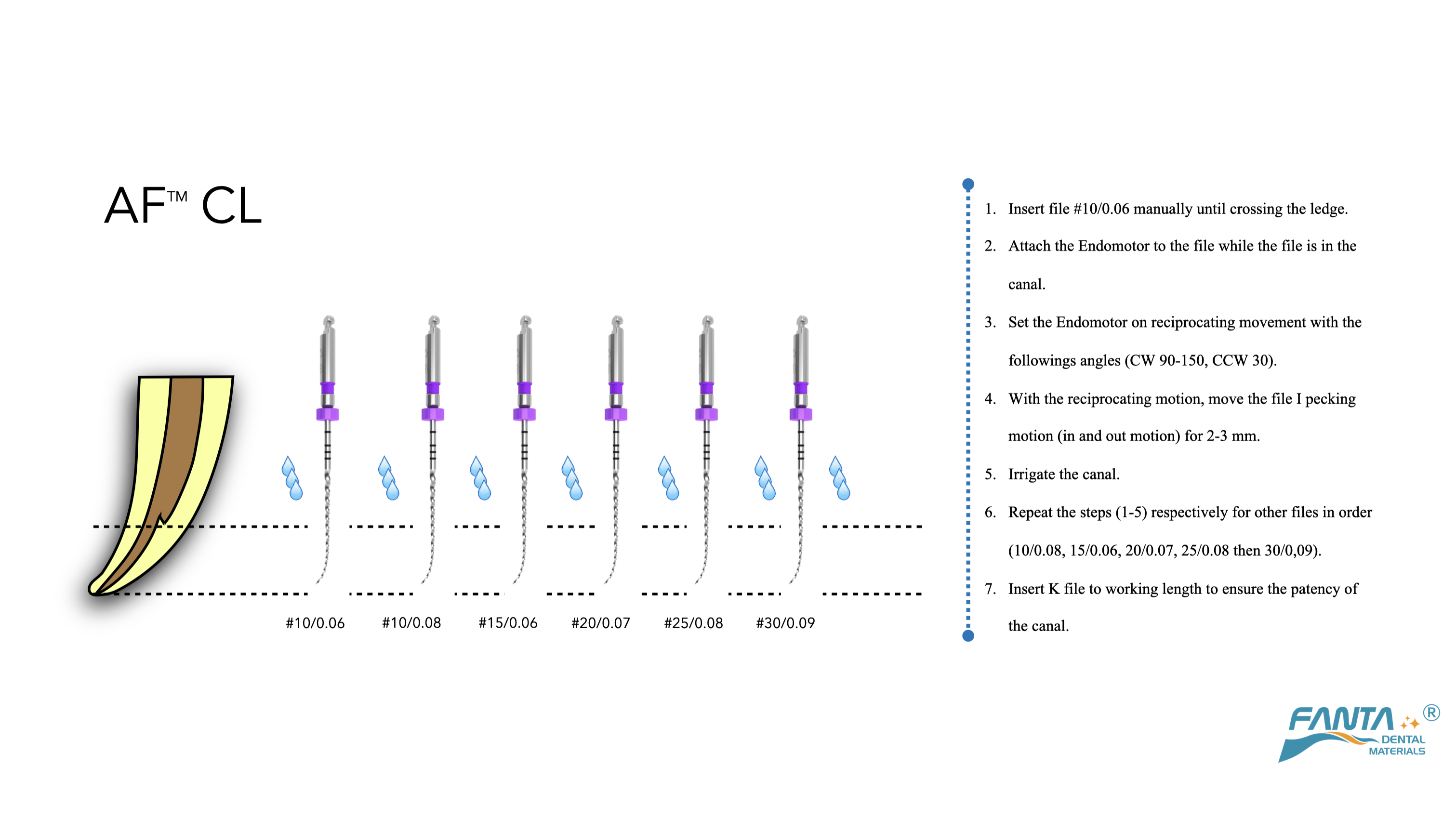
Fig. 7
Here is the protocol
The concept is to remove the ledge progressively into a straight pathway. This is done simply by inserting the pre-bend files manually to cross the ledge, then attach the Endomotor to the file using reciprocation motion.
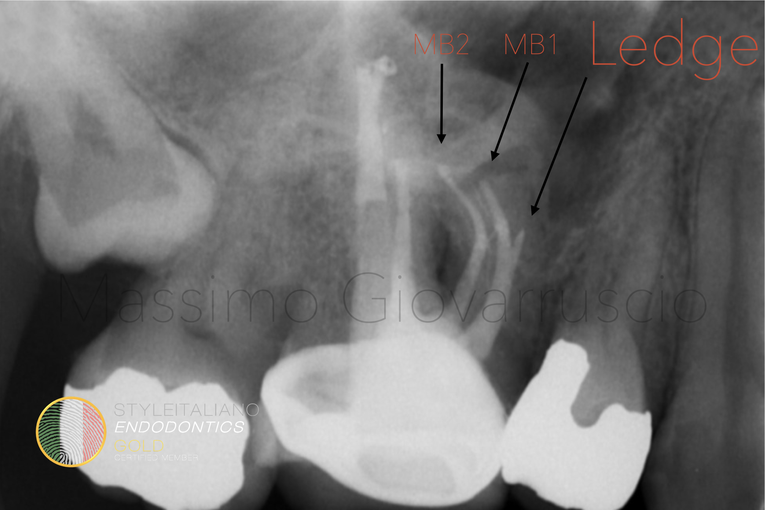
Fig. 8
The post operative X-ray shows the successful shaping of the tooth
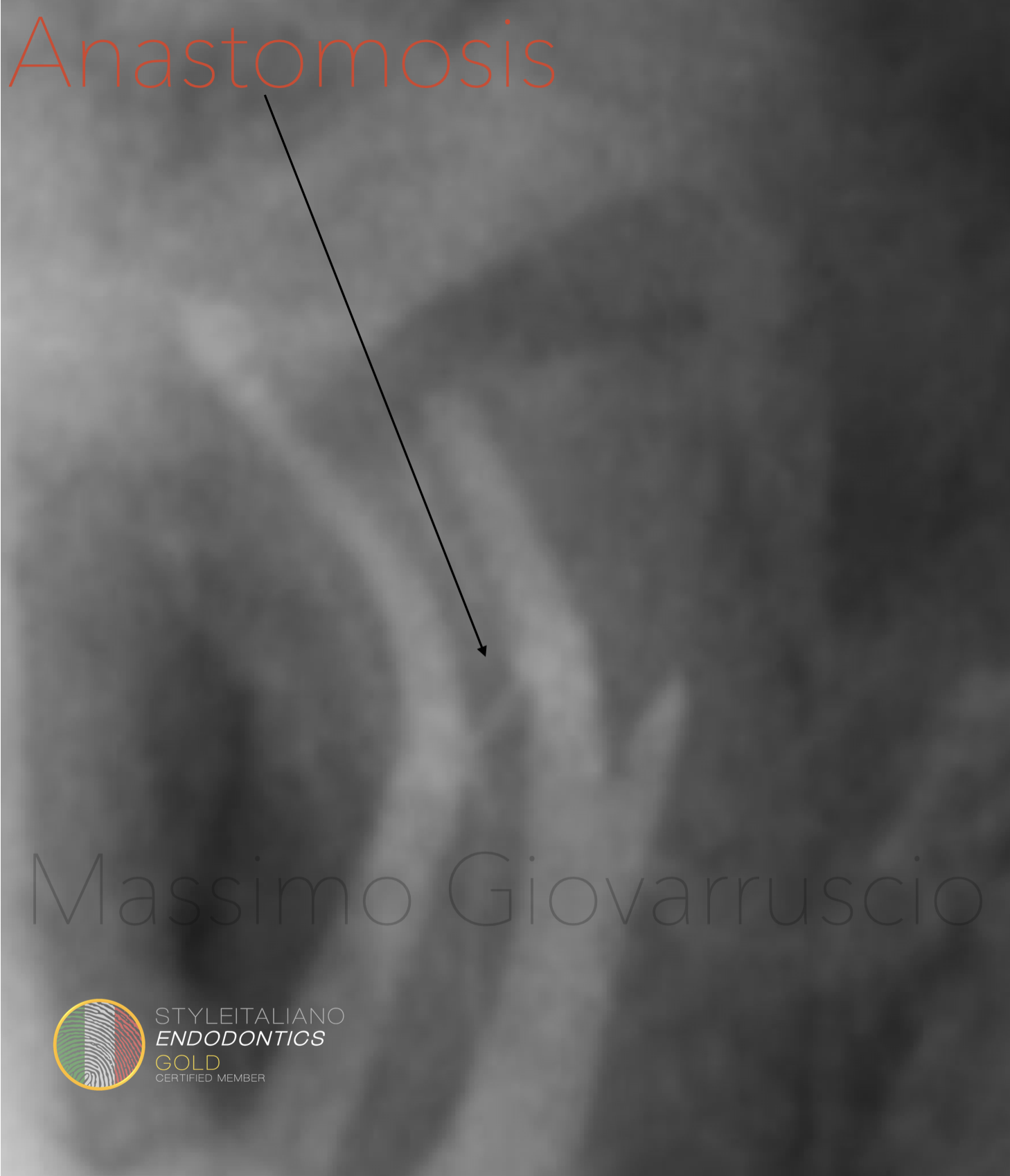
Fig. 9
A detail of the X-rays shows an anastomosis between the MB1 and MB2 canal
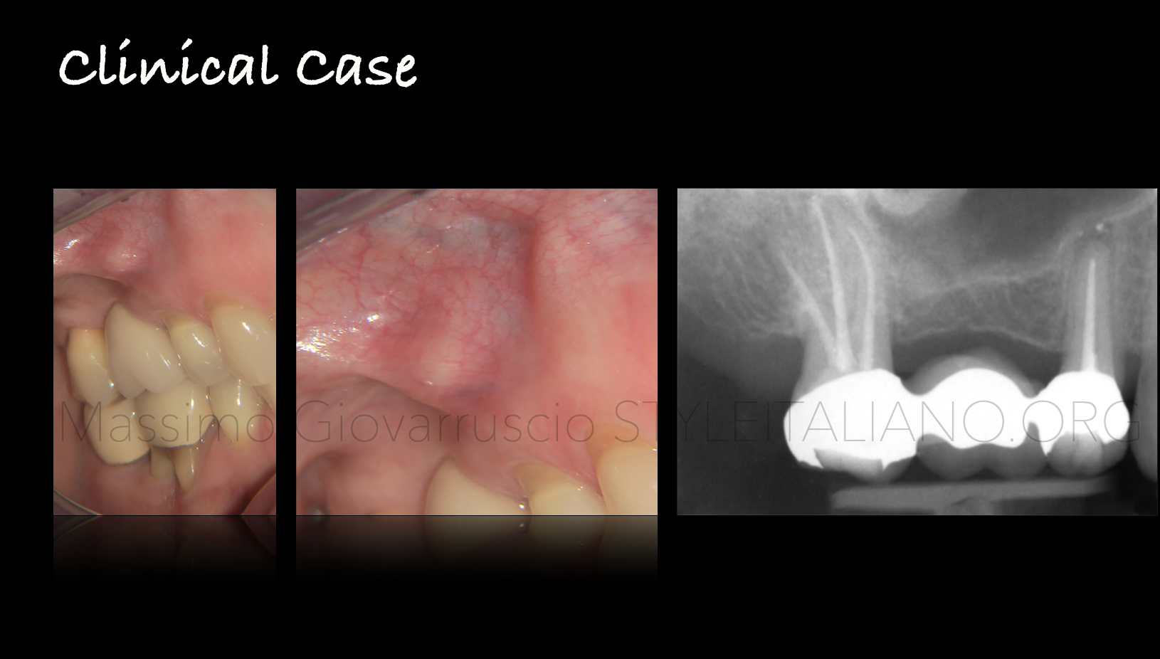
Fig. 10
Another cases solved in the same way shows a premolar with incomplete root canal therapy due to a ledge
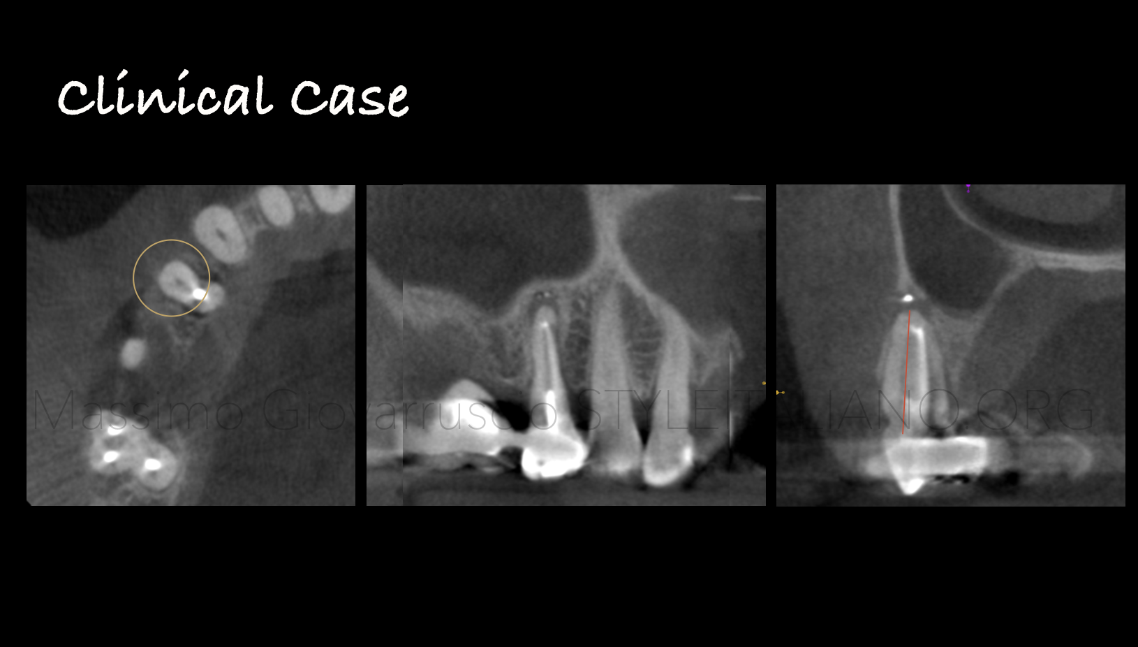
Fig. 11
The CBCT confirms the presence of an untouched part of the root canal system
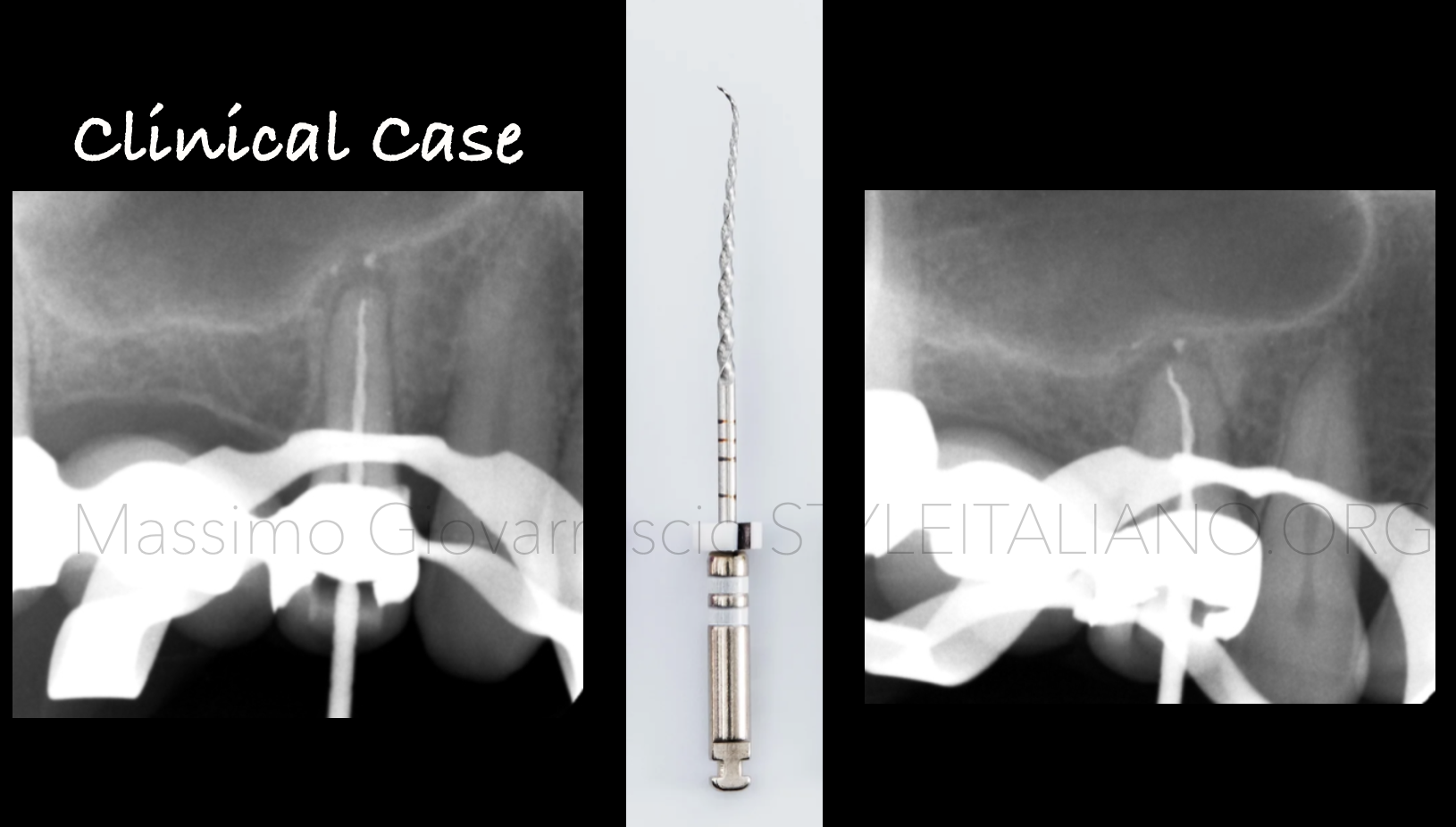
Fig. 12
The ledge is crossed with AF CL System
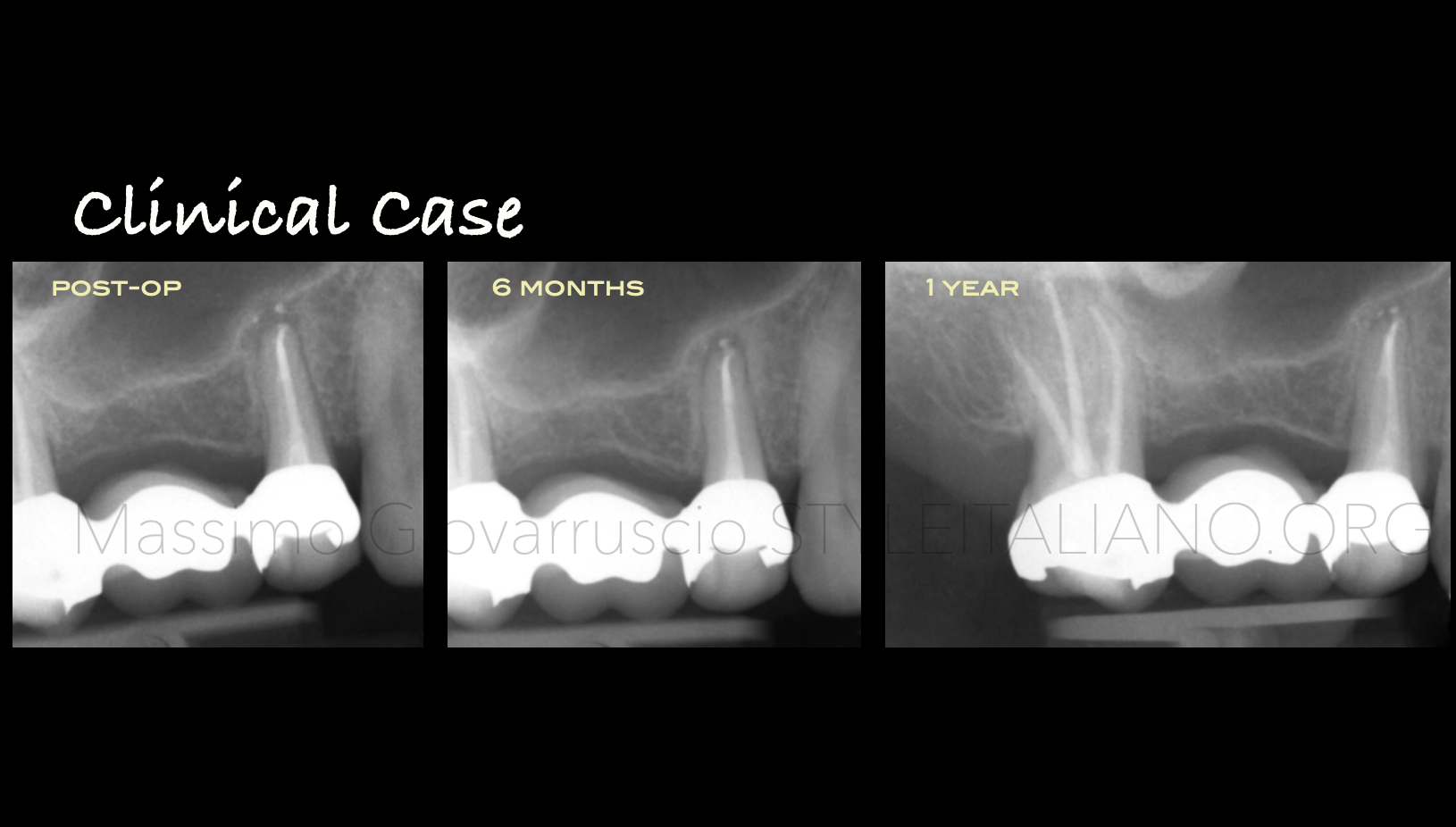
Fig. 13
The post operative X-rays and the follow up show the healing of the lesion
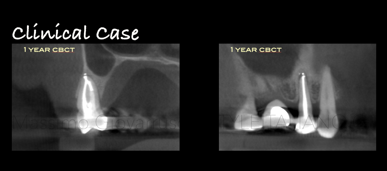
Fig. 14
Follow up CBCT
Conclusions
Conclusions
Ledge management in the past was really challenging and operator-dependant using a manual instrumentation: the shaping was really time consuming, invasive and with a high probability to create internal/external transportation.
Nowadays with the new pre-bended files, any ledge can be crossed without sacrifice dental canal tissue. A predictable procedures using a simple technique with a feasible, teachable and repeatable approach.
Bibliography
Peters OA.Current challenges and concepts in the preparation of root canal systems: a review. J Endod 2004;30:559–67
Hulsmann M, Peters OA, Dummer PMH. Mechanical preparation of root canals: shaping goals, techniques and means. Endod Top 2005;10:30–76.
Jafarzadeh H, Abbott PV. Ledge formation: review of a great challenge in endodon- tics. J Endod 2007;33:1155–62.
Green KJ, Krell KV. Cilinical factors associated with ledged canals in maxillary and mandibular molars.Oral Surg Oral Med Oral Pathol 1990; 70: 490-7.
Kaplas A, LambrianidisT. Factor Associated with root canal ledging during instrumentation. Endodo Dent Traumatol 2000; 16: 229-31.

