Single-Visit Management of a Dens Invaginatus Case
15/09/2025
Nuno Pinto
Warning: Undefined variable $post in /home/styleendo/htdocs/styleitaliano-endodontics.org/wp-content/plugins/oxygen/component-framework/components/classes/code-block.class.php(133) : eval()'d code on line 2
Warning: Attempt to read property "ID" on null in /home/styleendo/htdocs/styleitaliano-endodontics.org/wp-content/plugins/oxygen/component-framework/components/classes/code-block.class.php(133) : eval()'d code on line 2
Dens invaginatus, also referred to as dens in dente, is a rare developmental dental anomaly resulting from an infolding of the enamel organ into the dental papilla during the morphodifferentiation stage of odontogenesis. This invagination leads to a deep enamel-lined cavity that may extend variably into the crown or root of the affected tooth. The anomaly is most frequently observed in permanent maxillary lateral incisors, although it can affect other teeth as well.
Radiographically, dens invaginatus presents as a radiopaque invagination of enamel and dentin within the body of the tooth, giving rise to the characteristic “tooth within a tooth” appearance. Clinically, it may predispose the tooth to early pulp involvement due to the direct communication between the oral environment and the invaginated space, which can harbor bacteria and debris.
Oehlers (1957) classified dens invaginatus into three types based on the depth and extent of the invagination:
• Type I: The invagination is limited to the crown and does not extend beyond the cemento-enamel junction (CEJ).
• Type II: The invagination extends beyond the CEJ into the root but remains confined within the root canal, without communication with the periodontal ligament space. The pulp may or may not be involved.
• Type III: The invagination extends through the root, perforating the apical or lateral root surface and forming a second foramen. It communicates directly with the periodontal ligament and is usually not connected to the pulp:
• Type IIIa: The invagination extends laterally and exits at the side of the root.
• Type IIIb: The invagination exits through the apical foramen.
Due to the complex internal anatomy and high risk of pulpal and periapical pathologies, accurate diagnosis and individualized treatment planning are critical for successful management of teeth affected by dens invaginatus.
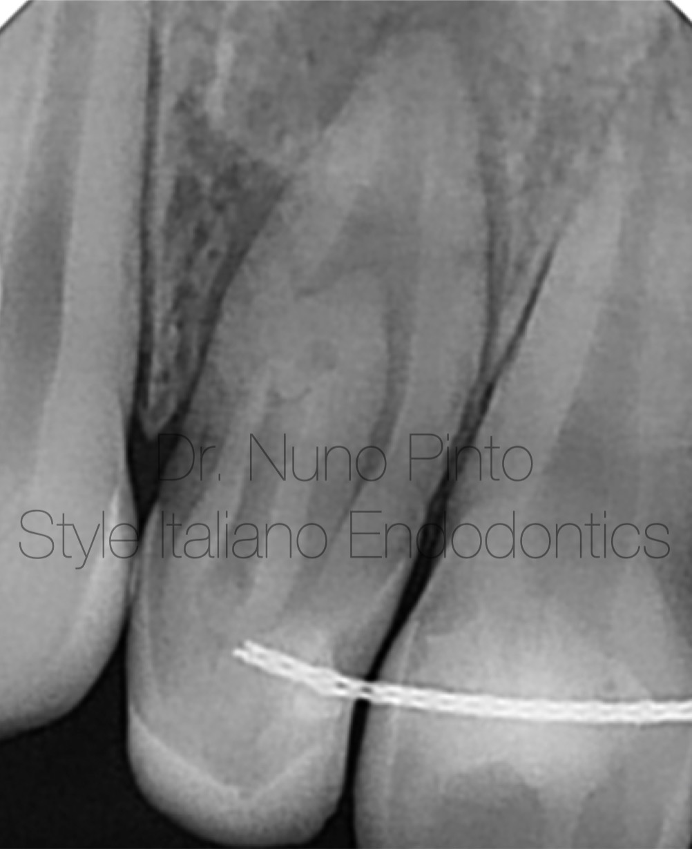
Fig. 1
The periapical X-ray highlights the complexity of the case, showing a chronic periapical abscess likely caused by either iatrogenic necrosis or trauma related to the use of orthodontic appliances.
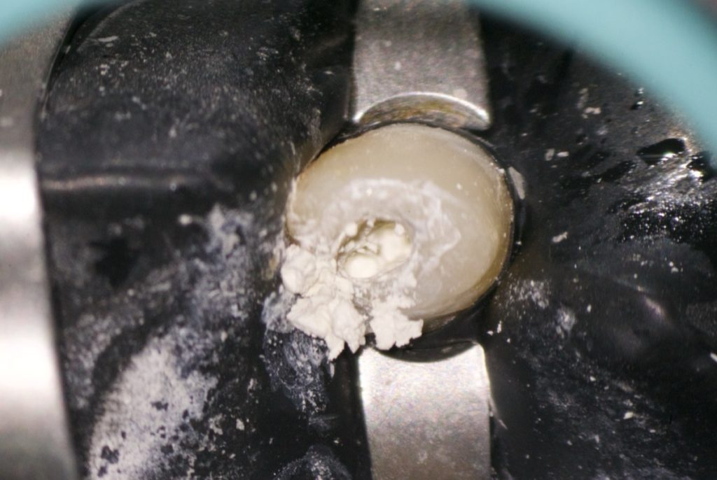
Access:
Performed using a long-shank bur and ultrasonic tips.
Shaping:
Carried out with Flash Glider 13.04 and Flash 25.06 files.
Irrigation:
Conducted with 8% sodium hypochlorite and 17% EDTA, activated with EDDY.
Obturation:
Done using Bio-C Repair (putty) for the apical seal, followed by backfill with warm gutta-percha.
Key Principle:
Simplifying each step is the key.
Conclusions
In this video, you can see each step in detail to complete the treatment of a dens invaginatus case in a single session. Case planning is the most critical phase, as it enables us to assess and define the most effective and predictable strategy. An equally important step is explaining the condition and treatment plan in detail to the patient, since their understanding and collaboration are essential—especially in complex anatomical situations.
The use of cone beam computed tomography (CBCT) played a fundamental role by providing a three-dimensional understanding of the invagination and root canal morphology, allowing for more accurate diagnosis and case planning, as well as clearer communication with the patient.
An essential part of the treatment protocol is the use of bioceramic materials, especially in cases involving complex anatomy, thin dentinal walls, or communication with the periodontal ligament space (Oehlers Type II and III). Bioceramics offer superior sealing ability, excellent biocompatibility, and the potential to promote periapical healing. Their bioactivity and dimensional stability make them ideal for obturation and apical sealing in invaginated canals or irregular spaces that cannot be predictably managed with conventional materials.
During the follow-up, clinical evaluation is always combined with periapical radiography and, when indicated, CBCT imaging to confirm long-term healing and the absence of pathology.
Bibliography
1. Oehlers, F. A. C. (1957). Dens invaginatus (dilated composite odontome). I. Variations of the invagination process and associated anterior crown forms. Oral Surgery, Oral Medicine, Oral Pathology, 10(11), 1204–1218.
2.Hülsmann, M. (1997). Dens invaginatus: Aetiology, classification, prevalence, diagnosis, and treatment considerations. International Endodontic Journal, 30(2), 79–90.
3.Alani, A., & Bishop, K. (2008). Dens invaginatus. Part 1: Classification, prevalence and aetiology. International Endodontic Journal, 41(12), 1123–1136.
4.Alani, A., & Bishop, K. (2008). Dens invaginatus. Part 2: Clinical, radiographic features and management options.International Endodontic Journal, 41(12), 1137–1154.