Shaping&Cleaning of the endodontic space: the Style Italiano Endodontic Philosophy and techniques
24/06/2016
Fabio Gorni
Warning: Undefined variable $post in /home/styleendo/htdocs/styleitaliano-endodontics.org/wp-content/plugins/oxygen/component-framework/components/classes/code-block.class.php(133) : eval()'d code on line 2
Warning: Attempt to read property "ID" on null in /home/styleendo/htdocs/styleitaliano-endodontics.org/wp-content/plugins/oxygen/component-framework/components/classes/code-block.class.php(133) : eval()'d code on line 2
Endodontic diagnosis, therapies and follow-up procedures can be the most complex of dental treatments; among these, shaping the root canals has always been considered as the basic and most important procedure. Various instruments and techniques have been developed in the history of endodontics, to deal with the almost infinite anatomical variations of root canals; this topic has been extensively debated elsewhere in this web site (http://styleitaliano.hime.host/history-of-endodontic-instruments). A relatively recent technological evolution has put forth a variety of endodontic files, more aggressively shaped, flexible but resistant to breakage, made from new alloys, with new surface treatments, mechanically driven with rotary or reciprocating motion. These instruments should be chosen and used according to the manufacturer’s instructions, in a precise sequence or sometimes as a single file, after evaluation of canal anatomy, curvature and gauge. This evaluation is either made with an initial scouting of the canal with small instruments, or even directly examining a periapical x-ray.
Obviously, each commercial brand of endodontic instruments proposes a different protocol, based upon the use of instruments of its own production. The possibility to choose instruments of different brands for different canals, or the use of simplified sequences, is ruled out by marketing rules.
StyleItalianoEndodontics watchword is simplification for excellence.
This article is the first of a series, in which the philosophy of root canal shaping will be thoroughly explained, along with clinical and practical examples that will show the best way to deal with anatomic complexities.
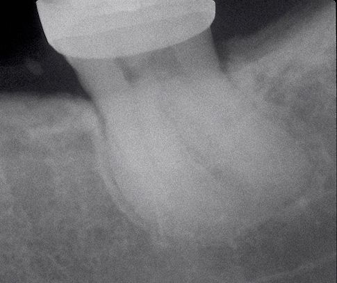
Fig. 1
Successful endodontic treatment depends mostly on how well the root canal is shaped and cleaned. Adequate shaping allows irrigants to go deeper in the root canal system, exerting their antimicrobial activity and dissolving organic debris. So, the concept of cleaning and shaping the root canal space should be changed, swapping the order of the words, changing their sequence in shaping & cleaning. The root canals should be efficiently shaped with instruments, to permit thorough and extended cleaning of the entire root canal system with irrigants.
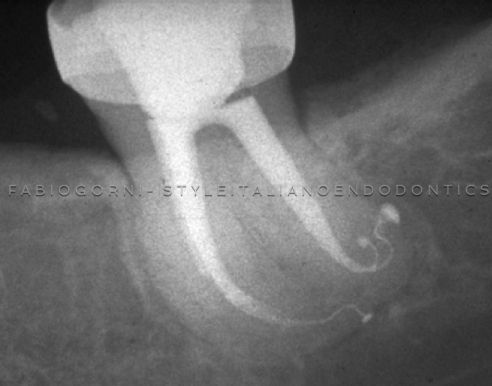
Fig. 2
Instruments shape, irrigants clean: and unique anatomies are revealed. Mechanical objectives for a good instrumentation, according to Schilder (1974), start from the respect of the anatomy of the root canal, and are focused towards the achievement of an optimal shape, allowing adequate cleaning and an effective three-dimensional filling. A continuously tapering preparation from the apex to the access, centered in the path of the original canal, respecting the apical foramen position without excessive enlargement should be obtained.
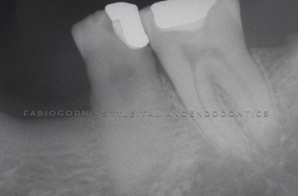
Fig. 3
The highly variable root canal anatomy cannot be shown by a preoperative X-ray.
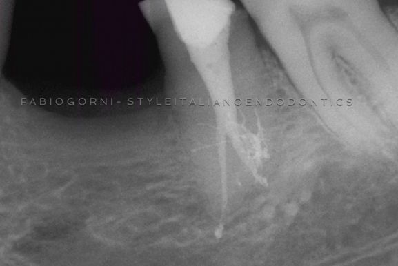
Fig. 4
Canal shaping opens the way for Sodium Hypoclorite. It digests the pulp tissue and promotes a deep cleaning in this complex anatomy. The final X-ray, luckily, looks like the anatomical drawings published by Hess in 1917, or like the micro-CT scans of today.
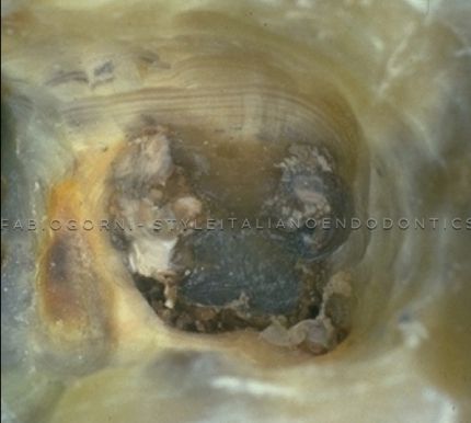
Fig. 5
Leaving aside the different techniques, visibility and accessibility are necessary to carry out an endodontic treatment. Hence, access cavity remains the most important step of the treatment.
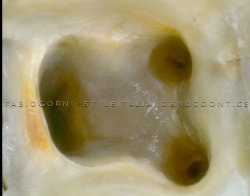
Fig. 6
Many different canal shaping techniques have been proposed. Basically, we can divide them into two philosophies: the Step-Back Technique, and the Step-Down Technique.
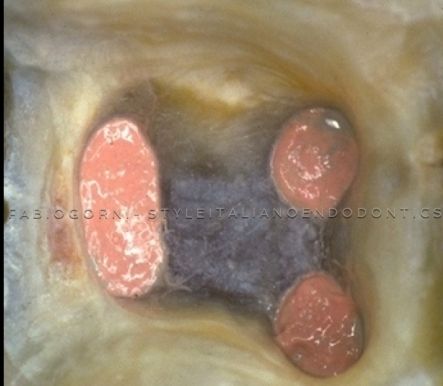
Fig. 7
Step-back technique has been described by Mullaney and others, including Clam, Weine, Martin and Walton. It is an apical to coronal technique, starting from the apex with fine instruments and working back up the canal to the crown with increasingly larger instruments. In the first phase, the apical part of the canal is enlarged, using files up to a final apical file size 25-30 or more; in the second phase, larger instruments are then inserted stepping back 1.0 or 0.5 mm from the working length, in a sequence. Refining and recapitulating (means using back smaller instruments), a continuous taper is formed. The step-back technique helped overcome the procedural errors of the previously used techniques in most anatomical situations, but in severely curved root canals or in very long roots problems still existed.
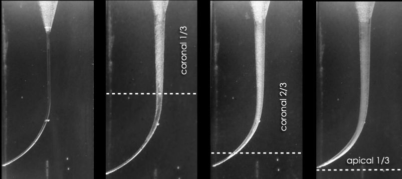
Fig. 8
Stepdown Technique
This method was first suggested by Schilder in 1974, and the technique was described in detail by Goerig in 1982. It has been followed by other similar techniques such as the double flared technique described by Fava in 1983 and the crown-down pressureless technique described by Morgan and Montgomery in 1984. The principle of these techniques is that the coronal aspect of the root canal is widened and cleaned before the apical part. We can divide the root into three thirds, coronal, middle and apical, thus simplifying the entire procedure into a series of smaller steps.
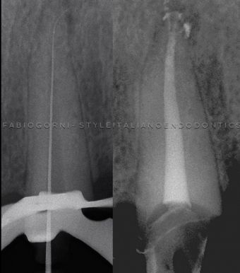
Fig. 9
The intra-op x-ray on the left shows clearly the space between the instrument and the canal wall in the coronal and middle thirds. This allows the pre-bent file to scout the apical anatomy.
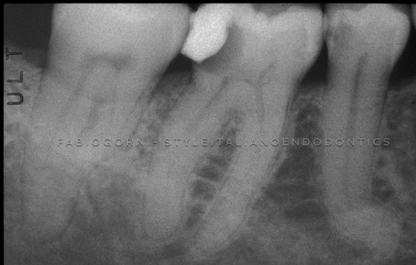
Fig. 10
STYLEITALIANO ENDODONTICS PHILOSOPHY
Our group, maintaining the advantages of this techinique, decided to follow this rationale since the 90s, with good success. Myself, in collaboration with Dr. Clifford Ruddle, edited an educational video, `The endodontic game´ about this early coronal enlargement technique. At that time, we only used manual files, but the approach was the same. In the following years we havent changed the technique, but only the instruments, moving towards rotary NiTi files. This means that we are talking about a universal technique, a real endodontic philosophy.
Clinically, the difference between step down and crown down isnt in the progression of the shaping, but in the sequence of the instruments. In general, when we are using the crown-down technique, we start to shape the coronal 2/3 of the anatomy with a greater taper, progressively reducing the size and taper of the instruments in the apical portion of the canal. By contrast, with the step down technique we start using smaller files, increasing their size during the treatment, but always from coronal to apical. Obviously, we can mix the sequences, but the most important rule is: never go to the apex first.
The power of this strategy is best appreciated in longer roots that hold more complicated anatomy, which exhibit significant calcification, challenging curvatures, deep canal divisions or delta.
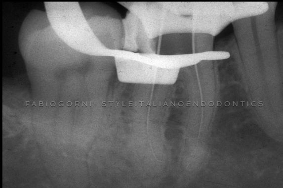
Fig. 11
Coronal and middle third enlargement allows apical scouting.
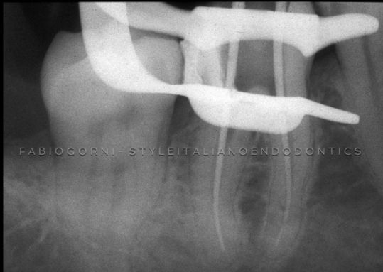
Fig. 12
Cone fit is checked radiographically.
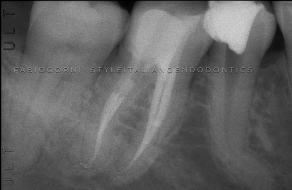
Fig. 13
On the left, the continuosly tapered preparation on the molar and on the right, in a double curved premolar, the pre-enlargement of the coronal two-thirds.
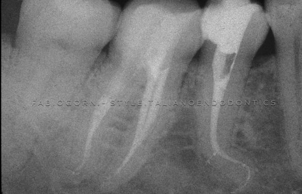
Fig. 14
Coronal and middle third of the canal are prepared first. This allows shaping & cleaning of the double curved canal.
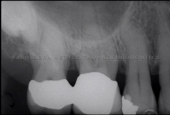
Fig. 15
ADVANTAGES
The advantages of these methods over the step-back can thus be listed:
I ) Step down technique permits straighter access to the apical region of the root canal. During the relocation of the canal orifices, widening the coronal part of the canal and removing the interferences allows the instruments to penetrate the canal with a better inclination and less torsional stress.
II ) Step down technique eliminates dentinal interferences found in the coronal two-thirds of the canal, allowing apical instrumentation to be accomplished quickly and efficiently, reducing the pressure from the coronal cutting flutes of the files.
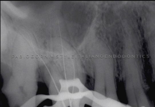
Fig. 16
The small file inserted in the canal, along the curvature, does not reach the foramen because of coronal interferences.
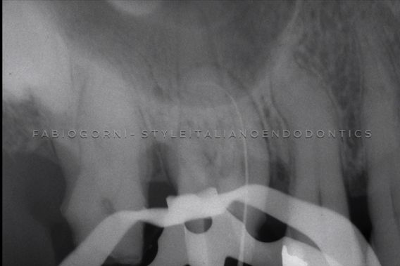
Fig. 17
By widening of the coronal two-thirds of the canal during preparation, the file can reach the apex. Note the better access of the file to the terminus.
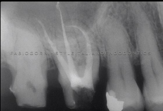
Fig. 18
The post-op x-ray shows the final shape and the canal filled correctly to the apex.
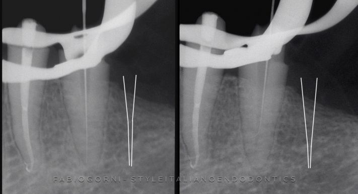
Fig. 19
III ) Step down technique, reducing the interferences with the pre-enlargement, gives to the clinician a better tactile control in every moment during the canal shaping. In particular, when we use a small pre-curved negotiating file into the apical third.
In this case, the increased coronal enlargement allows the pre-bent file to scout a lateral canal.
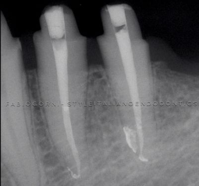
Fig. 20
Post op x-ray showing the root canal system filled trimensionally.
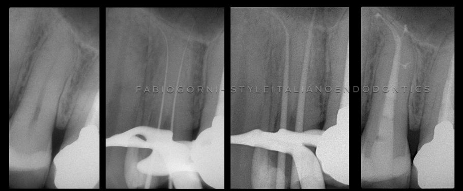
Fig. 21
IV ) Step down technique improves diagnostic. A pre-enlarged canal passively accepts a larger file into the apical third. This makes easier to visualize its extent radiographically. Electronic apex locators work better and are more accurate when utilized in pre-enlarged canals using bigger instruments. During the working length evaluation, the apical gauging will be more accurate, thanks to a more direct path to the terminus. The determination of the correct dimension of the apical foramen is one of the most important moments of the treatment.
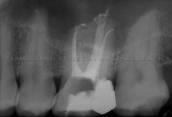
Fig. 22
V ) Step down technique increases the volume of the irrigants and encourages the elimination of the debris, enhancing the action of cleaning agents. A bigger volume of irrigant means better debris management. Narrow and more reduced preparations are dangerous; in this kind of preparation files work in virtually dry canals, increasing the risks of compacting debris apically with greater risks of blocks and ledges. A pre-flared canal accepts a greater volume of warm irrigant, which accelerates the apical and lateral dissolution of the pulp tissue. Pre-flared coronal and middle third of the canal costitutes a reservoir of irrigant which is more readly replenished in the root canal system. For all these reasons the pre-enlarged canals dramatically promote the removal of dentine mud improving pathway.
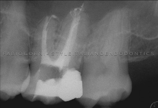
Fig. 23
In this clinical case we can see how the technique allows to shape, clean and fill the entire anatomy: apical delta in the palatal root, apical curvature in the distal root and a complex anatomy in the apical third of the mesial root.
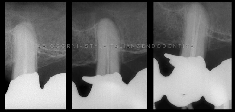
Fig. 24
VI ) Step Down Technique decreases post-treatment problems. Passing files through a cleaned, pre-enlarged canal means less debris inadvertently inoculated periapically. Passing files through underprepared canals coronally pushes more irritants periapically and generates more post-operative pain,
especially in necrotic teeth.
Upper premolar with a lesion of endodontic origin, single visit. In the x-rays is evident the the pre enlargement of the coronal 2/3.
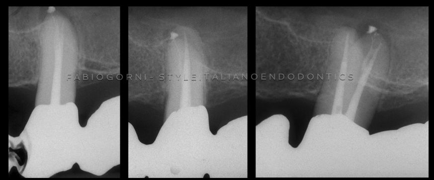
Fig. 25
Post op x-rays with different angulations. We can note the complex anatomy of the buccal root.

Fig. 26
12 years follow up shows complete healing of the lesion.

Fig. 27
Clifford J. Ruddle
THE TECHNIQUE
Step down technique is divided in some basic steps that are the following:
- Access cavity
- Scouting of the coronal two-thirds
- Glide path
- Pre-enlarging the coronal two-thirds
- Scouting of the apical one third
- Working leght
- Shaping
- Apical gauging
- FinishingThe steps are the same with manual or mechanical instruments. Using NiTi instruments, the rationale and the sequence is not changed, only the use of hand k-files is reduced.

Fig. 28
Manual files sequence

Fig. 29

Fig. 30
NiTi files sequence
Conclusions
Aim of this article was to summarize the reasons of the choice of the step-down technique as the favourite among StyleItalianoEndodontics members, and to outline the most important features of this technique. The next articles will describe the details of the various phases of the proposed technique.
Bibliography
- Schilder H. Cleaning and shaping the root canal, Dent Clin North Am 18:2, 269-296, April 1974.
- Mullaney TP. Instrumentation of finely curved canals. Dental Clinics of North America 1979; 23:575585.
- Clem WH. Endodontics: the adolescent patient. Dent Clin North Am. 1969 Apr;13(2):482-93.
- Walton RE, Torabinejad M. Principles and practice of endodontics. Philadelphia: WB Saunders Ed., 1989.
- Weine FS, Kelly RF, Lio PJ. The effect of preparation procedures on original canal shape and on apical foramen shape. J Endod. 1975 Aug;1(8):255-62.
- Morgan LF, Montgomery S. An evaluation of the crown-down pressureless technique. J Endod 1984;10:491-498.
- Goerig AC, Michelich RJ, Schultz HH. Instrumentation of root canals in molars using the step-down technique. J Endod 1982; 8:550-554.
- Fava LR. The double-flared technique: an alternative for biomechanical preparation. J Endod 1983;9:76-80.
- Ruddle CJ. Ch. 8, Cleaning and shaping root canal systems. In Pathways of the Pulp, 8th ed., Cohen S, Burns RC, eds. St. Louis: Mosby, pp. 231-291, 2002


