Retreatments and ReTreaty
28/12/2023
Ibrahim El Naggar
Warning: Undefined variable $post in /home/styleendo/htdocs/styleitaliano-endodontics.org/wp-content/plugins/oxygen/component-framework/components/classes/code-block.class.php(133) : eval()'d code on line 2
Warning: Attempt to read property "ID" on null in /home/styleendo/htdocs/styleitaliano-endodontics.org/wp-content/plugins/oxygen/component-framework/components/classes/code-block.class.php(133) : eval()'d code on line 2
The field of endodontic retreatment is wide and the cases are variable according to the mishaps included, in this case we have 2 separated instruments one apical and another coronal as well a ledge and short GP, so let’s see how RE TREATY will perform in this case.
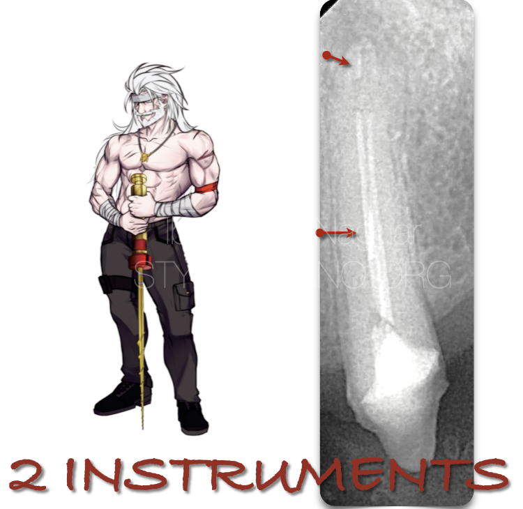
Fig. 1
Pre - Op Radiograph , showing upper premolar with 2 instruments & ledge.
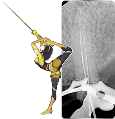
Fig. 2
Working length X Ray after reaching the full WL and the instruments one been retrieved and the other is bypassed.
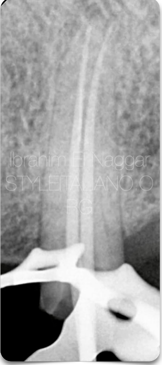
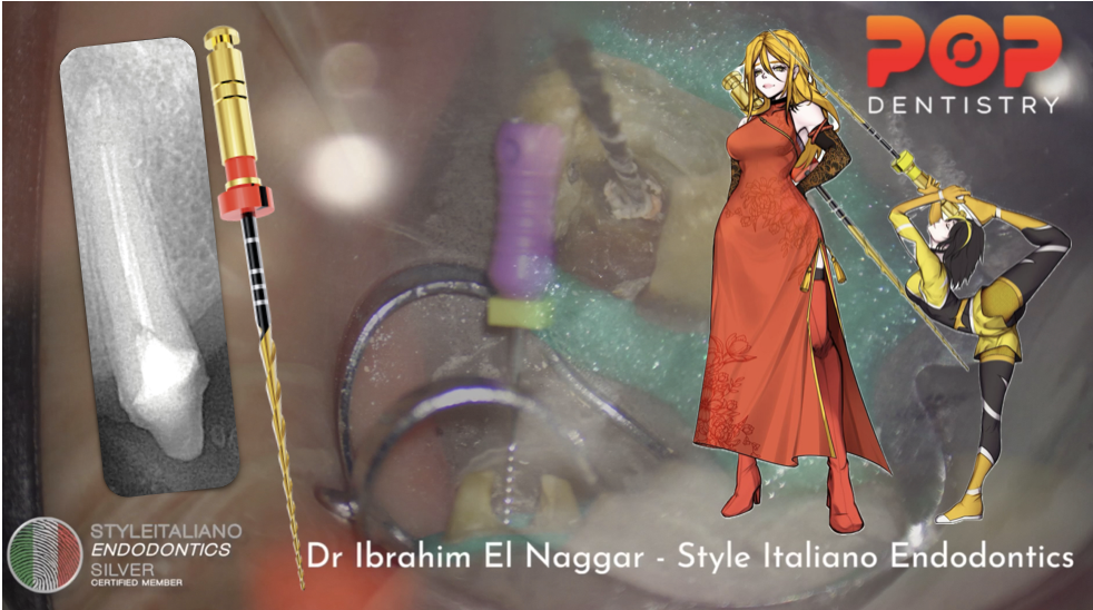
Fig. 3
The master cone X - Ray.
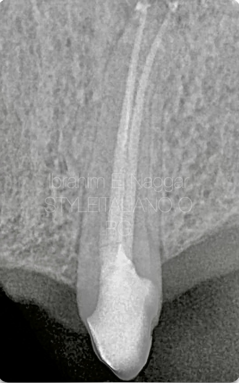
Fig. 4
Post-OP X Ray , the Picture show how conservative the treatment was.
THE VIDEO
Conclusions
The Clinician must have a knowledge about files , kinematics & metallurgy and to choose the best set for every situation , RE-TREATY kit presents an excellent option for every day rettt situations.
Bibliography
1. Endodontic Facts, American Association of Endodontists. Available at: http://www. aae.org/about-aae/news-room/endodontic-facts.aspx. Accessed June 1, 2015.
2. BorgesAH,BandecaMC,TonettoMR,etal. Portland cement use in dental root perforations: a long term followup. Case Rep Dent 2014;2014:637693.
3. TsesisI,FussZV.Diagnosisandtreatmentofaccidentalrootperforations.Endodontic Topics 2006;13:95–107.
4. Tsesis I, Rosenberg E, Faivishevsky V, et al. Prevalence and associated periodontal status of teeth with root perforation: a retrospective study of 2,002 patients’ medical records. J Endod 2010;36:797–800.
5. Gorni FG, Gagliani MM. The outcome of endodontic retreatment: a 2-yr follow-up. J Endod 2004;30:1–4.
6. Clauder T, Shin S-J. Repair of perforations with MTA: clinical applications and mechanisms of action. Endodontic Topics 2006;15:32–55.
7. Kvinnsland I, Oswald RJ, Halse A, Gronningsaeter AG. A clinical and roentgenological study of 55 cases of root perforation. Int Endod J 1989;22:75–84.