Re-treatment of Upper right Second Premolar
15/08/2020
Viresh Chopra
Warning: Undefined variable $post in /home/styleendo/htdocs/styleitaliano-endodontics.org/wp-content/plugins/oxygen/component-framework/components/classes/code-block.class.php(133) : eval()'d code on line 2
Warning: Attempt to read property "ID" on null in /home/styleendo/htdocs/styleitaliano-endodontics.org/wp-content/plugins/oxygen/component-framework/components/classes/code-block.class.php(133) : eval()'d code on line 2
Success of Endodontic therapy depends upon the triad of thorough cleaning and shaping, disinfection & adequate obturation up-to the calculated working length of the root canals. Failure to follow the protocol in any of the above steps can lead to failure of the endodontic treatment. Reading a preoperative periapical radiograph is of utmost importance. Though, they are 2 dimensional but do give us an idea of what approximate type of root canals we will be dealing with in a particular case. Cases with apically calcified canals can be very challenging. Many factors can lead to calcification of a or a part of root canal. Failure to negotiate the calcified part of the root canal system in the endodontic process might result in inadequate cleaning and shaping therefore resulting in short obturation and persistence of the diseased tissue.
This case report shows a Case of Symptomatic Apical Periodontitis (SAP) with chronic abscess associated with a previous root canal treatment with inadequate obturation in a maxillary premolar.
Description of the case
Patient information
- Age:29-year old
- Gender: Male
- Medical history: non-contributory
- Identification: Right maxillary second premolar (Tooth 15)
- Dental history :
Chief complaint: I am having pain when I eat or put pressure on my tooth. History of previous root canal treatment done on right upper maxillary second premolar.
Clinical examination findings:
The tooth 15 is tender to percussion and has an all-ceramic crown on it. Periodontal probing was within normal limits. Buccal vestibule had a slight intraoral swelling, which was tender to touch.
Radiographic findings:
The periapical radiograph revealed previous root canal treatment in 15. The obturation is short of the radiographic apex. The root is associated with periapical radiolucency. The apical part of the canals (which are unobturated), are suspected to be calcified
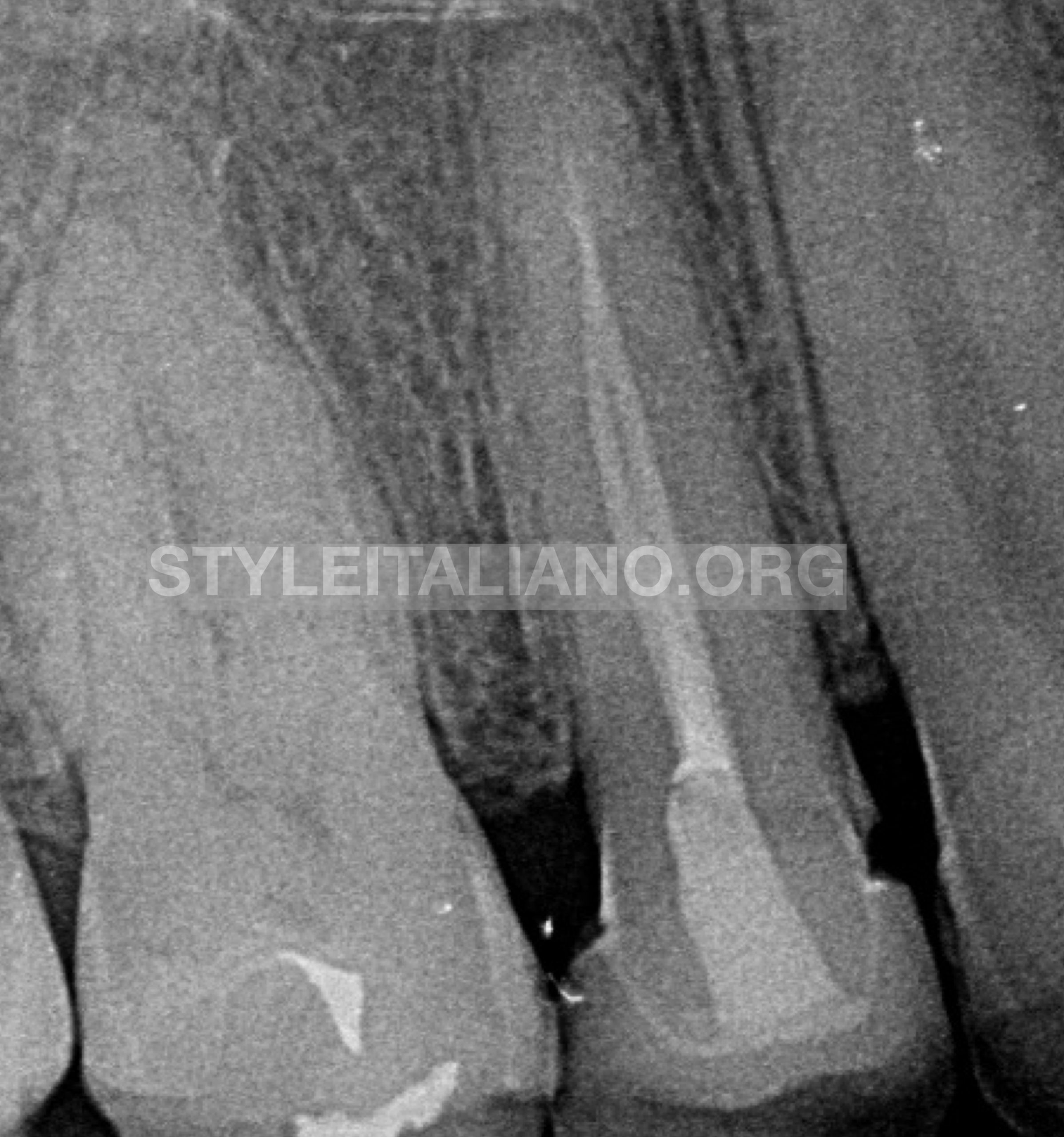
Fig. 1
Periapical radiograph inadequate root canal treatment in 15.
Periapical radiolucency associated with the root of 15 can also be seen.
Treatment plan:
Re-root canal treatment with the following options:
- Re-rct through the present all ceramic crown
- Re-rct after emoval of the all ceramic crown
Patient chose option 1.
However, it was made clear Incase All ceramic crown decemented during the retreatment procedure then a new all ceramic crown will have to be fabricated. Patient agreed and we started the re-rct through the present crown.
Step wise procedures in the first visit:
- Buccal infiltration anesthesia was administered, the tooth was isolated with rubber dam isolation and the tooth was seen under the microscope preoperatively.
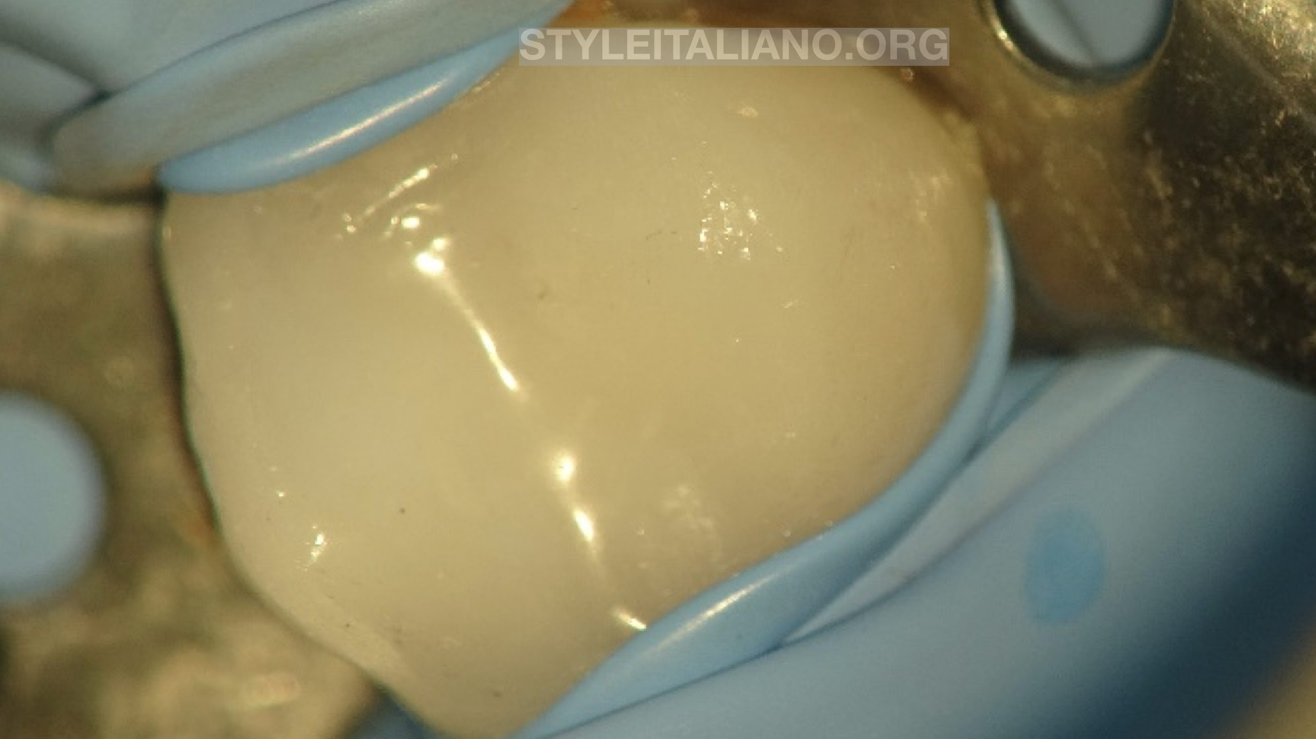
Fig. 2
Tooth 15 under isolation with all ceramic crown before starting with the retreatment procedure.
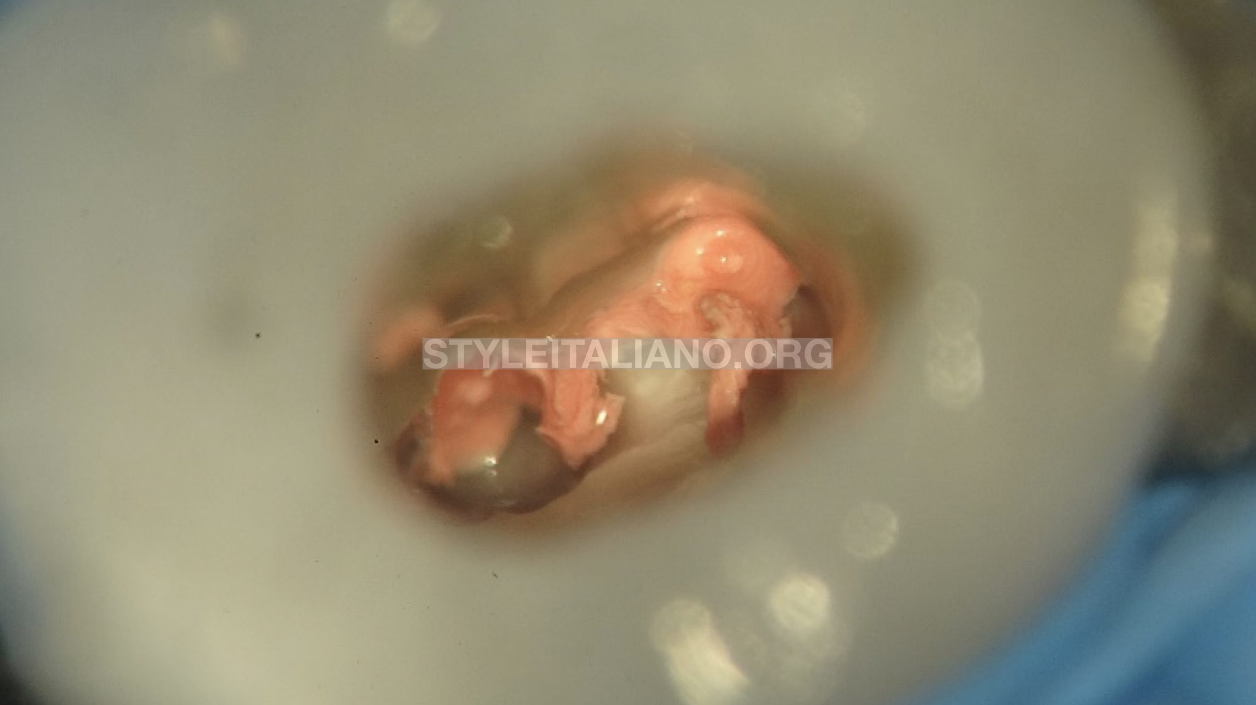
Fig. 3
Endodontic access was prepared through the ceramic crown and older gutta perchas located.
The coronal part of the gutta perchas in the pulp chamber were removed with ultrasonic scaler.
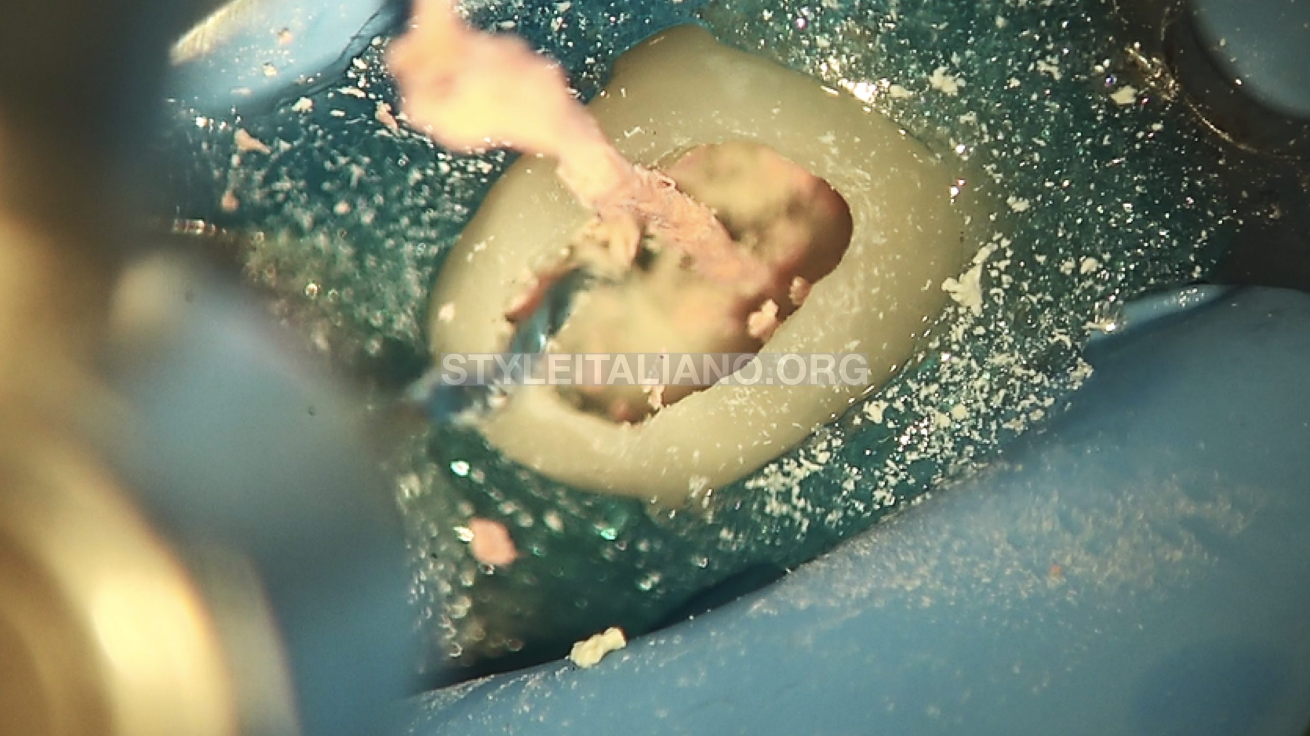
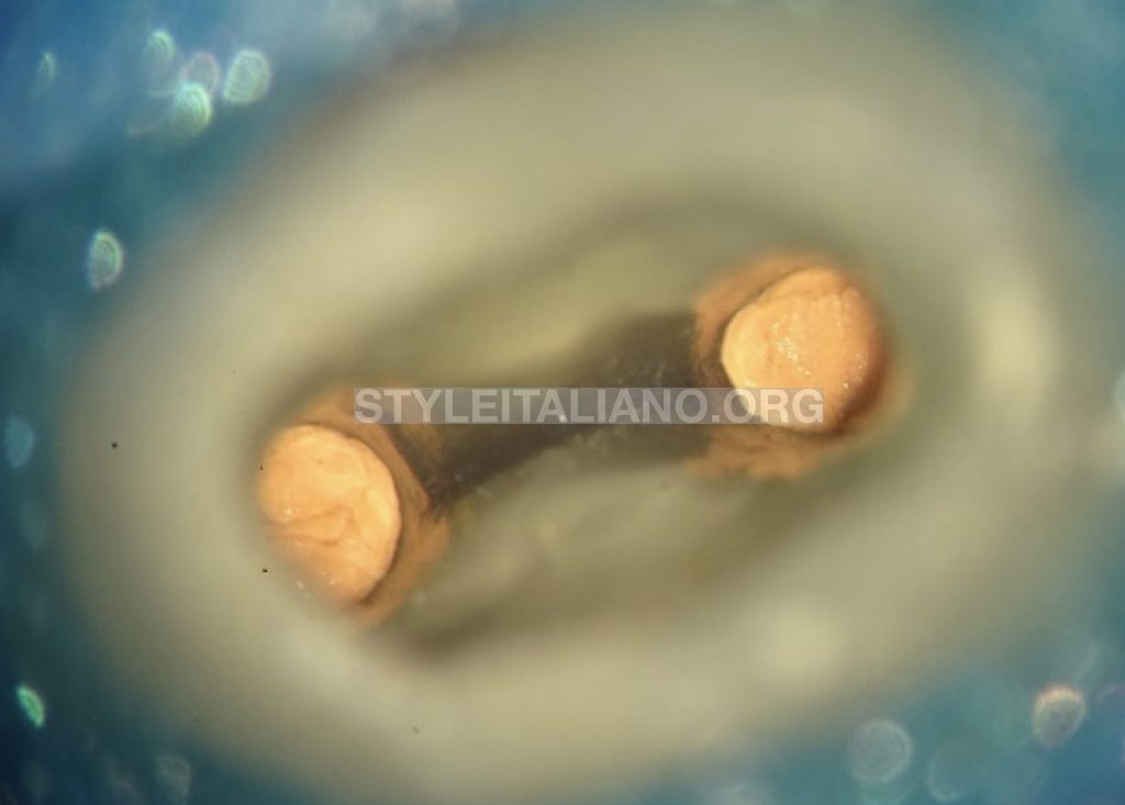
Fig. 4
Gutta percha inside the root canal was removed with the help a dedicated file used at 2500rpm.
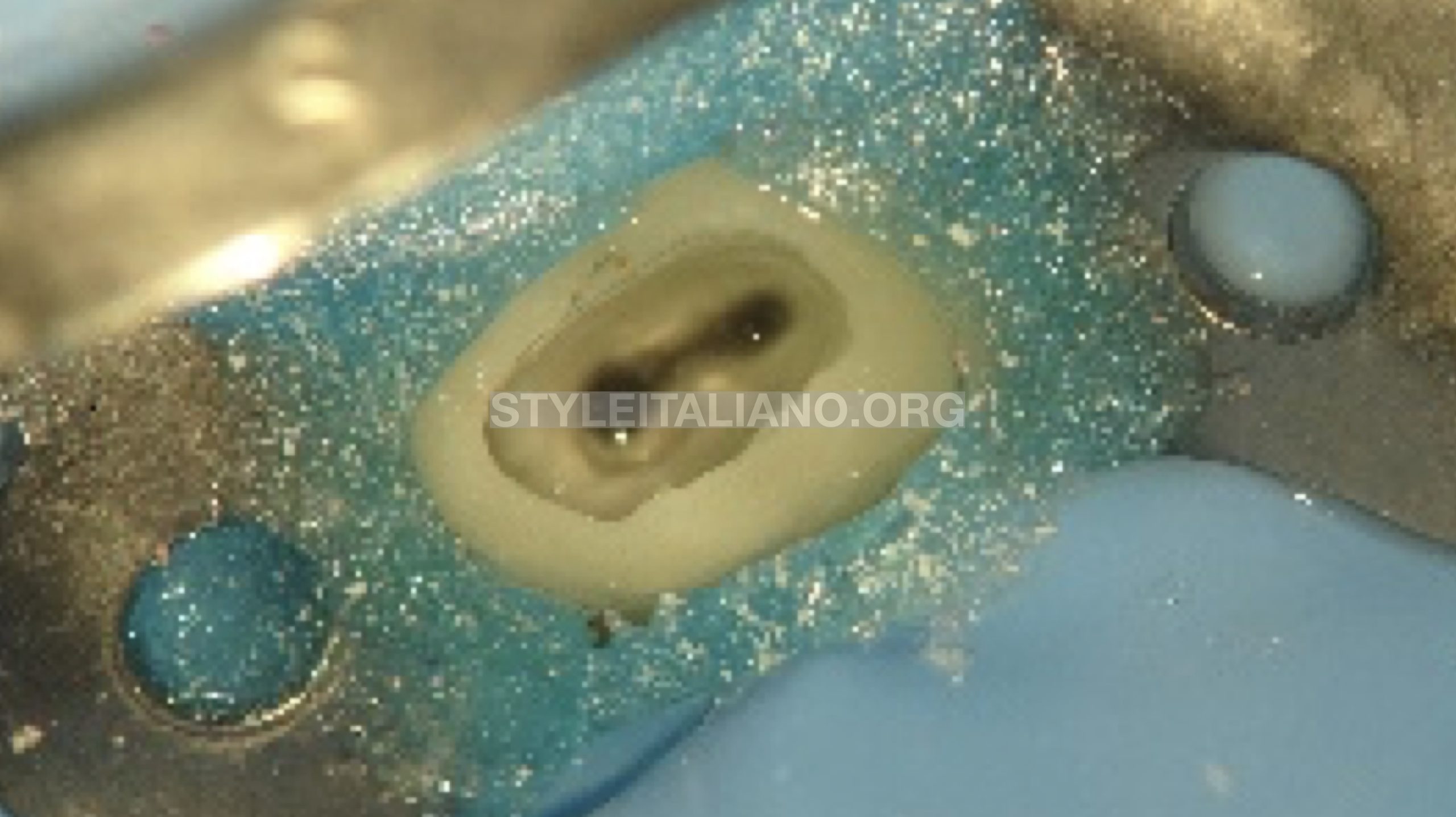
Fig. 5
Previous gutta percha was removed and intracanal medicament placed. The patient was recalled for second visit.
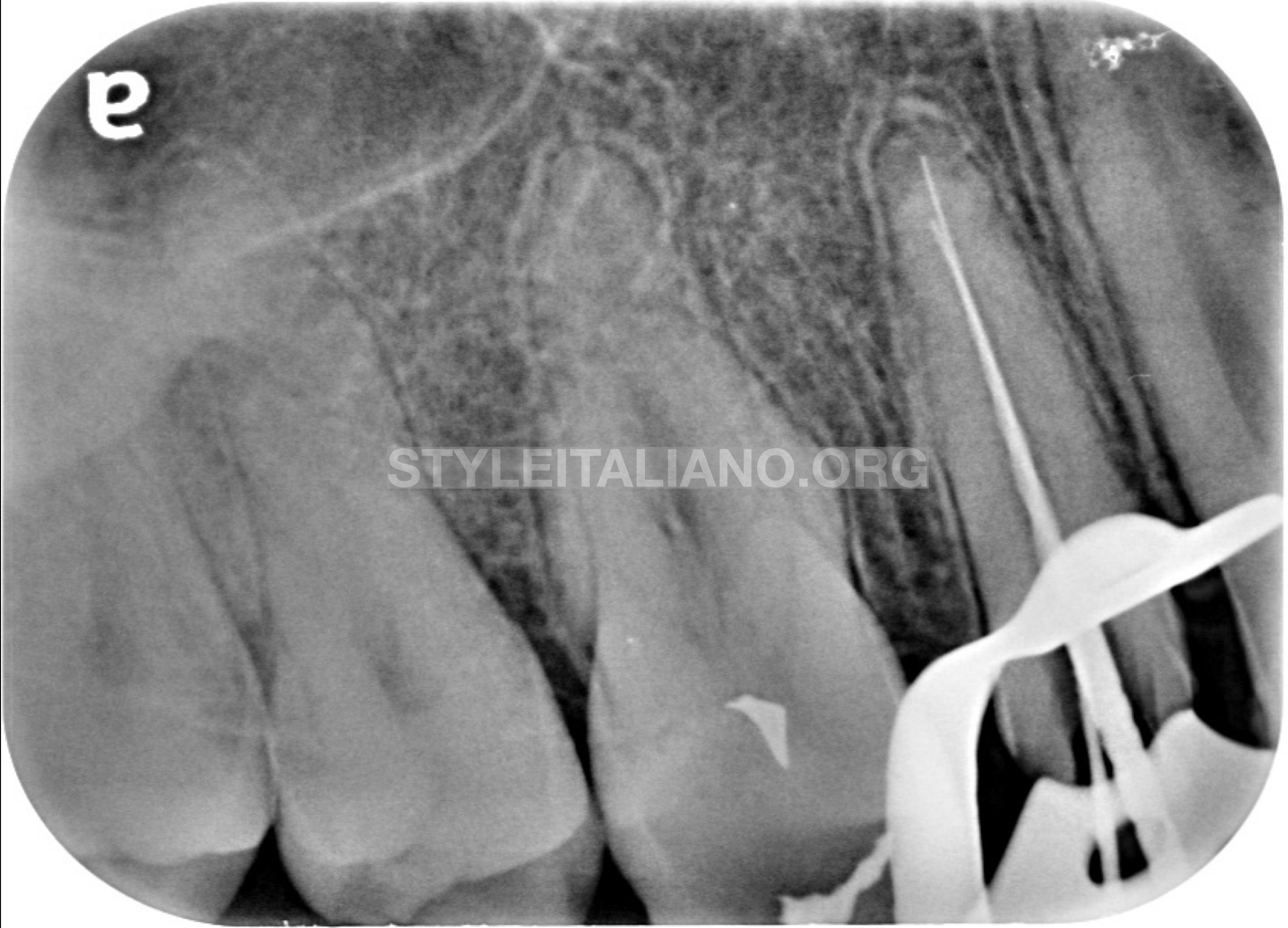
Fig. 6
While placing the clamp the all-ceramic crown popped out and the patient was informed. Irrigation was done to remove the Intracanal medicament from the canals. 08K and 10K endodontic files were pre curved and used in an attempt to open the calcified canals. C+ files were also used in order to open up the apically calcified part of the root canals. These files were used with passive motion in presence of the irrigants. Once the calcified part was negotiated, working length was determined with Electronic apex locator and verified with a periapical radiograph.
In the initial phase of cleaning and shaping, a rotary file was used with copious irrigation.
Cleaning and shaping under irrigation
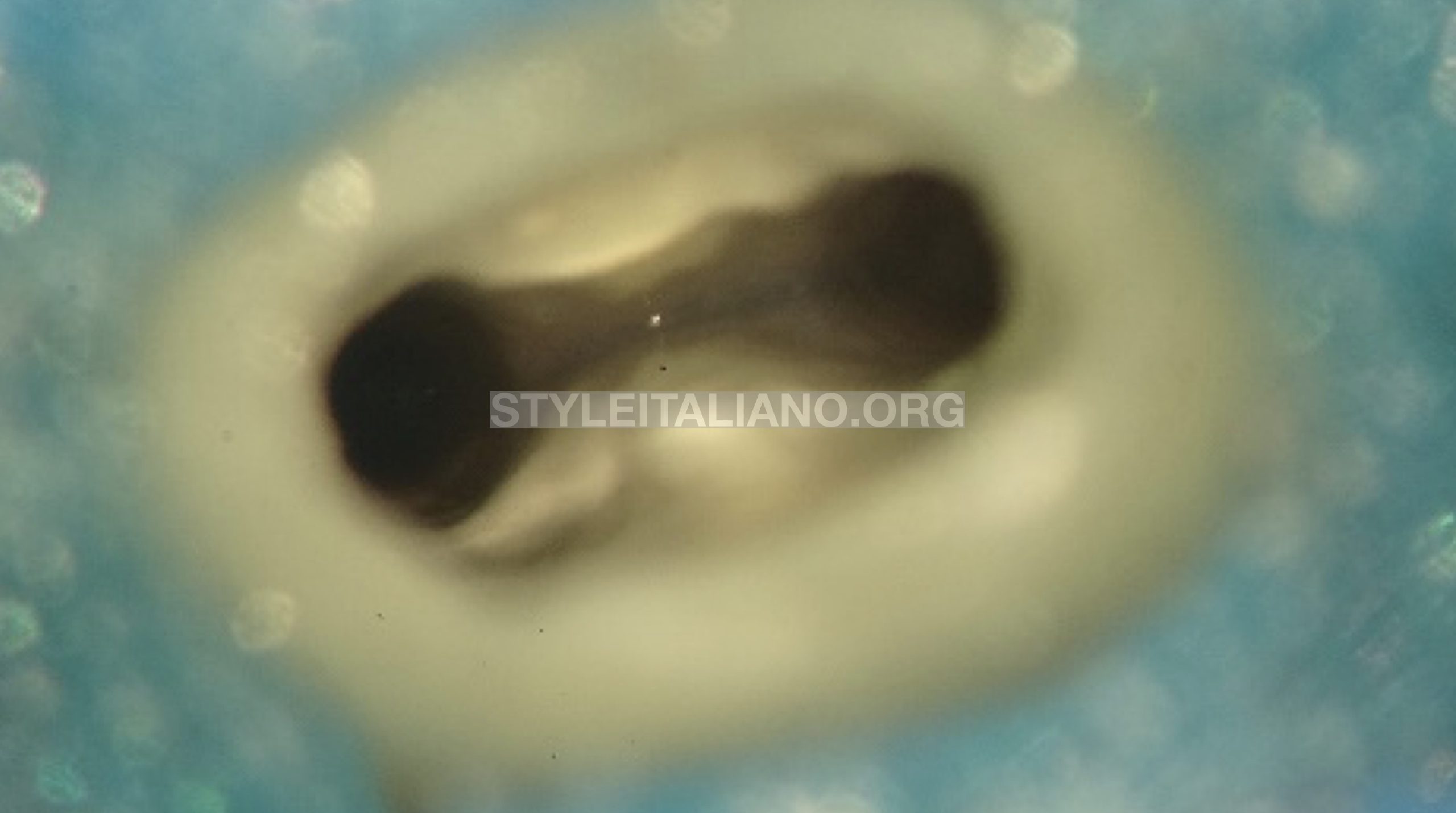
Fig. 7
Both the canals were finally prepared up to 25/04
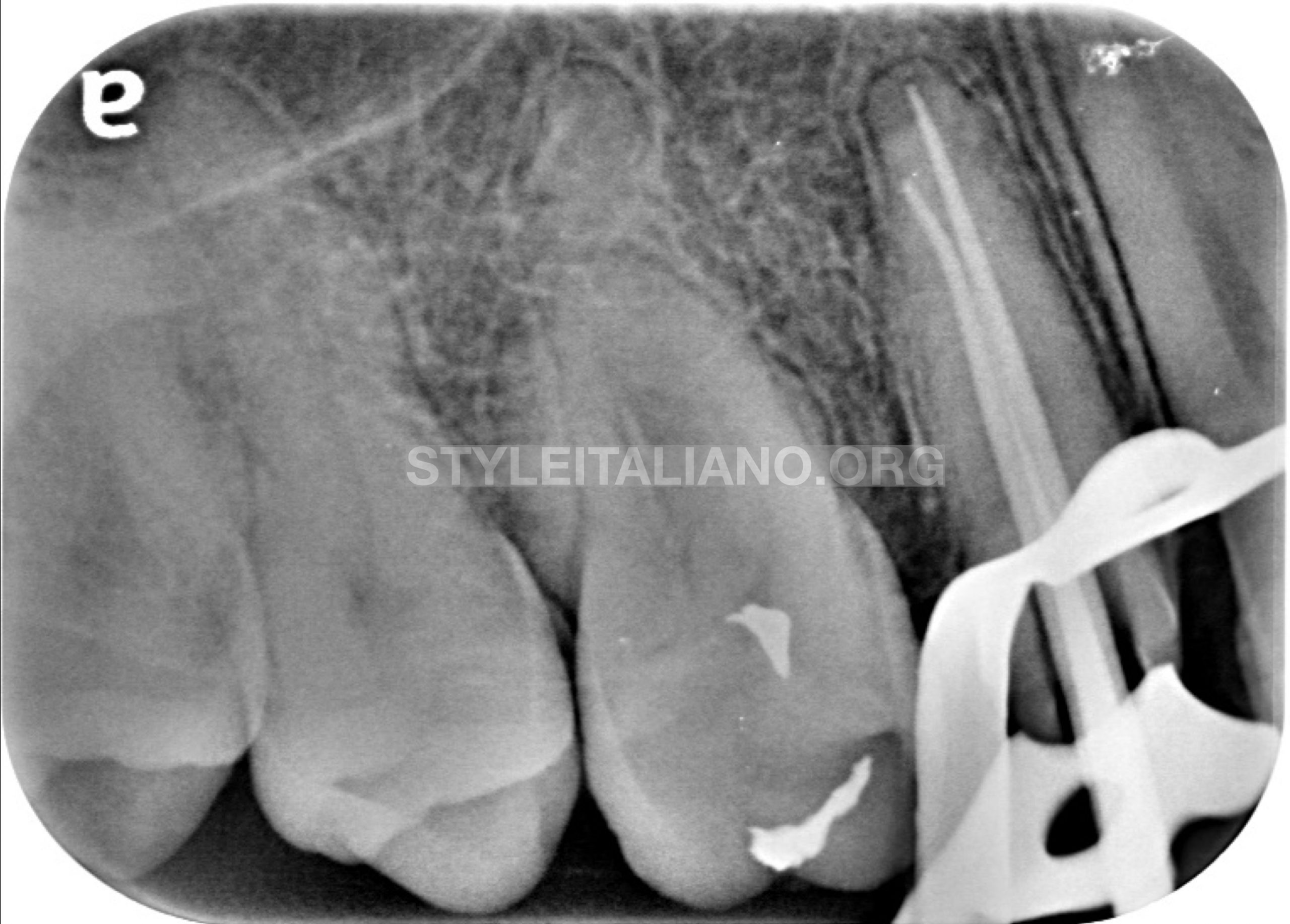
Fig. 8
Master cone verified for the cone fit. The master cones were verified on the periapical radiograph.
AH plus sealer was used and applied inside the canals. The master cones were coated with the sealer and placed inside the canals. Warm vertical condensation technique was used to obturate the canals.

Fig. 9
Both the canals were obturated and the pulp chamber cleaned of any gutta percha or sealer.
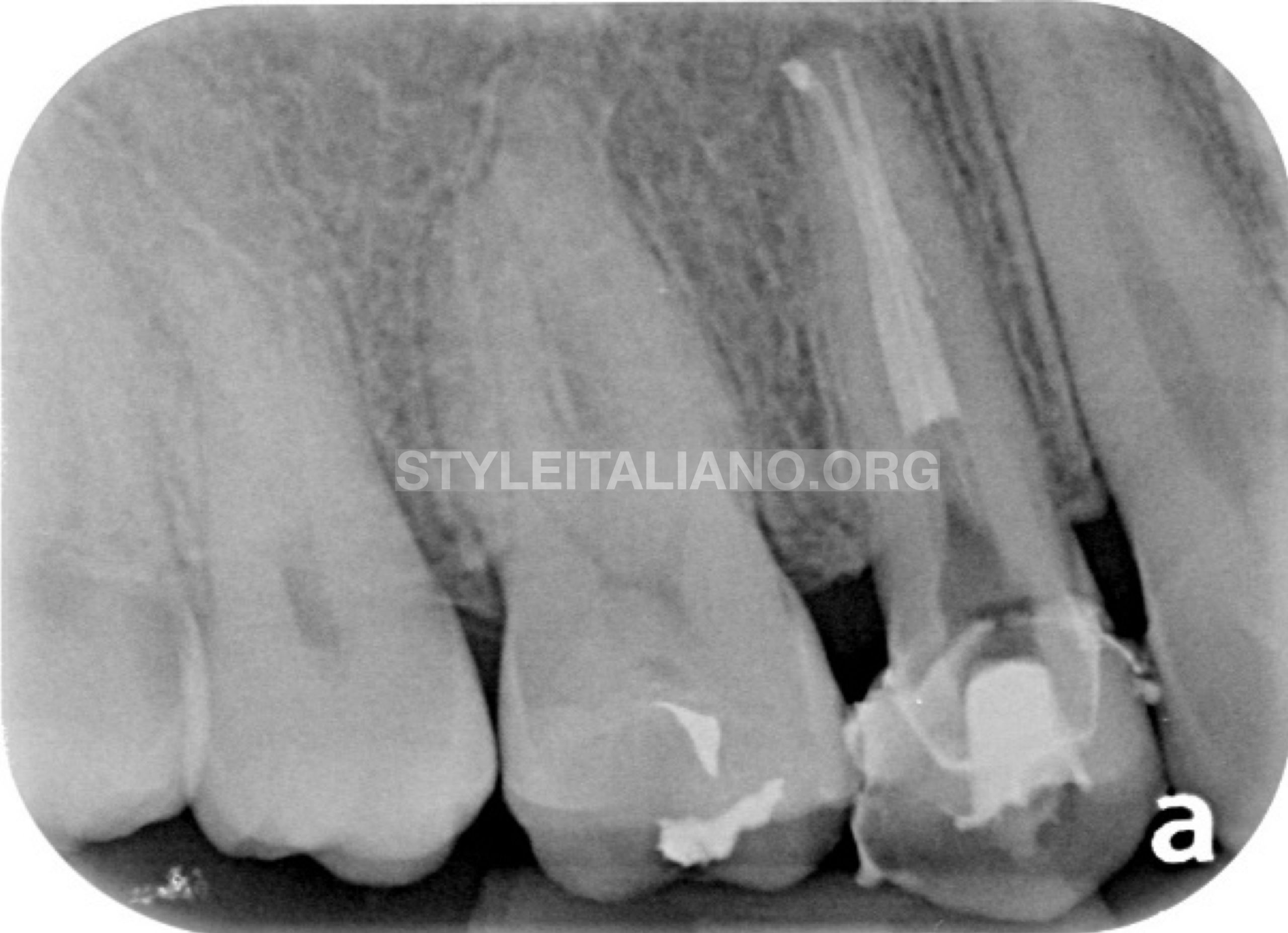
Fig. 10
Immediate postoperative radiograph was taken to verify the final obturation.
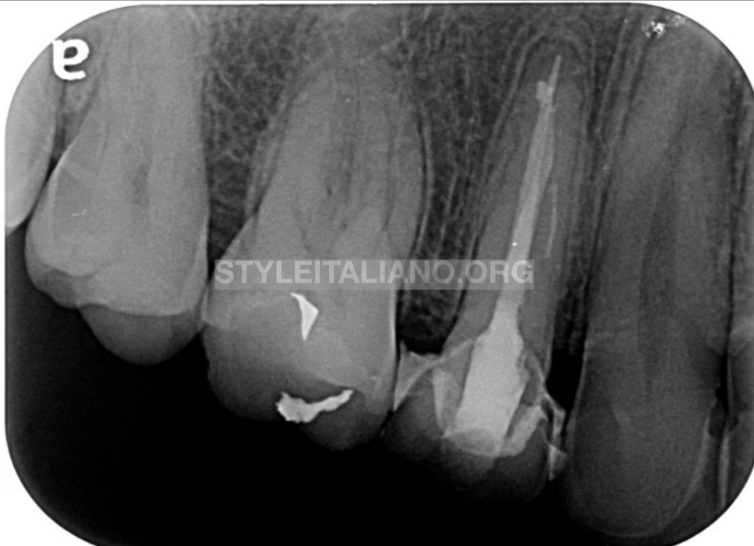
Fig. 11
New All ceramic crown was fabricated and cemented. 6- month recall of the case showed adequate healing and patient remained asymptomatic.
Conclusions
In order to achieve success in endodontic treatment, root canal system needs to be thoroughly cleaned, shaped, disinfected and obturated during the treatment.
Inability to negotiate the whole length of the canal will lead to inadequate cleaning and shaping leaving residual microorganisms behind which might cause failure of the root canal treatment as seen in this case.
Therefore, the root canals should be, coronally & apically sealed, properly up to the full working length in order to achieve success in endodontic treatment.
Bibliography
- Retreatment of endodontic fillings: Gunnar Bergenholtz Ulf Lekholm, Ralph Milthon, Gunnar Heden , Björn Ödesjö , Bure Engström; https://doi.org/10.1111/j.1600-0722.1979.tb00675.x
- The Outcome of Endodontic Retreatment: A 2-yr Follow-up: Fabio G.M.Gorni, Massimo M.Gagliani; https://doi.org/10.1097/00004770-200401000-00001
- Nonsurgical Retreatment: Clifford J.Ruddle;
- Determination of the Minimum Instrumentation Size for Penetration of Irrigants to the Apical Third of Root Canal Systems: AbbasaliKhademi, MohammadYazdizadeh, MahboobeFeizianfard; https://doi.org/10.1016/j.joen.2005.11.008