
Re-treatment Decoding : obturation dilemma
19/03/2024
Fellow
Warning: Undefined variable $post in /home/styleendo/htdocs/styleitaliano-endodontics.org/wp-content/plugins/oxygen/component-framework/components/classes/code-block.class.php(133) : eval()'d code on line 2
Warning: Attempt to read property "ID" on null in /home/styleendo/htdocs/styleitaliano-endodontics.org/wp-content/plugins/oxygen/component-framework/components/classes/code-block.class.php(133) : eval()'d code on line 2
Every failed root canal treatment has a reason, when facing non-surgical re-treatment the clinician must be aware of the reason of failures in order to deal with such cases in predictable way, reducing secondary risk of failure.
One of the most strategic factors effecting the non-surgical re-treatment is the apical seal, obturation material and way of obturation, every situation has its own scenario for an optimum outcome.

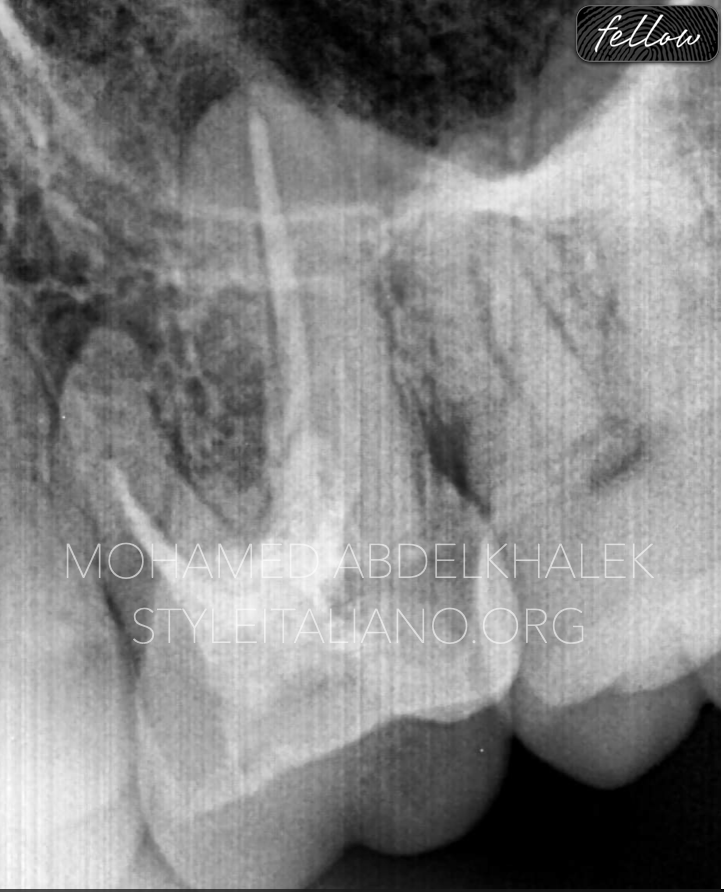
Fig. 1
A patient came to my office complaining about tenderness on chewing related to the upper first molar. We start with a pre-operative radiograph to diagnose in a proper way and find out the following.
1. Bad coronal seal
2. Short obturation related to the mesial system within a peri-apical lesion
3. Short obturation in the distal root
4.Peri-apical lesion related to the palatal canal due to improper seal with a blunt apex
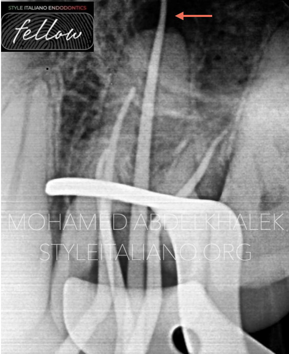
Fig. 2
After understanding the reason for failure we need to do the re-treatment in a predictable way for a long-term successful outcome. To start, the existing root canal filling material was removed and the root canal system was shaped with adequate ISO and taper for proper cleaning.
The mesial system show over-extended gutta-percha within 2 mm and type 2 canal configuration ( confluent MB1 and MB2 )
The distal system shows well-fitted master cone to the working length
Regarding the palatal canal, a gutta-percha point size 40 taper 4 was placed after removing the previous filling material. MTA apical plug will be our technique of choice to achieve proper seal.
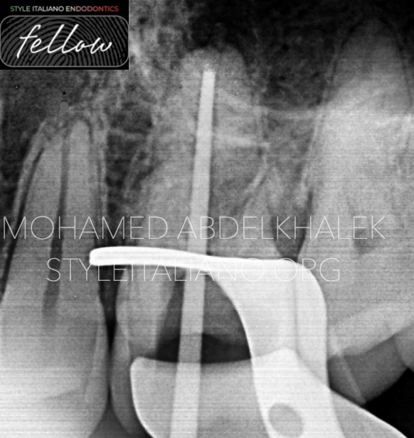
Fig. 3
A customized gutta-percha point, 2 mm shot to the working length of the palatal canal, was used as plugger for adequate MTA plug,
Endo ice is usually used to harden the gutta-percha for easy MTA packing in the apical part.
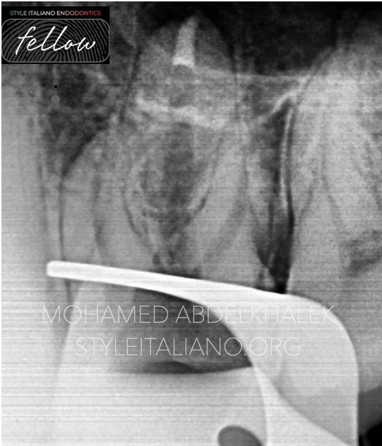
Fig. 4
A Radiograph was taken immediately after the plug: it shows that the MTA apical plug at the last 3 mm was adequately done (map system red-coded tip and customized gutta-percha).
In this video we are showing step by step the obturation technique related to every canal: with the right armamentarium and good knowledge it is possible to achieve a feasible and reproducible obturation.
Obturation was done with resin-based sealer using WVC technique in both medial and distal systems, while in the palatal canal a MTA apical plug followed by thermoplastized gutta-percha was done.
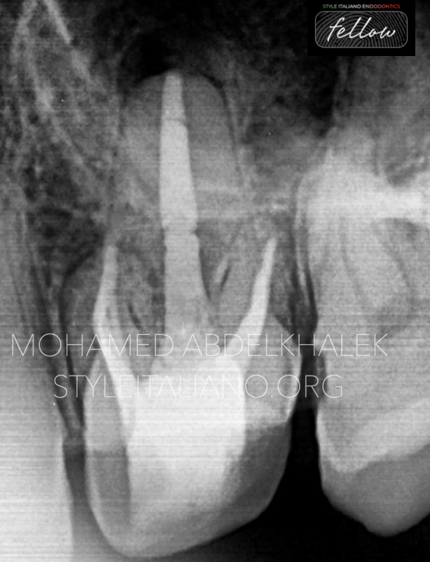
Fig. 5
Post-operative radio-graph showing good obturation adequate to the working length and coronal seal ready for post-endodontic restoration.

Fig. 6
About the author:
Mohamed Abdelkhalek
ABOUT ME
I'm a dentist was born and raised in Alexandri, I'm really passionate about endodontics and digital dentistry.
Proudly I'm providing elite services to my patient, also have been sharing knowledge with other colleagues inwebinars and local conference.
EDUCATION
2012- 2017
University of Pharos
WORK EXPERIENCE
Dental derma dreams
Full time conservative dentist related to micro-scopic and digital dentistry
Advanced endodontic in Mazen dental clinic
Microscopic dentist
Digital dentistry
ADA certificate in endodontics
Co-founder@dentaldermadreams.eg
Conclusions
Endodontic re-treatments are mainly strategy-based. Pre-operative assessment of the case with good knowledge of different filling materials and root canal anatomy associated with suitable armamentarium as map system, surgical microscope and WVC devices will enhance the success rate of the re-treatment
Bibliography
1. Martins JNR,Marques D, Mata A, Caramês J. Root and root canal morphology of the permanent dentition in a caucasian population: a cone-beam computed tomography study. Int Endod J 2017;50:1013–1026.
2. Studebaker B, Hollender L, Mancl L, Johnson JD, Paranjpe A. The Incidence of Second Mesiobuccal Canals Located in Maxillary Molars with the Aid of Cone-beam Computed Tomography. J Endod. 2018;44(4):565-70.
3. Hoen MM, Pink FE. Contemporary endodontic retreatments: an analysis based on clinical treatment findings. J Endod 2002;12:834–836.
4. Paul R. Cooper, Henry F. Duncan, Matthias Widbiller, Kerstin M. Galler. Treatment of Immature Teeth with Pulp Necrosis. Book Editor(s):Josette Camilleri. First published: 19 March 2021 https://doi.org/10.1002/9781119513568.ch3
5.G. Krastl, R. Weiger, A. Filippi, H. Van Waes, K. Ebeleseder, M. Ree, T. Connert, M. Widbiller, L. Tjäderhane, P. M. H. Dummer, K. Galler. Endodontic management of traumatized permanent teeth: a comprehensive review
6. E. Mandel, C. Bourguignon-Adelle. Endodontic retreatment: a rational approach to non‐surgical root canal therapy of immature teeth
7. European Society of Endodontology (ESE) developed by:, G. Krastl, R. Weiger, A. Filippi, H. Van Waes, K. Ebeleseder, M. Ree, T. Connert, M. Widbiller, L. Tjäderhane, P. M. H. Dummer, K. Galler. European Society of Endodontology position statement: endodontic management of traumatized permanent teeth
