Poor prognosis?...
28/06/2023
Fellow
Warning: Undefined variable $post in /home/styleendo/htdocs/styleitaliano-endodontics.org/wp-content/plugins/oxygen/component-framework/components/classes/code-block.class.php(133) : eval()'d code on line 2
Warning: Attempt to read property "ID" on null in /home/styleendo/htdocs/styleitaliano-endodontics.org/wp-content/plugins/oxygen/component-framework/components/classes/code-block.class.php(133) : eval()'d code on line 2
A case presentation of hemisectioning of the distal root of a LR6 due to fracture, and 6-year review.
The clinical aspects and considerations are discussed.
Occasionally, during our practice, we are required to offer patients a less common treatment modality.
Knowing all the possible treatment options available to us, we can provide our patients with better care.
This case demonstrates that even the apparently poor prognosis tooth may benefit from more conservative treatment other than extraction and be retained for longer.
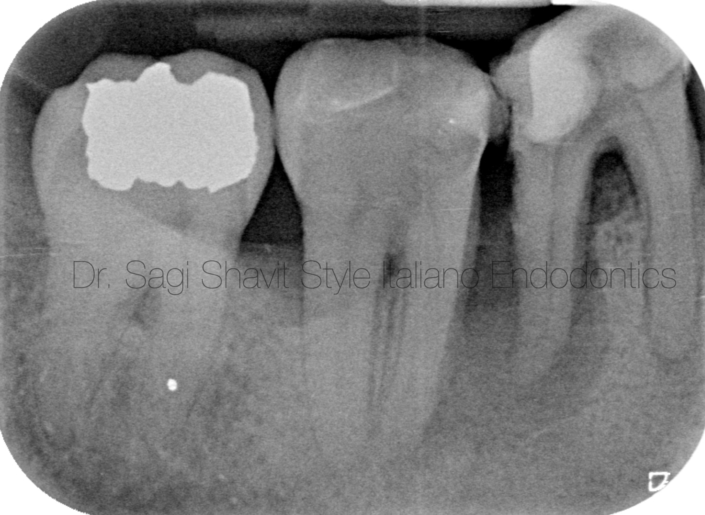
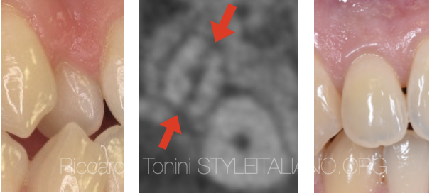
Fig. 1
Patient, male, in his late 60’s, with stable generalised chronic periodontitis, presented with swelling and pain from his LR6 (46) which appeared relatively recently.
Upon examination, the LR6 had defective coronal restoration, deep periodontal pocketing at the distal root, no excessive mobility and fluctuant buccal swelling. The tooth was not responsive to cold testing and was tender to pressure.
Periapical radiograph shows large radiolucency around the entire distal root and into the furcation, and small periapical radiolucency around the apex of the mesial root.
The diagnosis of pulp necrosis, chronic apical periodontitis and abscess was made.
Due to the ‘J’ shape nature of the lesion, perpendicular fracture was highly probable.
The patient was informed regarding the guarded overall prognosis of this tooth.
The treatment options of extraction or hemisection were presented to the patient, who was very keen to keep as much of his natural dentition and therefore elected to have hemisection.
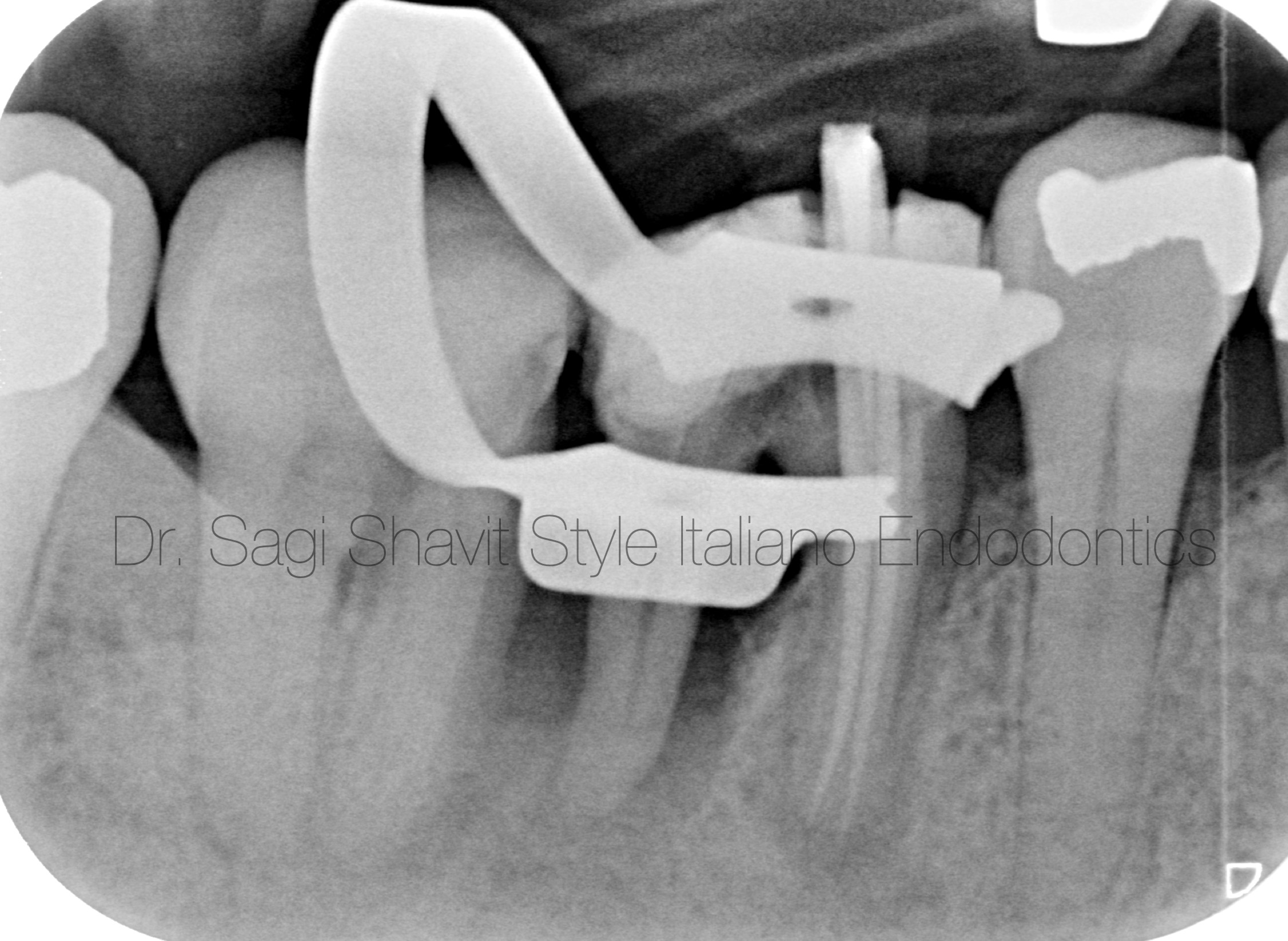
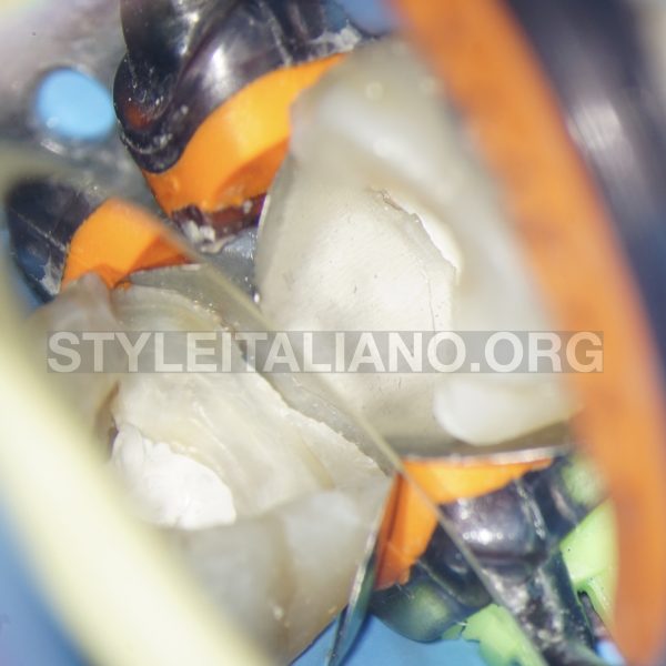
Fig. 2
After adequate local anaesthesia and rubber dam placement, access was gained into the 2 mesial canals, followed by chemo-mechanical treatment. Care was taken not to over enlarge the canals, in order to preserve as much dentine as possible.
Intra operative radiograph taken shows adequate length control.
The mesial canals were obturated with gutta percha and sealer , using the warm vertical technique.
Composite core was then placed.
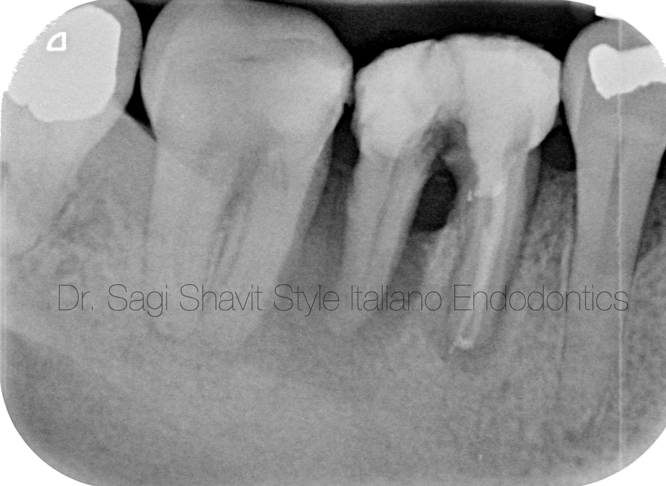
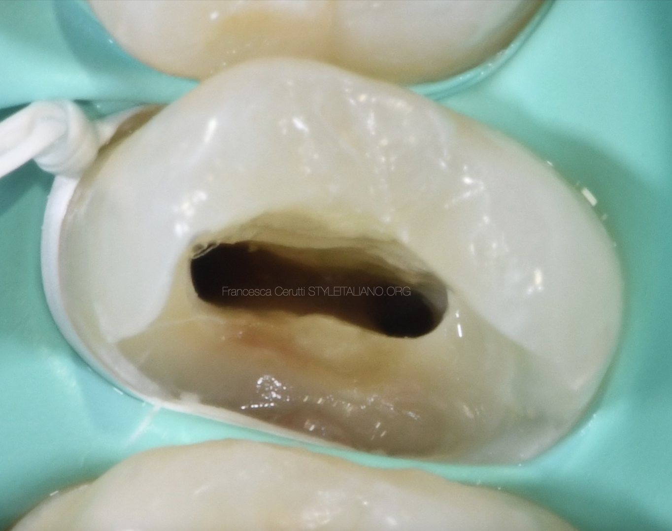
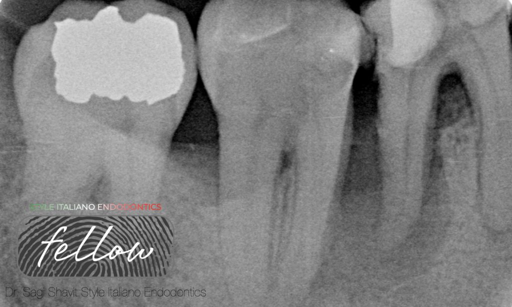
Fig. 3
Post operative radiograph shows adequate obturation.
Also, to note, the rapid deterioration and bone loss, in the few weeks that passed from the initial radiograph.
The perpendicular fracture into the furcation can be seen.
After the endodontic treatment has been completed,the LR6 was then sectioned at the furcation level and the distal root was removed carefully using luxators and forceps.
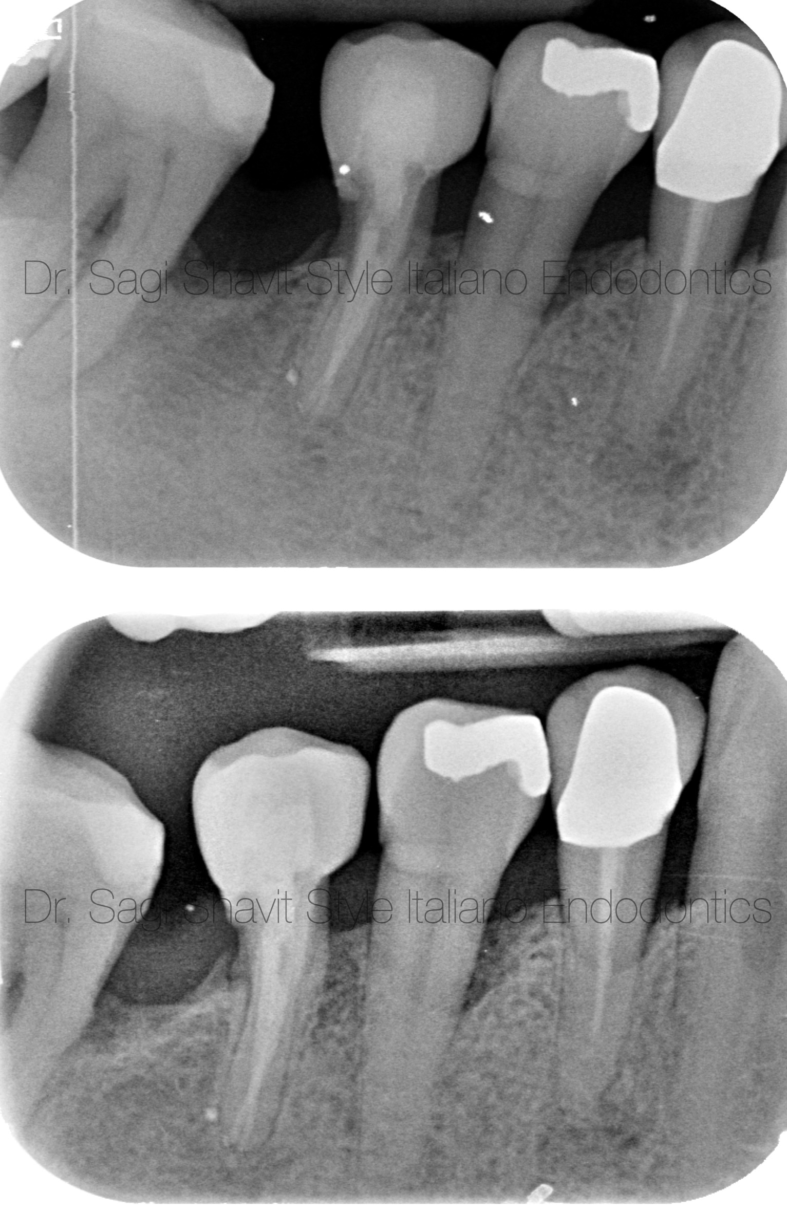
Fig. 4
After a few weeks of healing, the mesial root was restored with porcelain crown, by his referring dentist, Dr Jon Henley.
The patient returned for a follow up appointment after 6 months (top radiograph) and 1 year (bottom radiograph).
The tooth has been symptom free, and the periapical lesion has healed.
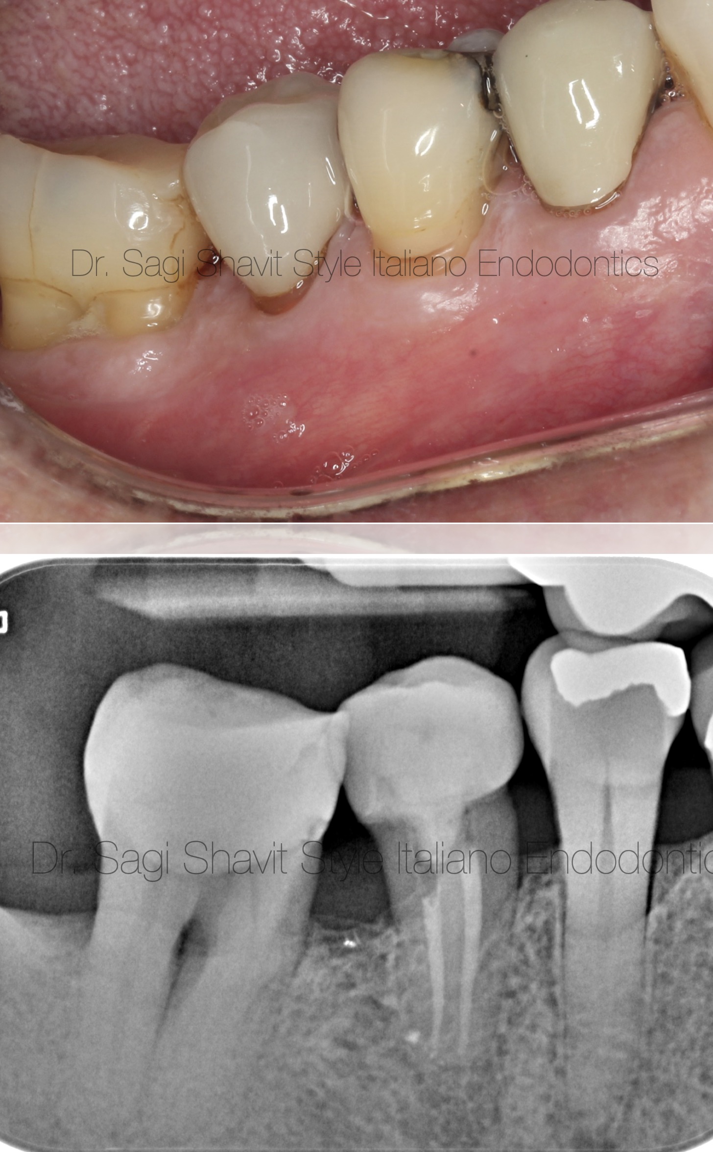
Fig. 5
The tooth was reviewed 6 years post operatively. It is functional and symptom free.
Clinically (top photo) the soft tissues look healthy, and the tooth looks aesthetically acceptable. Radiographically there are no signs of endodontic pathology (bottom).
Conclusions
Hemisection is a simple, effective and conservative modality of preserving part of the natural tooth.
It can be considered when one root of a multirooted tooth presents with severe bone loss due to vertical fracture, advanced periodontal lesion, deep caries, or inability to endodontically treat one of the roots (i.e., perforations).
The long-term prognosis of such cases varies from 70%-90% over a decade.
Several aspects must be considered prior to commencing treatment-
- The residual root and tooth structure- favourable crown to root ratio, sufficient tooth structure.
- Periodontal considerations- healthy periodontal tissue.
- Endodontic considerations- operable canals, conservative canal preparation to maintain as much tooth structure, no periapical pathology (if endodontic treatment already exists).
Bibliography
- Basten, C. H., Ammons, W. F., Jr, & Persson, R. (1996). Long-term evaluation of root-resected molars: a retrospective study. The International journal of periodontics & restorative dentistry, 16(3), 206–219.
- Bühler H. (1988). Evaluation of root-resected teeth. Results after 10 years. Journal of periodontology, 59(12), 805–810.
- Sharma, S., Sharma, R., Ahad, A., Gupta, N. D., & Mishra, S. K. (2018). Hemisection as a Conservative Management of Grossly Carious Permanent Mandibular First Molar. Journal of natural science, biology, and medicine, 9(1), 97–99.