Overfilling: Endodontic Failure Vs Clinical Success
21/05/2016
Calogero Bugea
Warning: Undefined variable $post in /home/styleendo/htdocs/styleitaliano-endodontics.org/wp-content/plugins/oxygen/component-framework/components/classes/code-block.class.php(133) : eval()'d code on line 2
Warning: Attempt to read property "ID" on null in /home/styleendo/htdocs/styleitaliano-endodontics.org/wp-content/plugins/oxygen/component-framework/components/classes/code-block.class.php(133) : eval()'d code on line 2
Many dentists believe that procedural errors in daily practice, such as over-filling, i.e. the contact of filling materials, generally gutta-percha and ZOE (Zinc Oxide Eugenol) sealer with the periradicular region in endodontically treated teeth are a direct cause of endodontic failure.
A number of Authors reported the negative impact of over-filling on the predictability of endodontic treatment and have highlighted the following:
1) The incidence of post operative pain as well as flare-ups
2) The toxicity of materials
3) Permanent irritation of periradicular tissues ranging from mild inflammations to foreign body reaction.
In contrast, other reports claim that, while overfilling should be avoided, its presence does not directly favor endodontic treatment failure by itself.
Ingle states that in Endodontics one achieves a high success rate in spite of overfilling.
Schilder said he has never found a case of failure specifically secondary to overfilling.
Weine states that luckily, since gutta-percha is so well tolerated by periapical tissue, it is a rare finding to see an over-filling in conjunction with a post-treatment failure. Most instances show no abnormal radiographic evidence, and, in some cases, there is an actual amputation of the overfilling with phagocitosys of the extra mass.
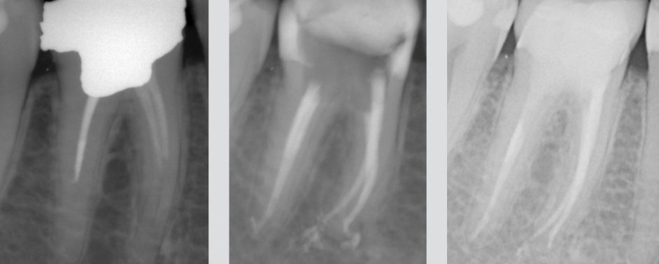
Fig. 1
First right mandibular molar with an infiltrated amalgam, and underfilled canal obturation.
After retreatment the Rx shows three-dimensional endodontics and slight surplus after filling on mesial canals obturated with Thermafil #25 and distal canal with warm vertical condensation.
After 5 years the excess has disappeared and periapical tissues are in healthy conditions.
These conflicting results can be attributed to differences in study methodology and to the diversity in composition of the extruded materials.
Generally, the most common causes are related to differences in apical anatomy hampering the performance of endodontic procedures in a variety of clinical situations.
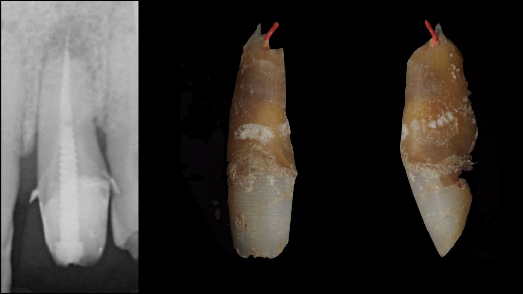
Fig. 2
Main factors for overfilling
- External apical inflammatory root resorption associated with apical lesion in teeth that have undergone previous intervention.
- Incompletely formed root.
- Overinstrumentation with deviation from the canals natural path and apical perforation.
- Inadequate manipulation of plasticized or non plasticized gutta-percha and sealers during obturation procedures.
It is critical to define and make a distinction between overfilling, which is the three-dimensionally cleaned, shaped, and packed root canal that exhibits surplus after filling Vs an obturation overextension with internal underfilling.
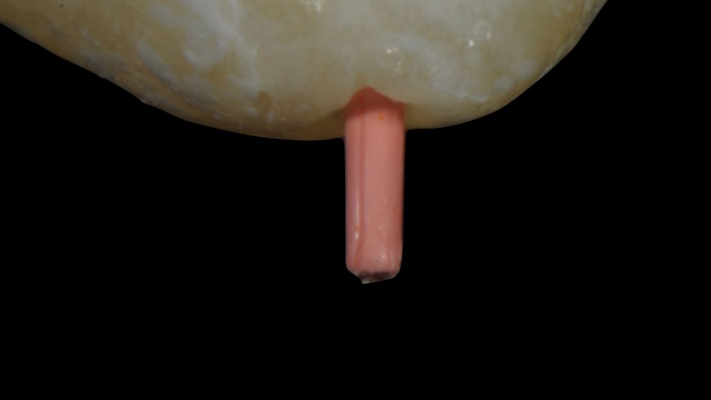
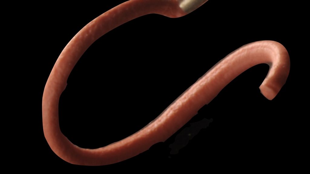
Fig. 3
Typical aspect of overfilling: note the scratch on the cone.
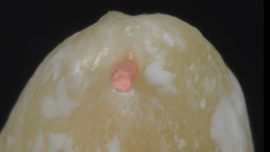
Fig. 4
Apical foramen is completely filled.
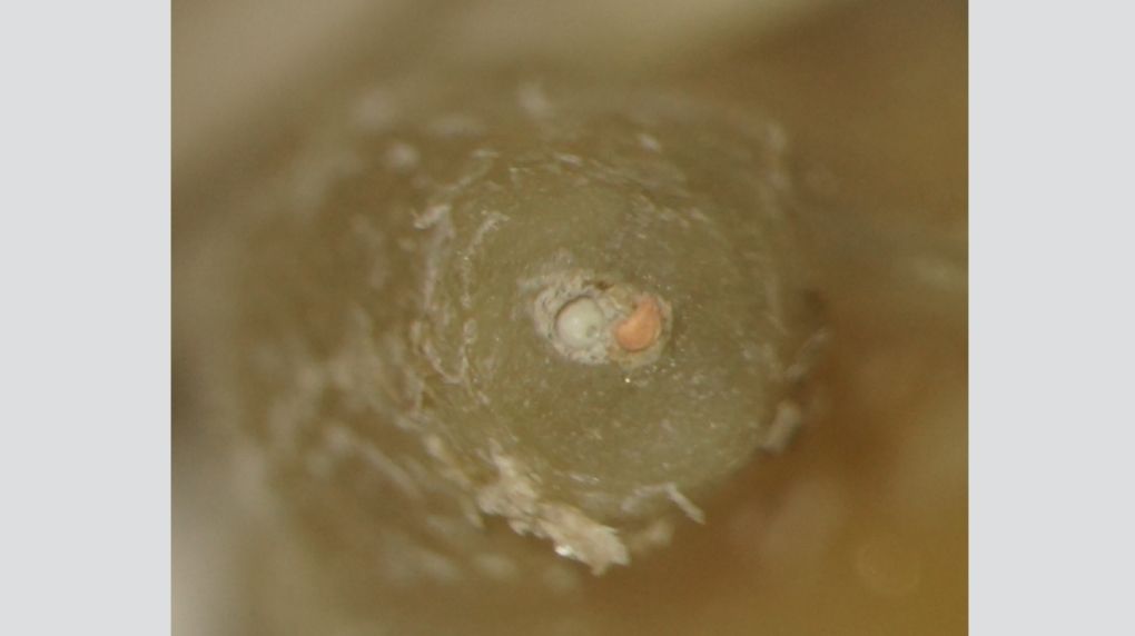
Fig. 5
Correct extension of the obturation but underfilling of canal. Notice the apical transportation during shaping procedure. Courtesy of Dr. M. Vitullo
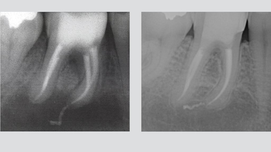
Fig. 6
Post-operative radiograph of a mandibular first molar reveals a large apical lesion of endodontic origin. Mesial canals were obturated with Thermafil. A 4-year recall demonstrates osseous repair. Filling excess has not prevented bone healing.
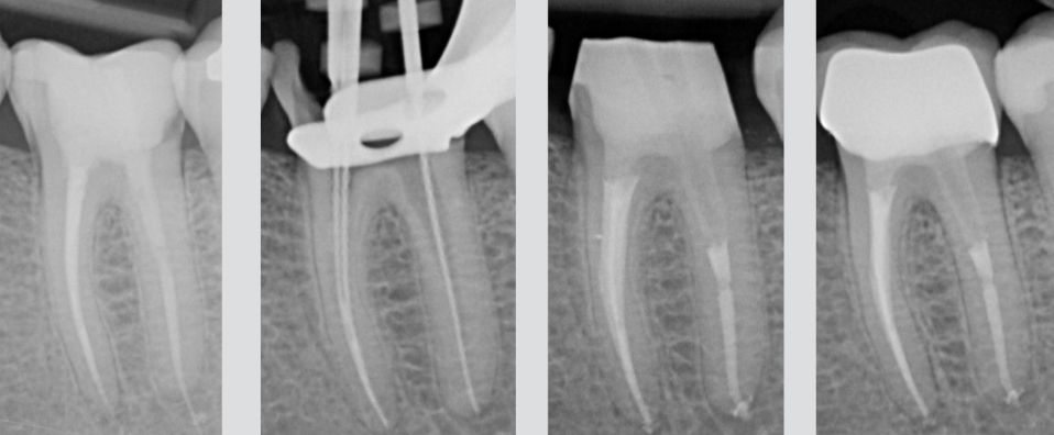
Fig. 7
Thermafil carrier overfilling on the distal root. Thermafil is a good technique for obturation as long as the protocol is respected. Notice the extrusion of the carrier and the obturation underselling the canal. After 6 months the lesion looks smaller than in the pre-op Rx and the patient reports disappearance of pain during function.
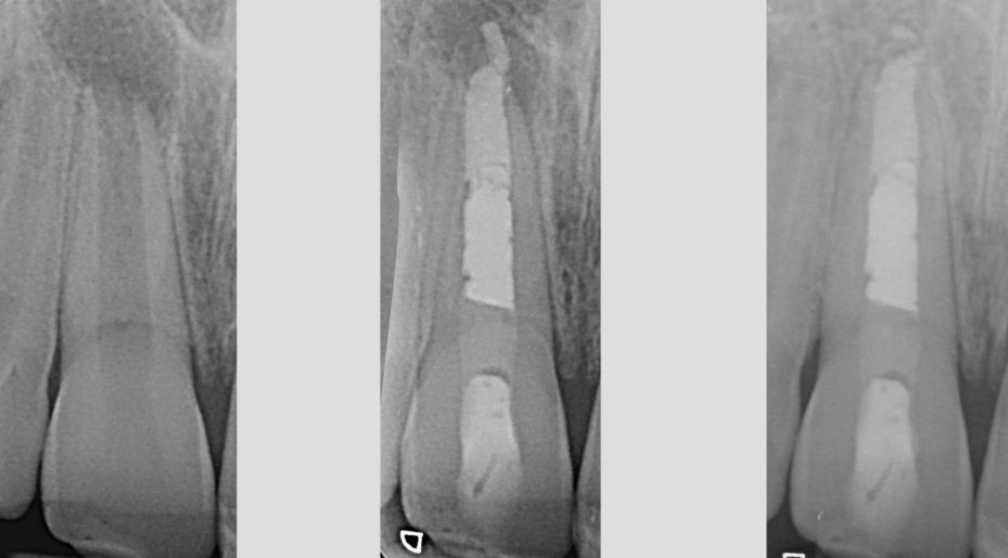
Fig. 8
Even MTA can be extruded into the periodontal tissues. In this case the rule is just the same as that of the previous cases: if the canal is clean, excess of MTA cannot prevent tissues from healing. 6 years follow-up, courtesy of Dr. R. Tonini.

Fig. 9
Obturation is the last step of an endodontic treatment: overfilling of the canals either with sealer or gutta-percha isn't necessarily a procedural error during obturation. It can however be due to an error during the shaping procedure, leading to a root canal with parallel walls instead of a tapered one, or maybe due to apical resorption or a widened apex.
Hence, we have to define the reasons for overfilling of a root canal: what is the direct cause? Is it an excess of material remained after the three-dimensional obturation or an overextension with canal underfilling? Surplus material does not constitute an endodontic goal, but it can be seen rather as a result of the hydraulics required to achieve a three-dimensionally packed root canal systems.
Conclusions
Despite of the overfilling, countless cases show clinical and radiographic long-term healing.
Bibliography
- West JD. Endodontic failures marked by lack of three- dimensional seal. The Endodontic Report Fall/Winter: 9,1987.
- Schilder H. Filling root canals in three dimensions. Dent Clin North Am 11: 723, 1967.
- Seltzer S. Ch. 11, Root canal failures. In: Endodontology: Biological Considerations in Endodontic Procedures. New York, NY: McGraw-Hill, 1971.
- Weine FS. Ch. 7, Intracanal treatment procedures. In: Endodontic Therapy. St. Louis, MO: C.V. Mosby Co, 1972. Chap 7.
- Ruddle CJ. Ch. 9, Three-dimensional obturation: the rationale and application of warm gutta percha with vertical condensa tion. In Cohen S, Burns RC, editors: Pathways of the Pulp. 6th ed. St. Louis, MO: Mosby Yearbook Co, 1994.
- Wolfson EM, Seltzer S. Reaction of oral connective tissue to some gutta-percha formulations. J Endod 1975;1:395-402.
- Seltzer S. Long-term radiographic and histological observations of endodontically treated teeth. J Endod 1999;25:818-22.
- Costerton JW, Stewart PS, Greenberg EP. Bacterial biofilms: a common cause of persistent infections. Science 1999;284:1318-22.
- Wolfson EM, Seltzer S. Reaction of oral connective tissue to some gutta-percha formulations. J Endod 1975;1:395-402.
- Castellucci A. Endodontics Editors: Il Tridente 2010.
- Ingle J.I. Root canal obturation. J Am Dent Assoc. 1956 Jul;53(1):47-55.