Non surgical endodontic management of periapical lesion with open apex
16/11/2024
The Community
Warning: Undefined variable $post in /home/styleendo/htdocs/styleitaliano-endodontics.org/wp-content/plugins/oxygen/component-framework/components/classes/code-block.class.php(133) : eval()'d code on line 2
Warning: Attempt to read property "ID" on null in /home/styleendo/htdocs/styleitaliano-endodontics.org/wp-content/plugins/oxygen/component-framework/components/classes/code-block.class.php(133) : eval()'d code on line 2
MTA is a bioactive cement that has gained immense popularity in endodontic treatments. It is composed of tricalcium silicate, dicalcium silicate, and bismuth oxide.
MTA possesses unique characteristics that make it an ideal material for various dental procedures. These properties include exceptional sealing ability, biocompatibility, good marginal adaptation, as well as its ability to promote tissue healing and mineralization.
The advantages of using MTA as a root canal obturation material include superior sealing ability, improved fracture resistance, repair and regeneration of supporting tissues, and resolution of resorptive defects.
This article describes a refer case in which 13 years old boy presented with open access cavity accompanied by pain and bad taste in his mouth, According to his parents; when he was 7 years old, he had a trauma to this tooth.
A crown fracture had occurred and the patient did not experience any discomfort.
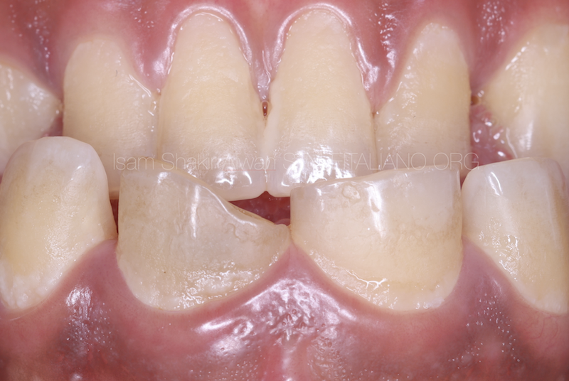
Fig. 1
Preoperative image show labial view of of tooth 21.

Fig. 2
Preoperative image show occlusal view of tooth 21.
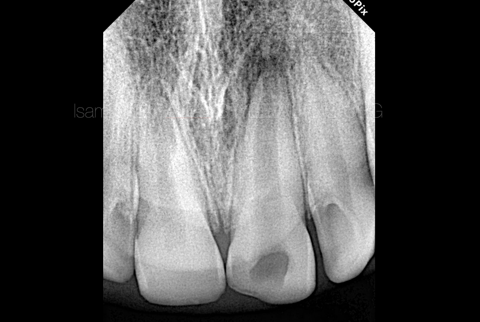
Fig. 3
Preoperative radiographic showing incompletely developed apex as a sign of periapical lesion.
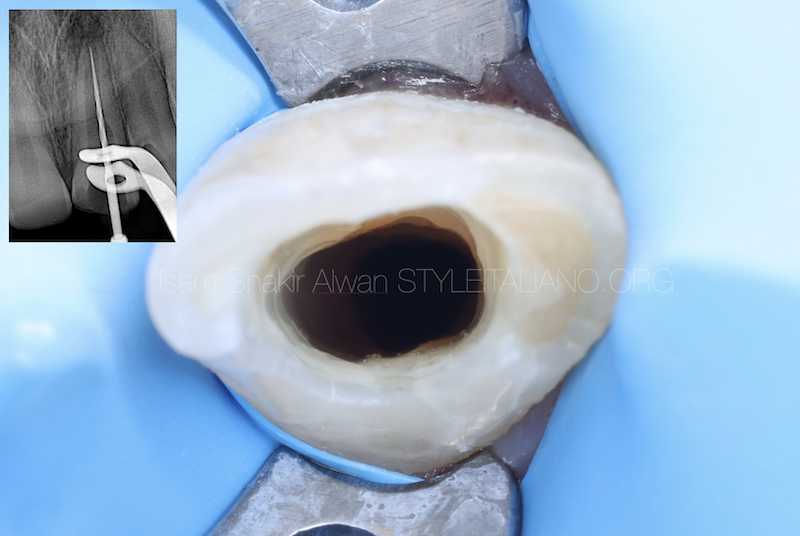
Fig. 4
After local anesthesia administration, rubber dam isolation and access cavity was done from the palatal side of the tooth.
The unsupported tooth structure removed, Arbitrary WL determination was done with apex locator and confirmed with IOPA radiography.
Since 50 size K file was freely passing through the apex, a decision for non surgical endodontic treatment was made.
This photography allows to see the canal after mechanically instrumentation.
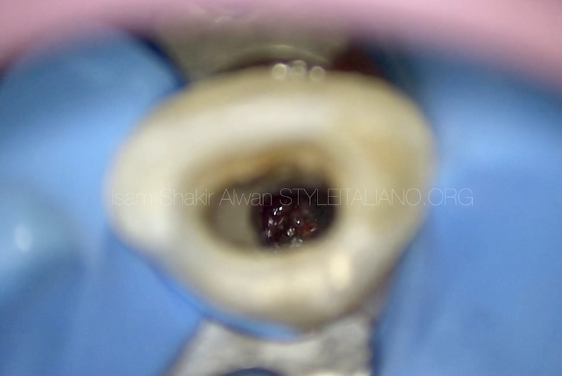
Fig. 5
Microscopic image allows to see directly the immature apex.
The canal was then irrigated with 2.5% sodium hypochlorite and EDTA17% using a 30 gauge side vented needle to avoid its apical extrusion and walls of the canal were cleaned using a circumferential filing motion and sonic activation.
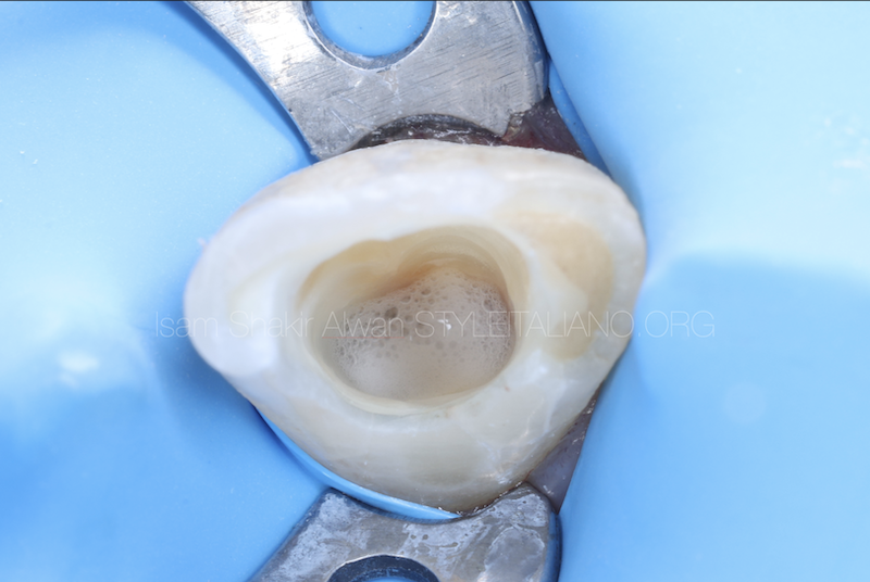
Fig. 6
The image shows the tooth immediately after sodium hypochlorite activation.
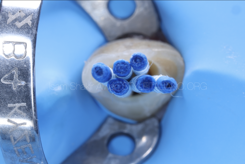
Fig. 7
The root canal was dried with paper points.
Clinical video step by step for the obturation with MTA.

Fig. 8
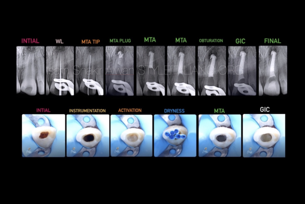
Complete obturation with MTA.
The extruded material (MTA) does not negatively affect the healing of the periapical tissues.
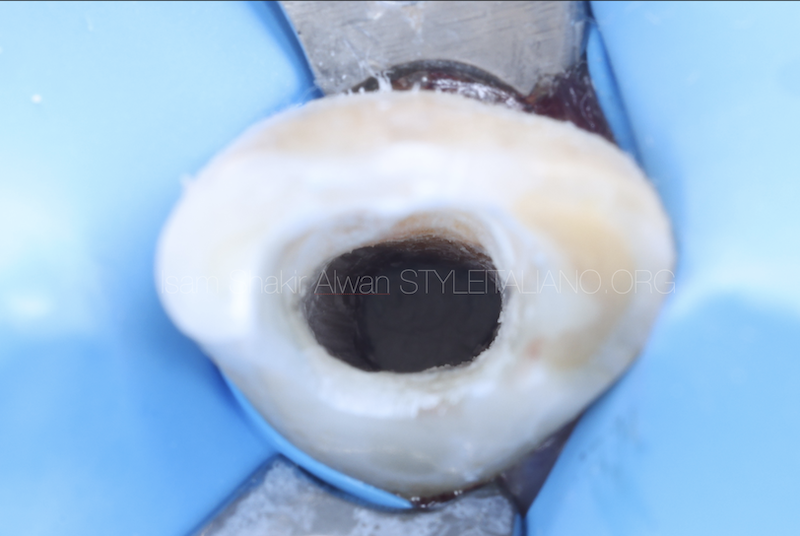
Fig. 9
Clinical image after complete the obturation.
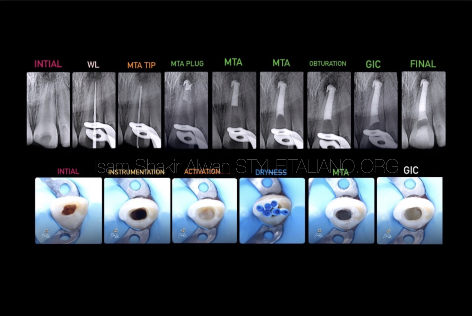
Fig. 10
Non surgical endodontic management of periapical lesion with open apex.

Fig. 11
About the author:
Dr. Isam Shakir Alwan
. Graduated with BDS from University of Alkafeel in 2021.
. My passion in endodontics started during my training at university.
. Have cetificate many courses in basic, advanced endodontics and endodontics microsurgery.
. Key Opinion Leader for MARUCHI.
. My work is committed to microscopic endodontics and aesthetic dentistry.
Conclusions
The management for immature root requires 3D seal to the root canal system.
This might represent a challenge to operators. But sometimes the most complicated cases can be managed in an easy and conservative way using the magnification (microscope) and using biocompatible filling materials like MTA or bioceramics that provides superior seal.
Bibliography
1. Mineral Trioxide Aggregate: a comprehensive literature review--Part I: chemical, physical, and antibacterial properties. Parirokh M, Torabinejad M. J Endod. 2010 Jan;36(1):16–27.
2. MTA: A clinical review Peter Z Tawil, DMD, MS, FRCD, Dip. ABE, Derek J. Duggan, BDentSc, MS, Dip. ABE, and Johnah C. Galicia, DMD, MS, PhD. Compend Contin Educ Dent. 2015 Apr; 36(4): 247–264.
3. Sealing properties of Mineral Trioxide Aggregate orthograde apical plugs and root fillings in an In vitro apexification model. JOE 2007 Mar;33(3):272-5. doi: 10.1016/j.joen.2006.11.002.
4. Comparative Evaluation of Mineral Trioxide Aggregate Obturation Using Four Different Techniques—A Laboratory Study. 2021 Jun; 14(11): 3126. Published online 2021 Jun 7. doi: 10.3390/ma14113126.
5. Long-term observation of the mineral trioxide aggregate extrusion into the periapical lesion: a case series.2013 Apr; 5(1): 54–57. Published online 2013 Apr 5. doi: 10.1038/ijos.2013.16.
