Multiple errors retreatment
14/05/2023
Ibrahim El Naggar
Warning: Undefined variable $post in /home/styleendo/htdocs/styleitaliano-endodontics.org/wp-content/plugins/oxygen/component-framework/components/classes/code-block.class.php(133) : eval()'d code on line 2
Warning: Attempt to read property "ID" on null in /home/styleendo/htdocs/styleitaliano-endodontics.org/wp-content/plugins/oxygen/component-framework/components/classes/code-block.class.php(133) : eval()'d code on line 2
In this article i am presenting a case with multiple errors came symptomatic while biting.
Lack of knowledge in the endodontic field may lead to some errors during the treatment if we are not having the right techniques combined with the right tools.
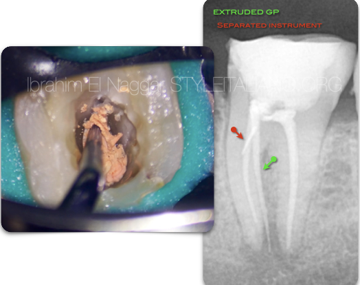
Fig. 1
Pre - Op Radiograph , showing a lower 1st molar with a long extended GP at the perforation and a separated instrument on the entrance of the missed canal covered by composite restoration.
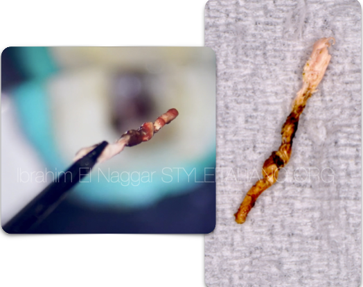
Fig. 2
Removal of the extruded GP , You can see the Granulation Tissue around.
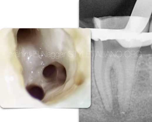
Fig. 3
Disassembling of all the canal filling was done and checked by X-Ray (Detailed Technique Showing in the Video)
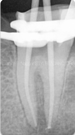
Fig. 4
Master Cone
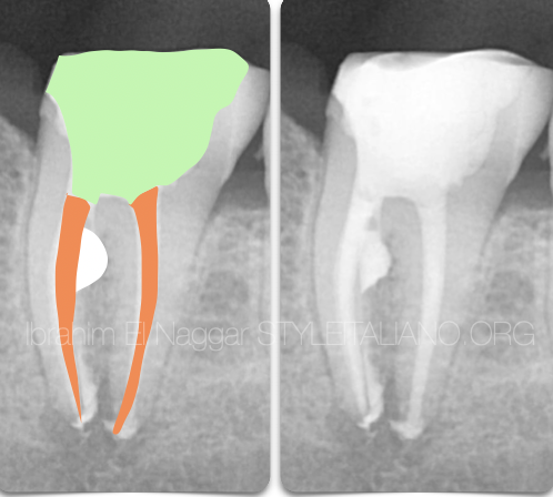
Fig. 5
Post Op
White : MTA
Orange : Gutta Percha
Green : Composite
Clinical Video
Conclusions
The Clinician must have enough knowledge about the root canal anatomy , techniques and to know how to deal with errors if happened.
Bibliography
1. Endodontic Facts, American Association of Endodontists. Available at: http://www. aae.org/about-aae/news-room/endodontic-facts.aspx. Accessed June 1, 2015.
2. Borges AH, Bandeca MC, Tonetto MR,et al. Portland cement use in dental root perforations: a long term followup. Case Rep Dent 2014;2014:637693.
3. TsesisI,FussZV.Diagnosisandtreatmentofaccidentalrootperforations.Endodontic Topics 2006;13:95–107.
4. Tsesis I, Rosenberg E, Faivishevsky V, et al. Prevalence and associated periodontal status of teeth with root perforation: a retrospective study of 2,002 patients’ medical records. J Endod 2010;36:797–800.
5. Gorni FG, Gagliani MM. The outcome of endodontic retreatment: a 2-yr follow-up. J Endod 2004;30:1–4.
6. Clauder T, Shin S-J. Repair of perforations with MTA: clinical applications and mechanisms of action. Endodontic Topics 2006;15:32–55.
7. Kvinnsland I, Oswald RJ, Halse A, Gronningsaeter AG. A clinical and roentgenological study of 55 cases of root perforation. Int Endod J 1989;22:75–84.