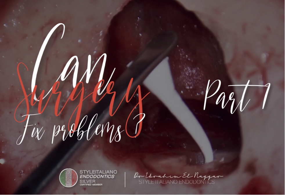
MISSED CANAL , SURGICAL APPROACH
24/10/2024
Ibrahim El Naggar
Warning: Undefined variable $post in /home/styleendo/htdocs/styleitaliano-endodontics.org/wp-content/plugins/oxygen/component-framework/components/classes/code-block.class.php(133) : eval()'d code on line 2
Warning: Attempt to read property "ID" on null in /home/styleendo/htdocs/styleitaliano-endodontics.org/wp-content/plugins/oxygen/component-framework/components/classes/code-block.class.php(133) : eval()'d code on line 2
Lack of knowledge in the endodontic field may lead to some errors during the treatment if we are not having the right techniques combined with the right tools as well the knowledge about the internal anatomy.
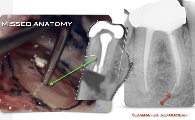
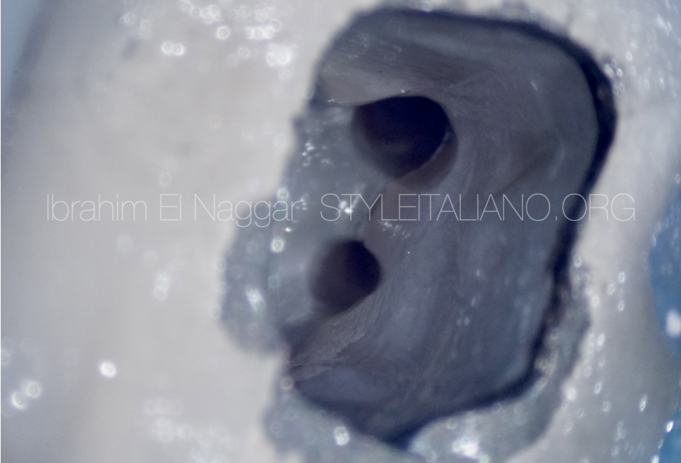
Fig. 1
Pre - Op Radiograph & CBCT , showing a lower 1st molar with a separated instrument & a separated instrument and having a prosthetic crown which makes the non surgical retreatment step difficult.
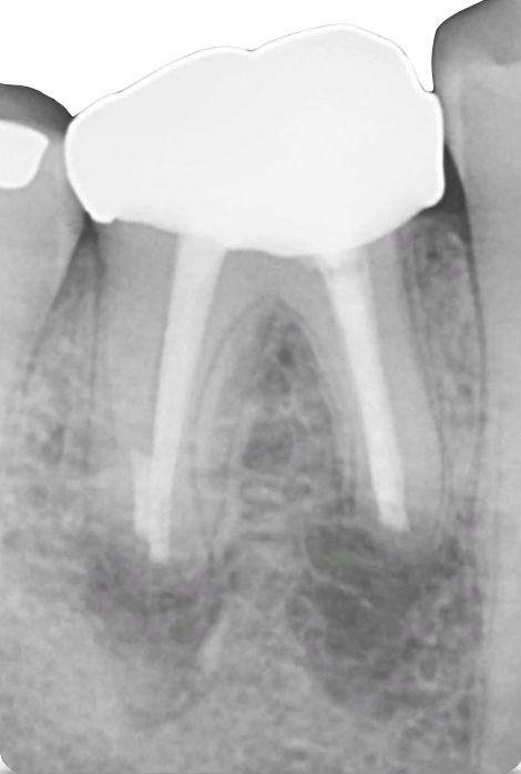
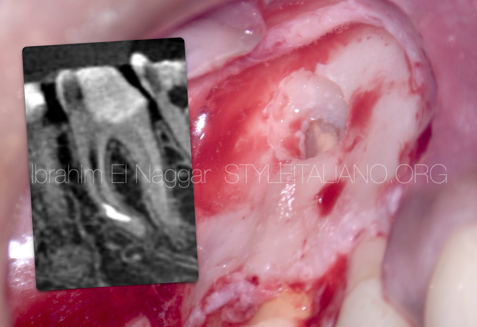
Fig. 2
POST OP , Resection of mesial root with separated instrument & Distal Root with Missed Anatomy.
Clinical Video
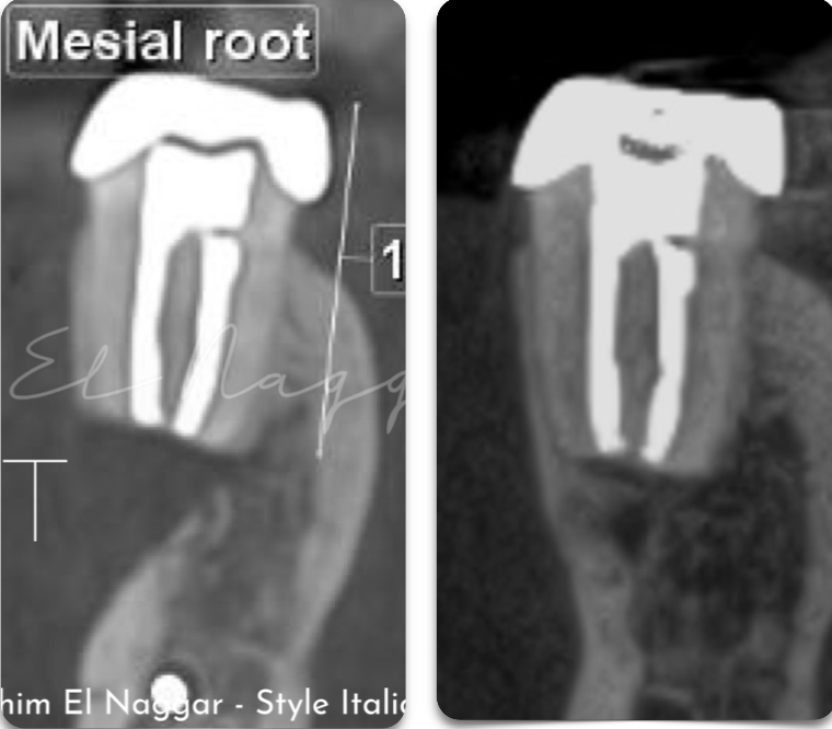
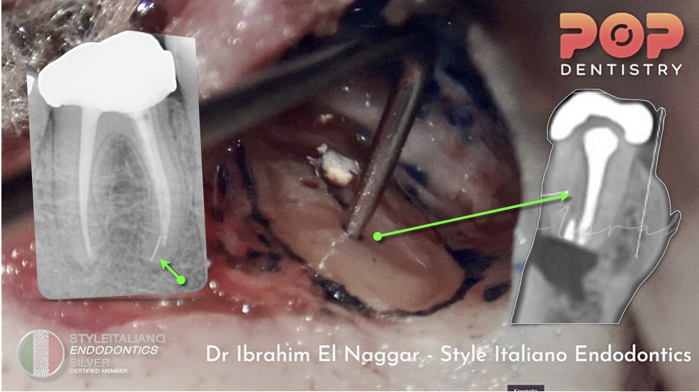
Fig. 3
Post Op and 3 YEARS Follow Up CBCT
Bone regeneration at surgical site
Conclusions
The Clinician must have enough knowledge about the root canal anatomy , techniques and to know how to deal with errors if happened as well to know the limitation of surgical and non surgical treatment.
Bibliography
1. Endodontic Facts, American Association of Endodontists. Available at: http://www. aae.org/about-aae/news-room/endodontic-facts.aspx. Accessed June 1, 2015.
2. Borges AH, Bandeca MC, Tonetto MR, et al.Portland cement use in dental root perforations: a long term followup. Case Rep Dent 2014;2014:637693.
3. TsesisI,FussZV.Diagnosisandtreatmentofaccidentalrootperforations.Endodontic Topics 2006;13:95–107.
4. Tsesis I, Rosenberg E, Faivishevsky V, et al. Prevalence and associated periodontal status of teeth with root perforation: a retrospective study of 2,002 patients’ medical records. J Endod 2010;36:797–800.
5. Gorni FG, Gagliani MM. The outcome of endodontic retreatment: a 2-yr follow-up. J Endod 2004;30:1–4.
6. Clauder T, Shin S-J. Repair of perforations with MTA: clinical applications and mechanisms of action. Endodontic Topics 2006;15:32–55.
7. Kvinnsland I, Oswald RJ, Halse A, Gronningsaeter AG. A clinical and roentgenological study of 55 cases of root perforation. Int Endod J 1989;22:75–84.