Middle mesial canal (MMC) in lower molars
20/12/2023
Fellow
Warning: Undefined variable $post in /home/styleendo/htdocs/styleitaliano-endodontics.org/wp-content/plugins/oxygen/component-framework/components/classes/code-block.class.php(133) : eval()'d code on line 2
Warning: Attempt to read property "ID" on null in /home/styleendo/htdocs/styleitaliano-endodontics.org/wp-content/plugins/oxygen/component-framework/components/classes/code-block.class.php(133) : eval()'d code on line 2
The main canals in the mesial root of lower molars are the mesiobuccal and mesiolingual canals.
A middle mesial canal (MMC) sometimes is present in the developmental groove between the other mesial canals.
The presence of middle mesial canal in the range of 4-28 % is recorded and divided into three types: INDEPENDENT, CONFLUENT, AND FIN.
Thus, magnification is a must , either loupes or microscope to detect any hidden anatomy.
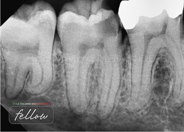
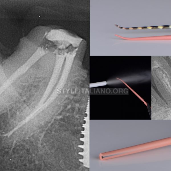
Fig. 1
PRE-OPERATIVE X-RAY
Shows tooth 47 with deep caries mesially involving the pulp chamber
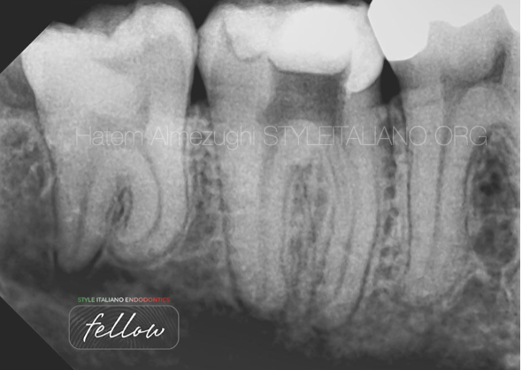
Fig. 2
Build up mesially was achieved which turned from class II to class I , to obtain proper isolation so it acts like reservoir for NaOCl.
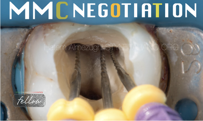
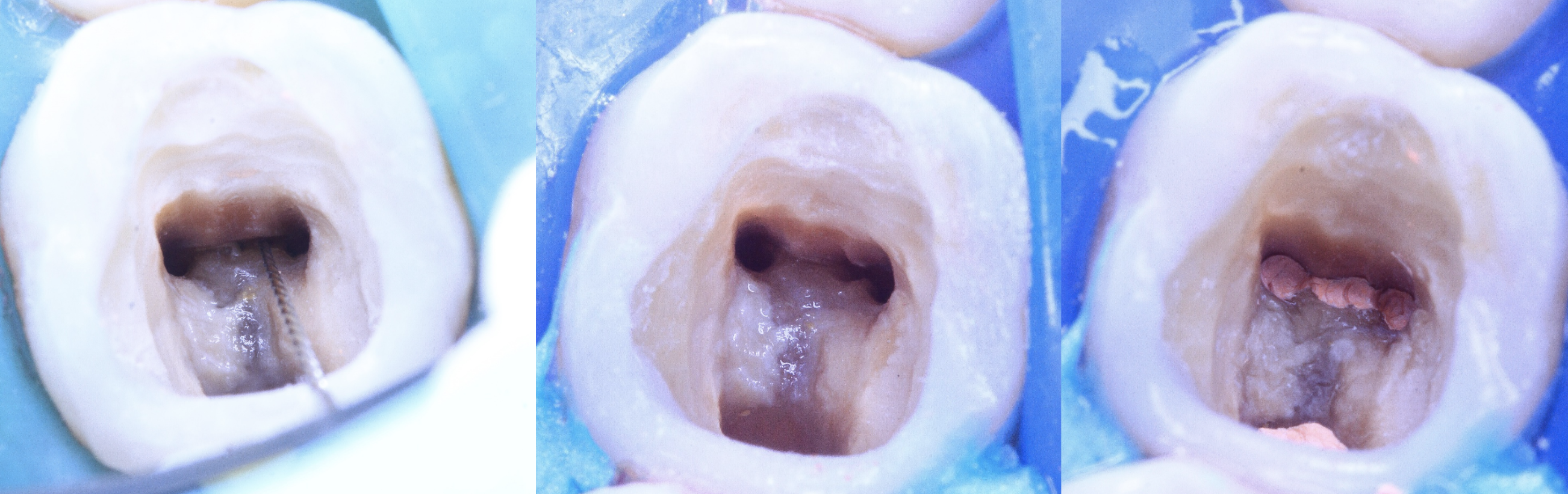
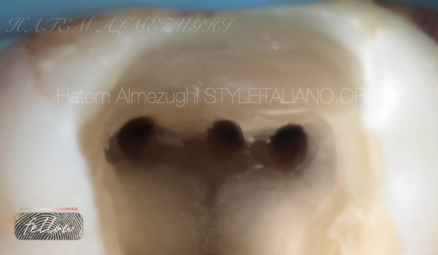
Fig. 3
Negotiating MB, ML, and MMC to obtain glide path, patency then have the canals ready for shaping and cleaning
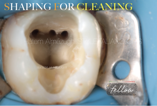
Fig. 4
Now all canals shaped, ready for cleaning and chemical activation
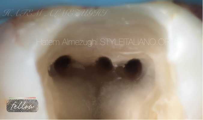
Fig. 5
Zoomed in mesial side after cleaning ready for obturation
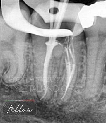
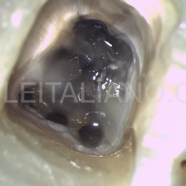
Fig. 6
first stage of down packing in mesials done.
Note: in distal seemingly if I go for down packing , there might be over extended fill, thus there's two options either cutting a little from the end of GP or changing the size to a larger one, to obtain apical control
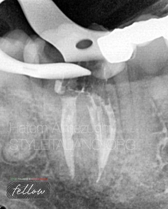
Fig. 7
Obturation done, just a bit more of hydraulic pressure on distal, to fill any unnecessary voids
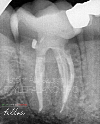
Fig. 8
Final obturation done, all voids filled , followed by placing permanent restoration

Fig. 9
About the author:
Dr Hatem Almezughi
Was graduated from Tripoli university, Libya.
Local and international speaker in different conferences and forums
instructor in many endodontic courses.
Dental practice limited to endodontics and restorative .
Fellow member in style Italiano endodontics.
Conclusions
Troughing into the groove between the mesiobuccal and mesiolingual orifices may be indicated to identify MMC canal ( use MAGNIFICATION).
Cleaning isthmus is crucial to reveal such a canal if its available, not to mention missing this canal and not detecting it may lead to future endodontic failure, especially if its independent one
instrumentation is one of the key factors in the success of endodontic therapy.
Bibliography
M. A. Versiani, R. Ordinola-Zapata, A. Keleş et al., “Middle mesial canals in mandibular first molars: a micro-CT study in different populations,” Archives of Oral Biology, vol. 61, pp. 130–137, 2016.
O. A. Sherwani, A. Kumar, R. K. Tewari, S. K. Mishra, S. M. Andrabi, and S. Alam, “Frequency of middle mesial canals in mandibular first molars in North Indian population-an in vivo study,” Saudi Endodontic Journal, vol. 6, no. 2, pp. 66–70, 2016.
A. Nosrat, R. J. Deschenes, P. A. Tordik, M. L. Hicks, and A. F. Fouad, “Middle mesial canals in mandibular molars: incidence and related factors,” Journal of Endodontics, vol. 41, no. 1, pp. 28–32, 2015.
K. M. De toubes, M. I. Côrtes, M. A. Valadares et al., “Comparative analysis of accessory mesial canal identification in mandibular first molars by using four different diagnostic methods,” Journal of Endodontics, vol. 38, no. 4, pp. 436–441, 2012.