Managing Calcified Canals: Techniques and Strategies for Successful Endodontic Treatment
06/04/2024
Fellow
Warning: Undefined variable $post in /home/styleendo/htdocs/styleitaliano-endodontics.org/wp-content/plugins/oxygen/component-framework/components/classes/code-block.class.php(133) : eval()'d code on line 2
Warning: Attempt to read property "ID" on null in /home/styleendo/htdocs/styleitaliano-endodontics.org/wp-content/plugins/oxygen/component-framework/components/classes/code-block.class.php(133) : eval()'d code on line 2
A 32-year-old man with a symptomatic maxillary right central incisor came to the private clinic. The patient reported that the incisor had been traumatized previously. The medical history was nonsignificant, and extraoral evaluation revealed normal soft-tissue structures with no apparent pathosis. Upon oral examination, no mobility or probing defect was observed. The tooth was sensitive to percussion and palpation. Furthermore, it responded negatively to vitality tests, and pulp status was diagnosed as necrosis. Radiographic evaluation revealed periapical lesion as well as calcification in the canal pathway.
Calcified canals present a unique challenge in endodontic therapy, often complicating the process of root canal treatment. These calcifications can impede access to the root canal system, making it difficult to clean, shape, and fill the canal effectively. However, with the right techniques and strategies, managing calcified canals can lead to successful outcomes in endodontic procedures. In this article, we will explore the management of calcified canals, including the challenges they present and the approaches that can be employed to overcome them.
Understanding Calcified Canals
Calcifications within the root canal system can manifest as various degrees of mineralized tissue deposition along the walls of the canal. These calcifications can be classified into different categories based on their location, extent, and density. Understanding the type and location of calcifications is crucial in devising an effective treatment plan for managing calcified canals.
Challenges in Managing Calcified Canals
Calcified canals pose several challenges during endodontic procedures, including:
Limited Access: Calcifications can obstruct access to the root canal system, making it challenging to locate and negotiate the canals.
Instrument Fracture: The increased risk of instrument fracture is a concern when attempting to navigate through calcified canals.
Incomplete Cleaning: Calcifications can prevent proper disinfection and cleaning of the canal system, leading to persistent infection and treatment failure.
Compromised Seal: Inadequate shaping and filling due to calcifications can compromise the seal of the root canal, increasing the risk of reinfection.
Strategies for Managing Calcified Canals
Preoperative Assessment
Advanced Imaging: Utilize advanced imaging techniques such as cone-beam computed tomography (CBCT) to assess the extent and location of calcifications.
Magnification: Use magnification tools like dental loupes or microscopes to enhance visualization and improve access to calcified canals.
Access Opening
Strategic Access: Create a conservative and strategic access cavity to maximize visibility and facilitate navigation through calcified canals.
Ultrasonic Tips: Use ultrasonic tips to remove calcified dentin while preserving the integrity of the tooth structure.
Canal Negotiation
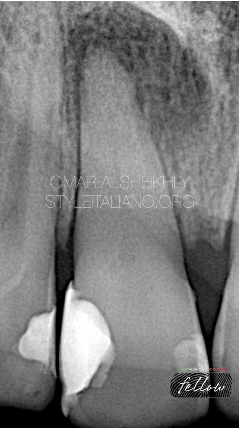
Fig. 1
X-ray examination was done and on radiographic examination calicification occurs at coronal and apical of canal of tooth No.11 .
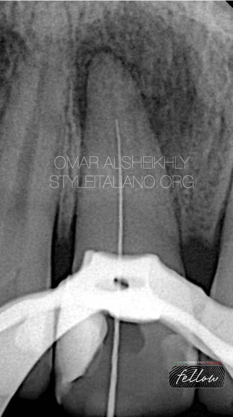
Fig. 2
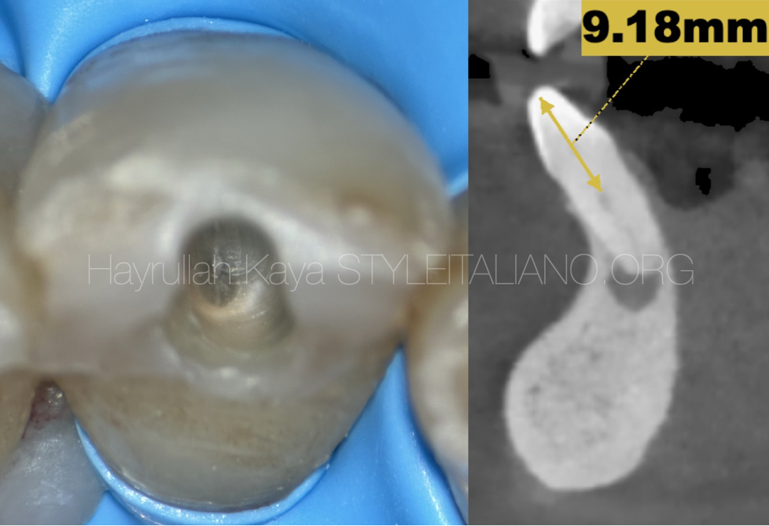
Access preparation was done with high speed round bur and modified using EX24 bur. No canal orifice was visible upon access opening initially. On exploration the orifice was found more towards the palatal aspect. File. No. 10-Dfinder file was inserted to confirm the opening of canal. File was in canal but not up to working length. File was 6-7mm short of working length due to calcification of canal.
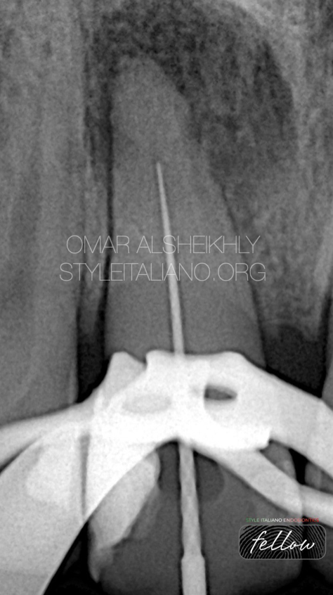
Fig. 3
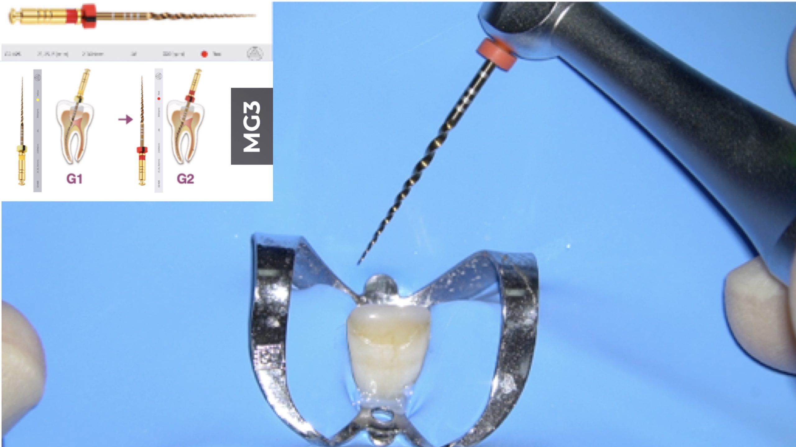
After file no 10-Dfinder was used, the working length was taken. The rotary file 15.04 was inserted
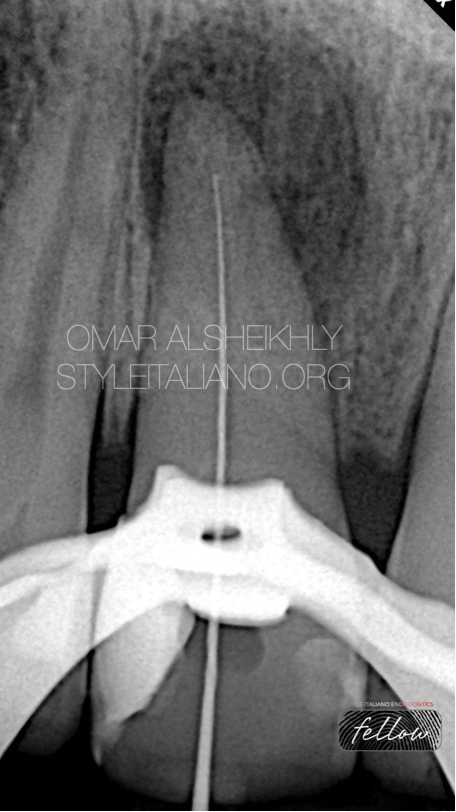
Fig. 4
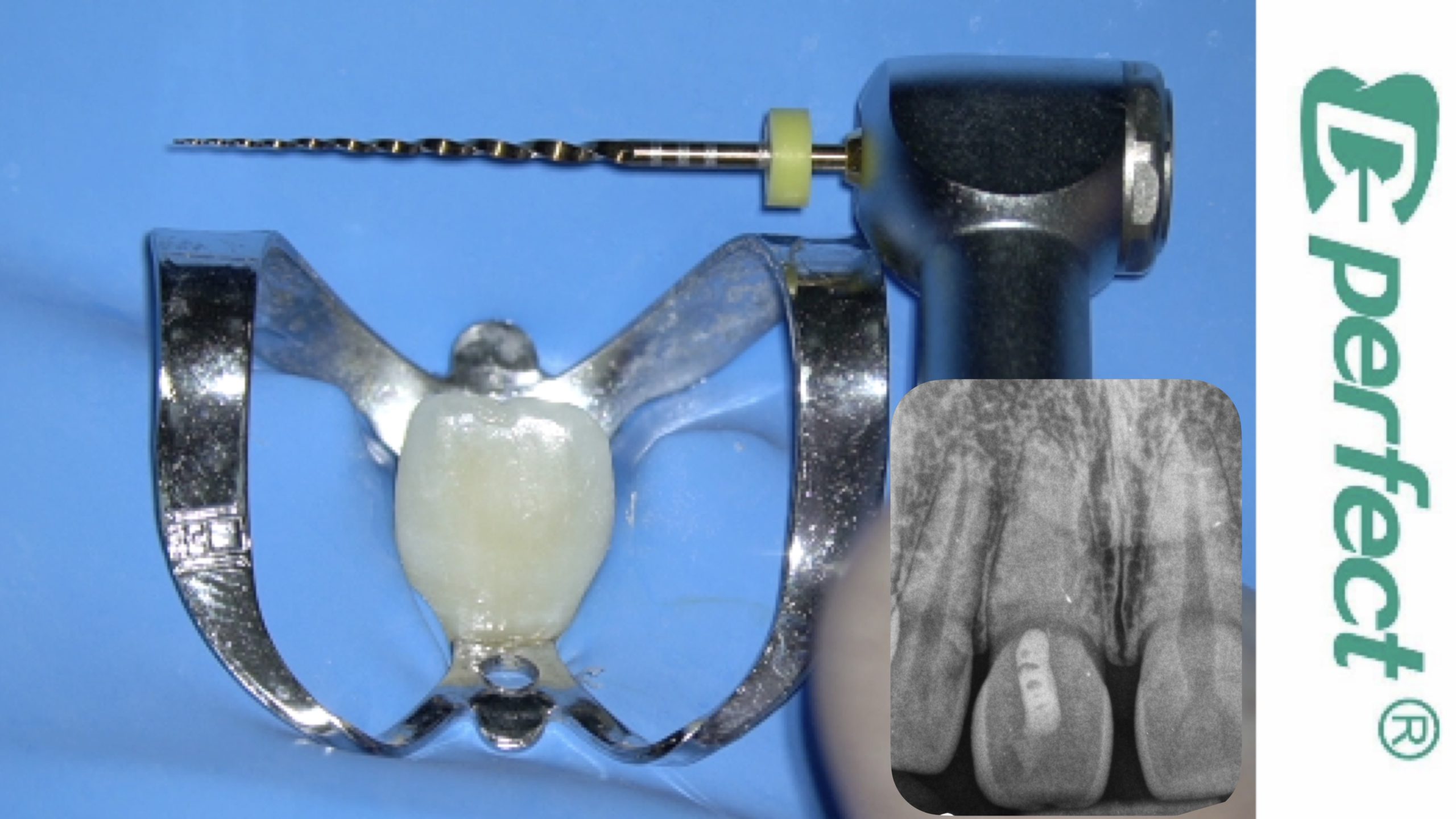
Again the canal was negotiated with D-finder but not up to working length. File was 2-3mm short from the working length
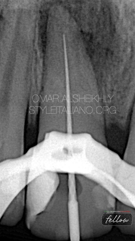
Fig. 5
After new working length taken, the rotary file 15.04 was inserted again
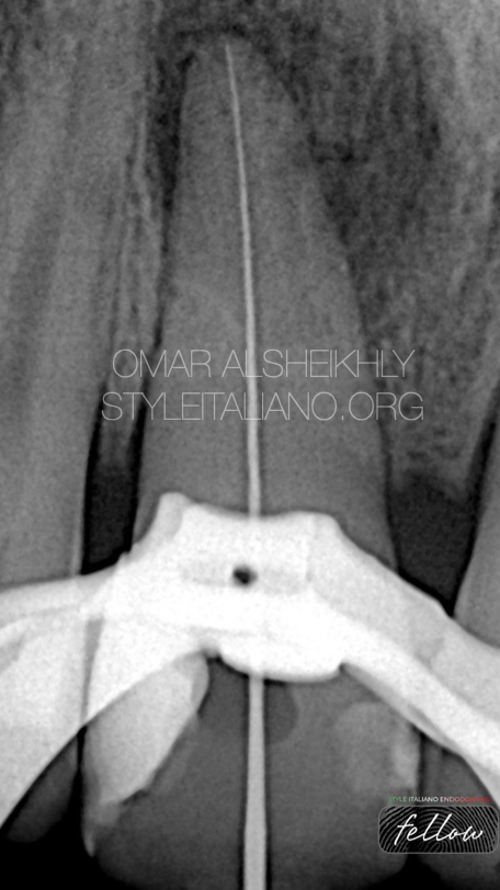
Fig. 6
Full working length , D-finder No10
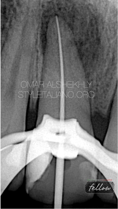
Fig. 7
Full instrumentation with 20.04 rotary file with the final working length
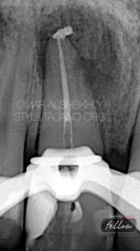
Fig. 8
Down packing
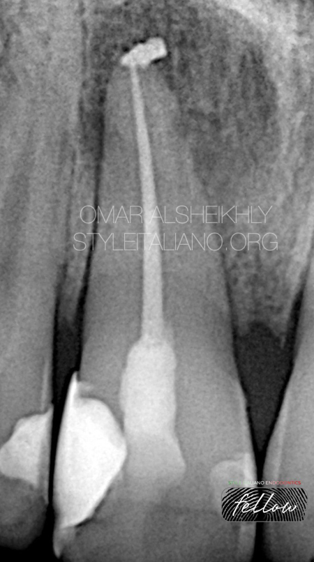
Fig. 9
Obturation with final restoration

Fig. 10
About the Author:
OMAR ALSHEIKHLY
Bachelor of dental surgery (BDS) Ivan Horbachevsky Ternopil State Medical University UKRAIN 2011-2016
Awarded the certificate of equivalency for the diploma Faculty of Dentistry \University of Baghdad IRAQ 2019
MSc master in advanced Endodontics University of Siena \ ITALY 2021\2023
Fellow of Style Italiano Endodontics
Conclusions
Managing calcified canals requires a combination of skill, technique and strategy to overcome the challenges presented by these complex anatomical variations. By employing advanced imaging, strategic access opening, effective negotiation techniques, and tailored cleaning and obturation methods, endodontists can enhance the success rate of root canal treatments in cases involving calcified canals. Through a comprehensive and meticulous approach to managing calcified canals, practitioners can achieve optimal outcomes, ensuring the long-term health and preservation of the natural dentition for their patients.
Bibliography
1. Calcified Canals – A Review 1Bernice Thomas, 2ManojChandak, 3Adityavardhan Patidar, 4Bharat Deosarkar, 5Harshit Kothari
2. NON-SURGICAL ROOT CANAL TREATMENT OF CALCIFIED CANAL Ziad Salim Abdul Majid1, Ranya Faraj Elemam2
3. Management of Calcified Root Canal: A Case Report Samrudhi Khatod1, Anuja Ikhar2, Pradnya Nikhade3, Arpan Jaiswal4
4. Thomas B et al. Calcified Canals – A Review, IOSR Journal of Dental and Medical Sciences (IOSRJDMS),13(50),2014,38-43.
5 Michael H, Edgar S, editors. Problems in Endodontics: Etiology, Diagnosis and Treatment. 1st ed. Germany: Quintessence publishing Co Ltd; 2009:145-72.


