Management of a deep bifurcation in a lower first premolar by perfect file
15/02/2021
Warning: Undefined variable $post in /home/styleendo/htdocs/styleitaliano-endodontics.org/wp-content/plugins/oxygen/component-framework/components/classes/code-block.class.php(133) : eval()'d code on line 2
Warning: Attempt to read property "ID" on null in /home/styleendo/htdocs/styleitaliano-endodontics.org/wp-content/plugins/oxygen/component-framework/components/classes/code-block.class.php(133) : eval()'d code on line 2
Mandibular premolars have been considered to be the most challenging teeth to be treated endodontically, especially when they present with multiple roots or canals. Their propensities for anomalous variations, narrow mesiodistal dimensions and the ensuing narrow access to canals, lack of visibility, and bifurcations and deltas are factors that further compound the difficulty for clinicians.
Preoperative radiograph identification, root canal access opening, intraoperative identification, location, instrumentation, debridement, disinfection, obturation are improtant for successful endodontic therapy

Fig. 1
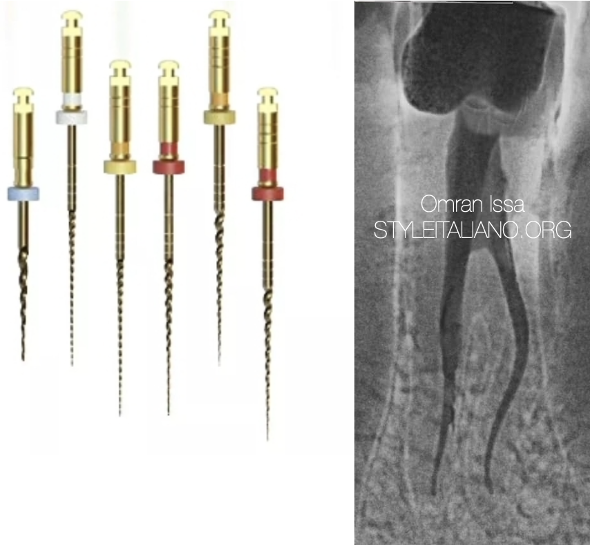
The pre-operative X-ray shows a large decay; we can also notice a deep split (two roots and two canals).

Fig. 2
Measuring the working lenght by using elctronic apex locator EAL , some difficulty was faced while obtaining glide path in a split canal due to curvature as seen on the radiograph

Fig. 3
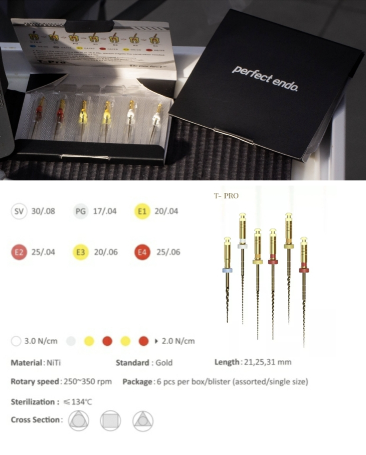
The tooth was shaped with Perfect Files T-Pro that were picked thanks to their improved cutting efficiency and fracture resistance. The processing with heat treatment allows these files to be pre-bent.

Fig. 4
PG file has a very high cyclic fatigue resistance, which makes it perfect to deal with curve canals. The tooth treated in this clinical case has a double stress area in the lingual canal, nevertheless it was successfully managed with the use of PG file.
The shaping was carried put using also E1 and E2 files.

Fig. 5
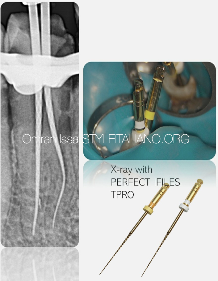
The X-ray shows the cone fit: the shaping obtained by using Perfect files respected the original anatomy of the canals

Fig. 6
The picture, taken with the operative microscope at 25X magnifications, shows the bifurcation of the tooth.
Conclusions
The treatment of such complex anatomy cases needs a lot of patience, as prolonged and multiple appointments are very much a certainty. The time taken in treating such cases of variable tooth anatomy depends on the clinical skill, and the armamentarium available to obtain remarkable results.
It goes without saying that the risk of missing anatomy during root canal treatments is high due to the complexity of the root canal system in mandibular premolars, therefore we may have to have CBCT to estimate the situation .
Bibliography
1: Vertucci FJ. Root canal Vertucci FJ. Root canal morphology and its relationship to endodontic procedures. Endod Topics. 2005;10:23–9.
2: England MC Jr, Hartwell GR, Lance JR. Detection and treatment of multiple canals in mandibular premolars. J Endod. 1991 Apr;17(4):174-8. doi: 10.1016/S0099-2399(06)82012-1. PMID: 1940736.
3: Slowey RR. Root canal anatomy. Road map to successful endodontics. Dent Clin North Am 1979;23:555–73.
