
Fully Calcified Canals : Is it a Challenge ??
19/05/2022
The Community
Warning: Undefined variable $post in /home/styleendo/htdocs/styleitaliano-endodontics.org/wp-content/plugins/oxygen/component-framework/components/classes/code-block.class.php(133) : eval()'d code on line 2
Warning: Attempt to read property "ID" on null in /home/styleendo/htdocs/styleitaliano-endodontics.org/wp-content/plugins/oxygen/component-framework/components/classes/code-block.class.php(133) : eval()'d code on line 2
A 30 years old female patient been referred to retreat her lower first molar patient was suffering from food packing in the area between Molar and second premolar Clinical examination showed that there was very bad amlgam Restoration with missing walls Radiography examination show that the tooth was fully calcified . So we start to treat this tooth
Since people are living longer and keeping their teeth longer, we need to be able to deal with the endodontic implications, and this includes treating an increasing number of calcified root canals. It would be a shame to recommend extraction of a perfectly functional and restoratively sound tooth because of concern over, or the inability to manage, calcified roots. Another cause of calcification because of using pulpotec like the case here
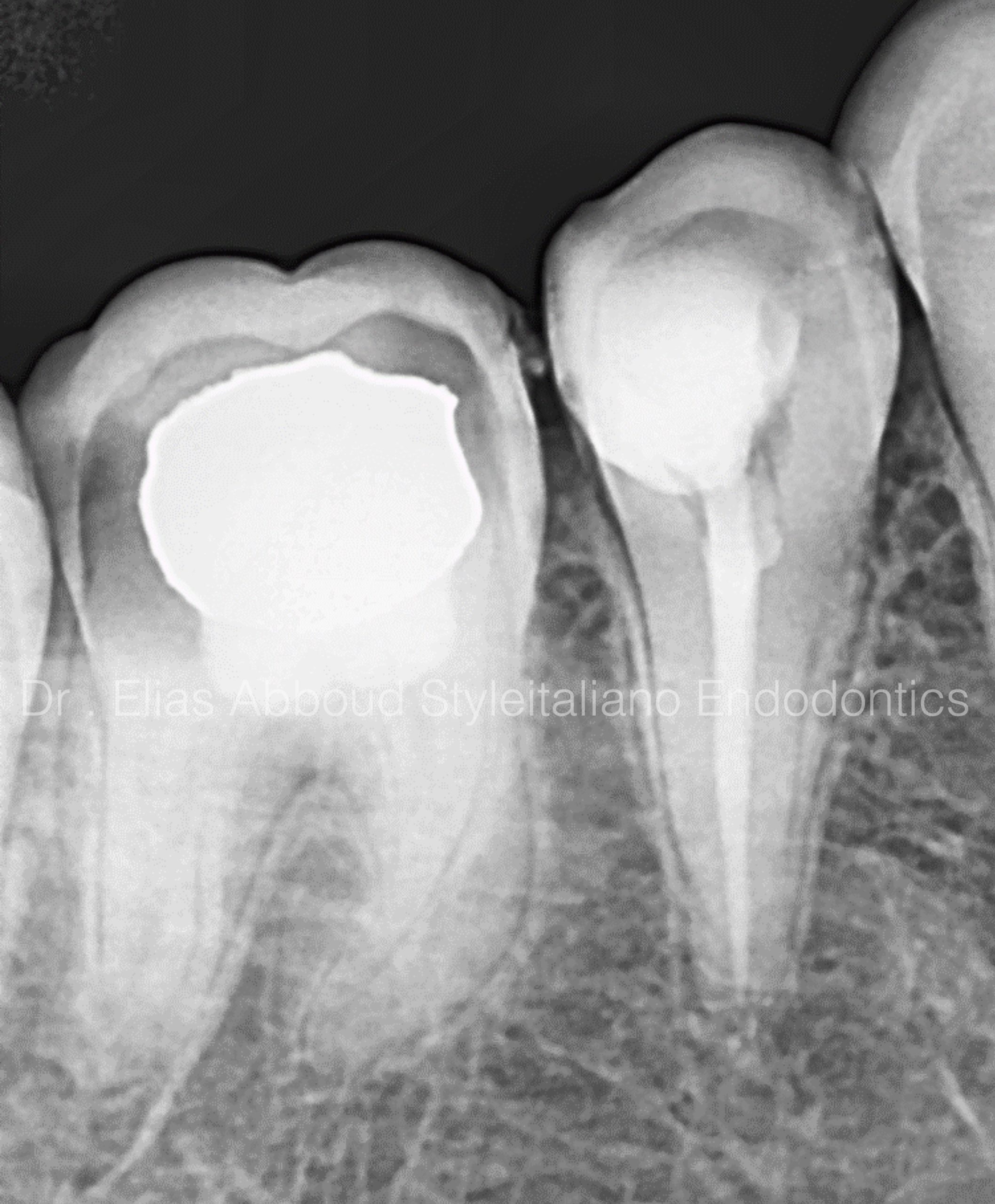
Fig. 1
Once calcified canals are identified radiographically the clinician should keep in mind the following fundamental protocols pertaining to instrumentation: locating the canals with proper magnification; instrumenting with stiff, pre-curved stainless steel hand files, followed by narrow and flexible mechanized NiTi files; and, all the while, ensuring the canals are kept lubricated during the filing process. In turn, instrumenting calcified canals comes down to having the proper armamentaria and techniques
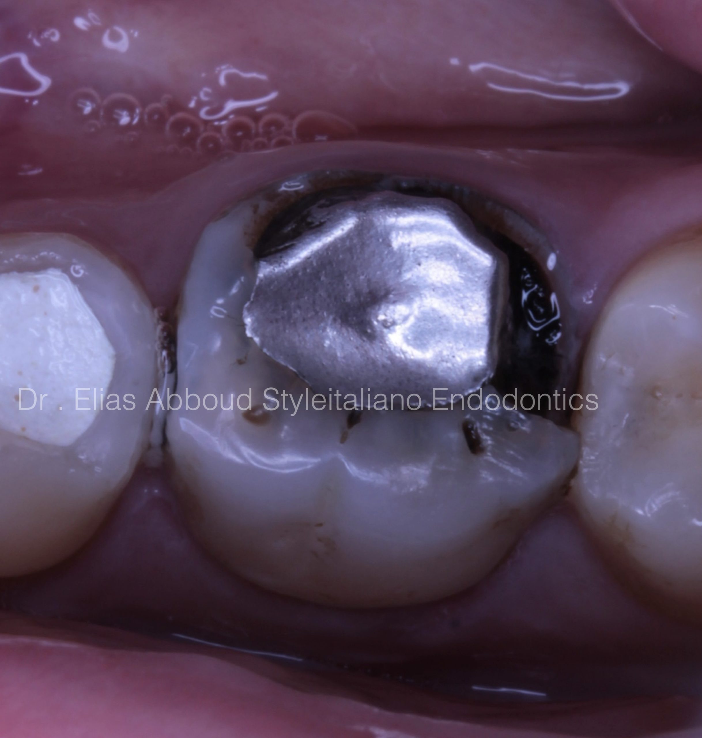
Fig. 2
Pre operative situation We notice a very bad amalgam restoration. That cause of fracture the walls
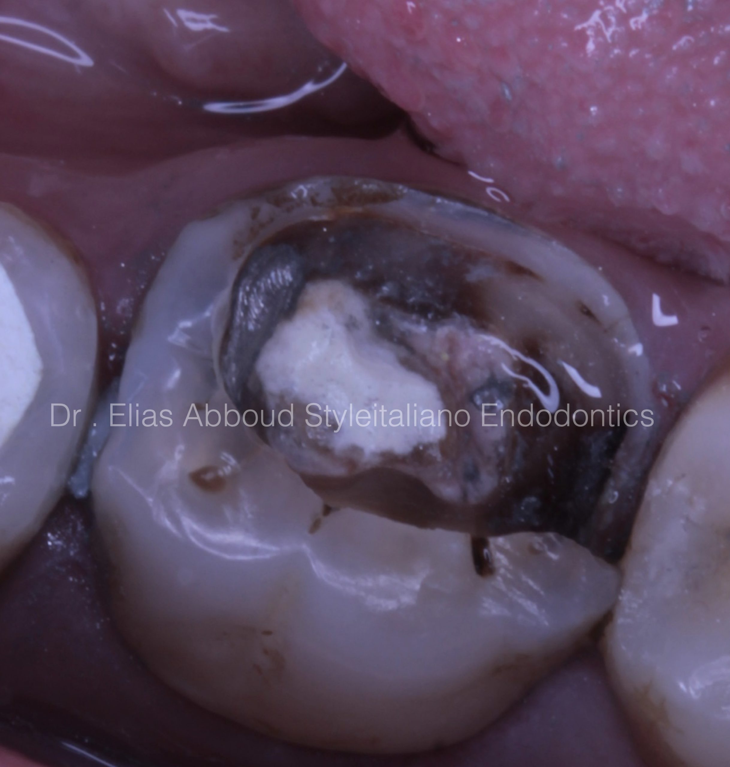
Fig. 3
The presence of pulpotec in The cavity was seen clearly. First of all we have to put the rubber dam then complete removing all the materials and inflamed tissue
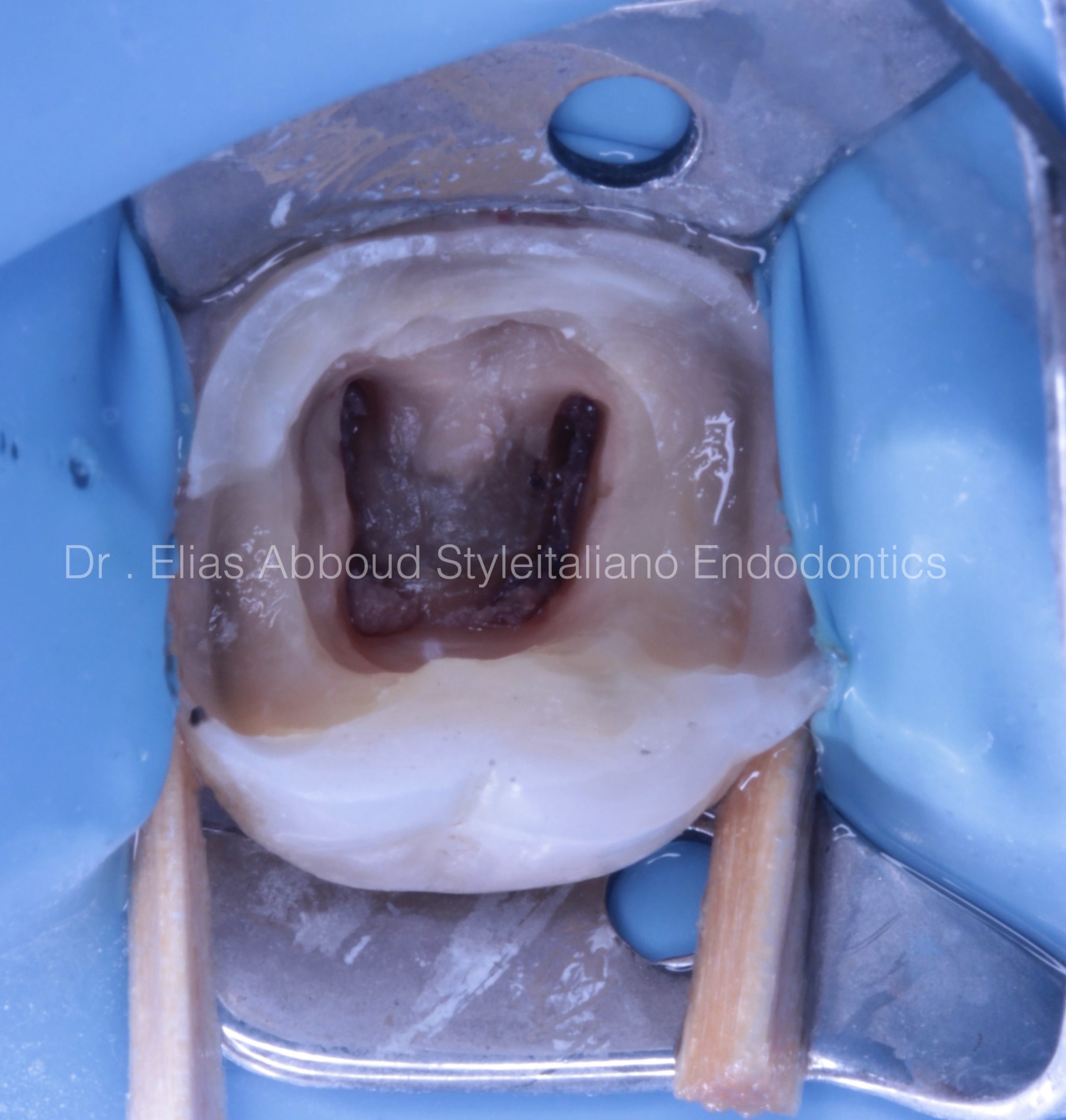
Fig. 4
After isolation we can notice necrotic pulp Tissue in the canals Most studies show that the best way to treat necrotic cases to use calcium hydroxide in the first appointment to reduce flare up. So we prepared coronal third then we use intracanal medication (calcium hydroxide )
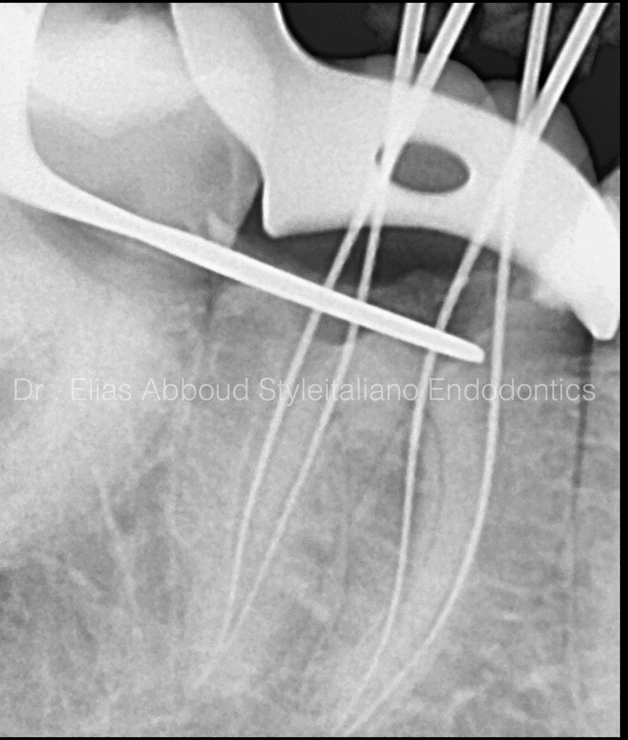
Fig. 5
The next visit
The canals were scouting with stanless still
Manual files
Start with #6 #8 #10 #15
That manual glide path facilitate
the preparation With rotary files and reduce
the possibility of files fracture
Then E Flex files were used
#19 2%
#20 4%
#25 4%
#30 4%
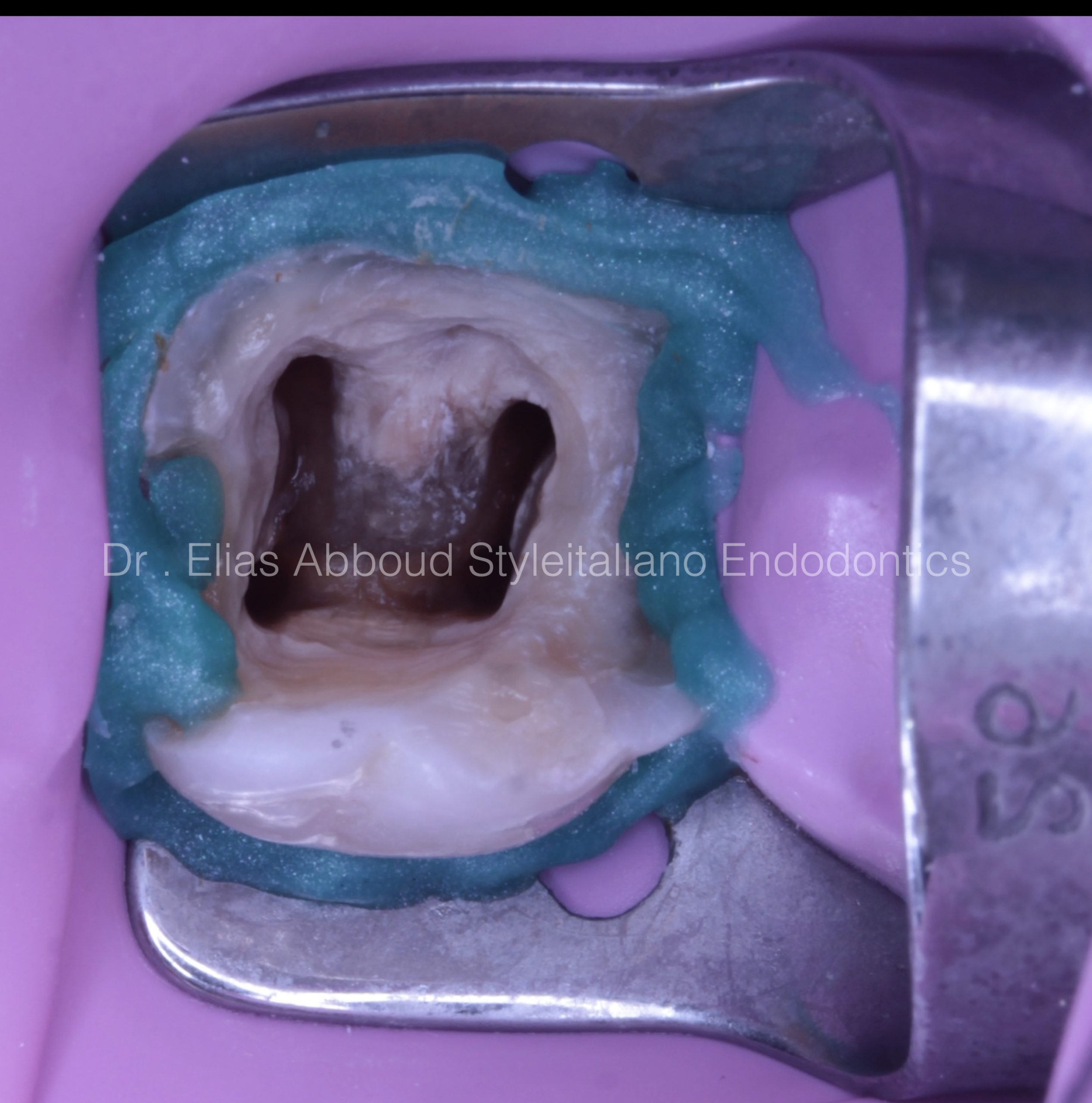
Fig. 6
After cleaning and shaping
And irrigation with
Sodium hypochloride 5.25 %
Edta 17 %
Chlorohexiden 2%
And saline between each irrigant
The canals were ready for obturation
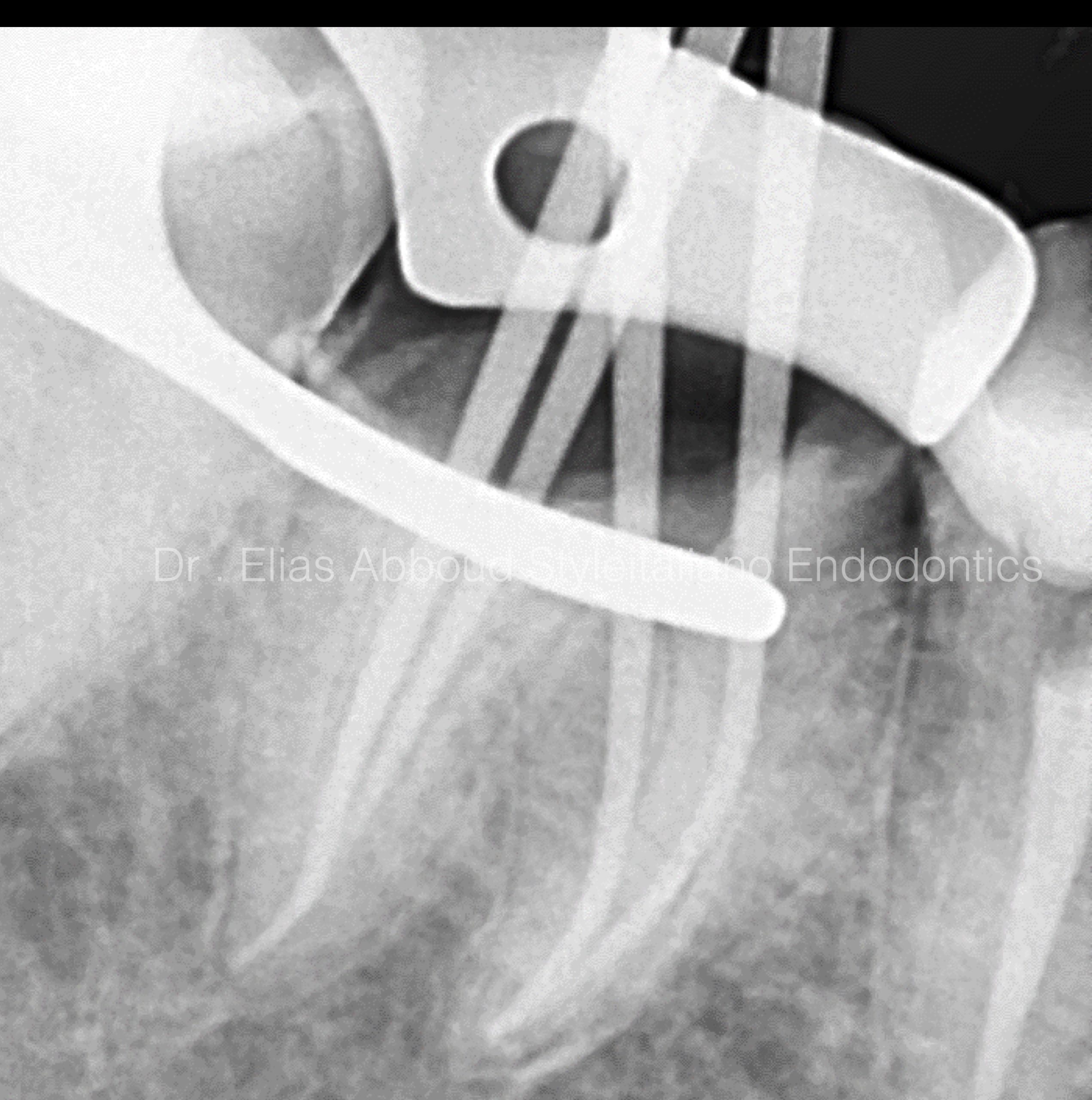
Fig. 7
Master cone radiography Is the most important image in hole treatment because here we can make an adjustment before obturation
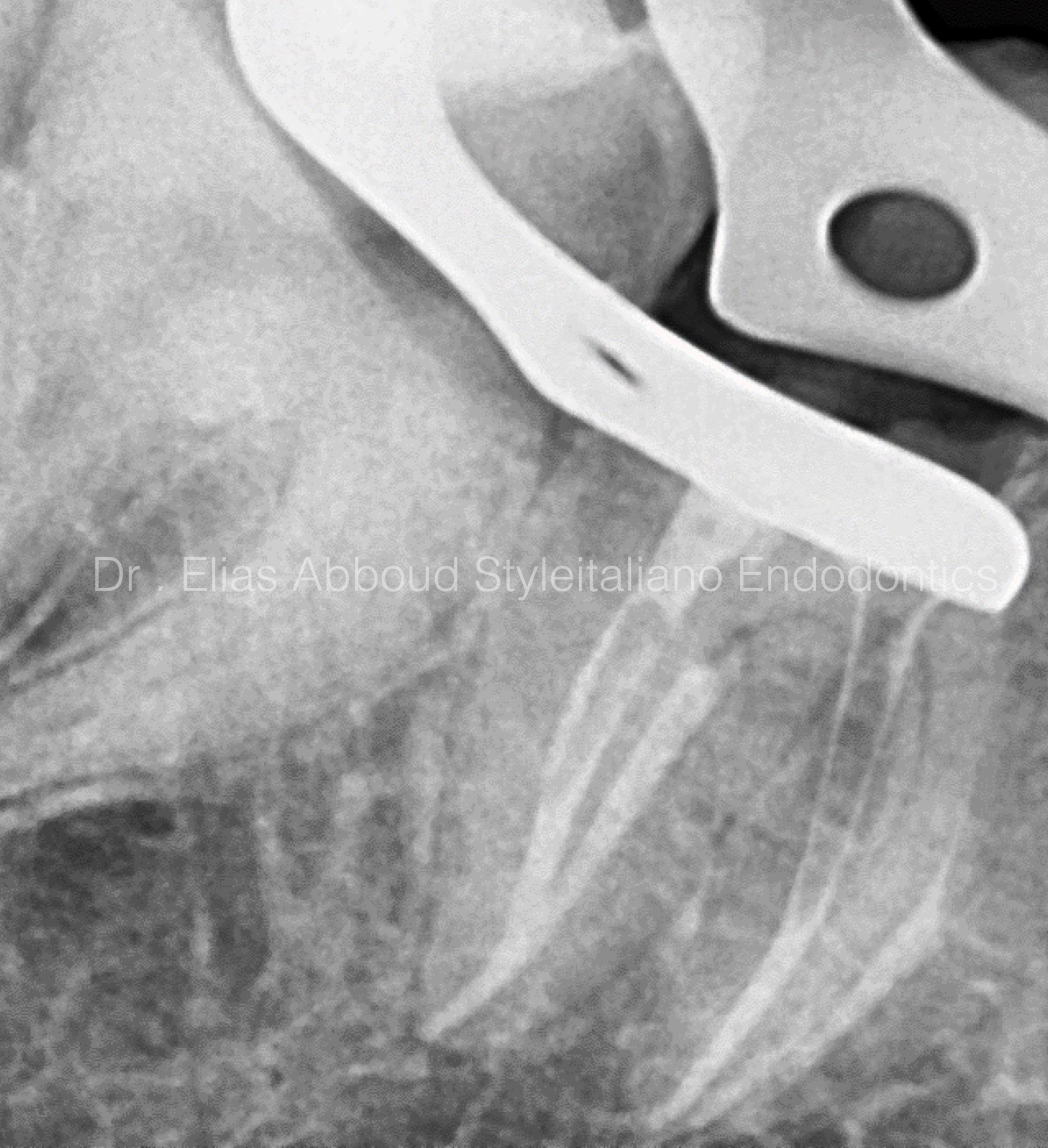
Fig. 8
Warm vertical compaction technique Was used Down pack with fast pack
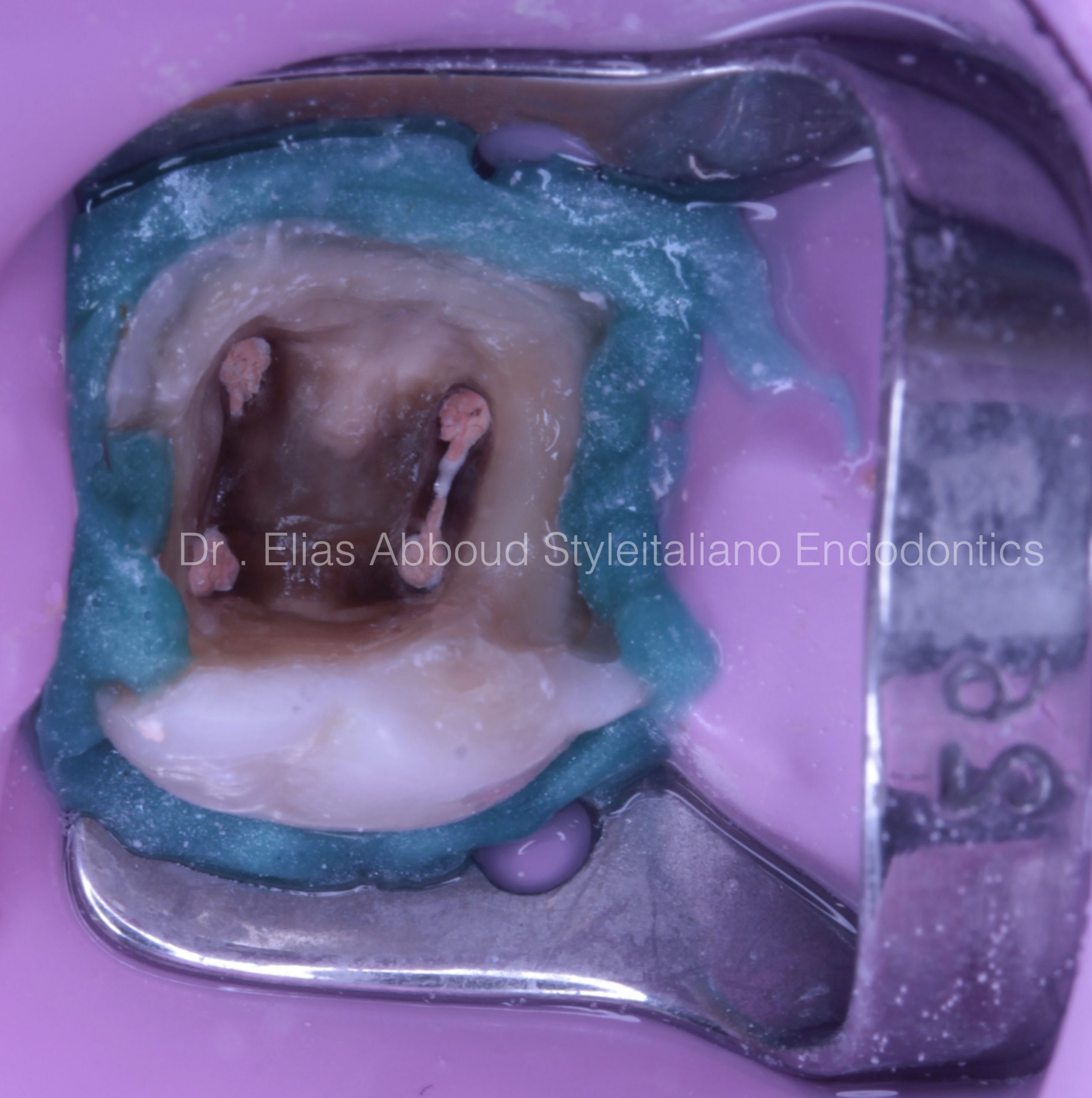
Fig. 9
Post op clinical view We have to clean the access cavity very well after obturation when we use resin sealer the using of alcohol and sandblasting.
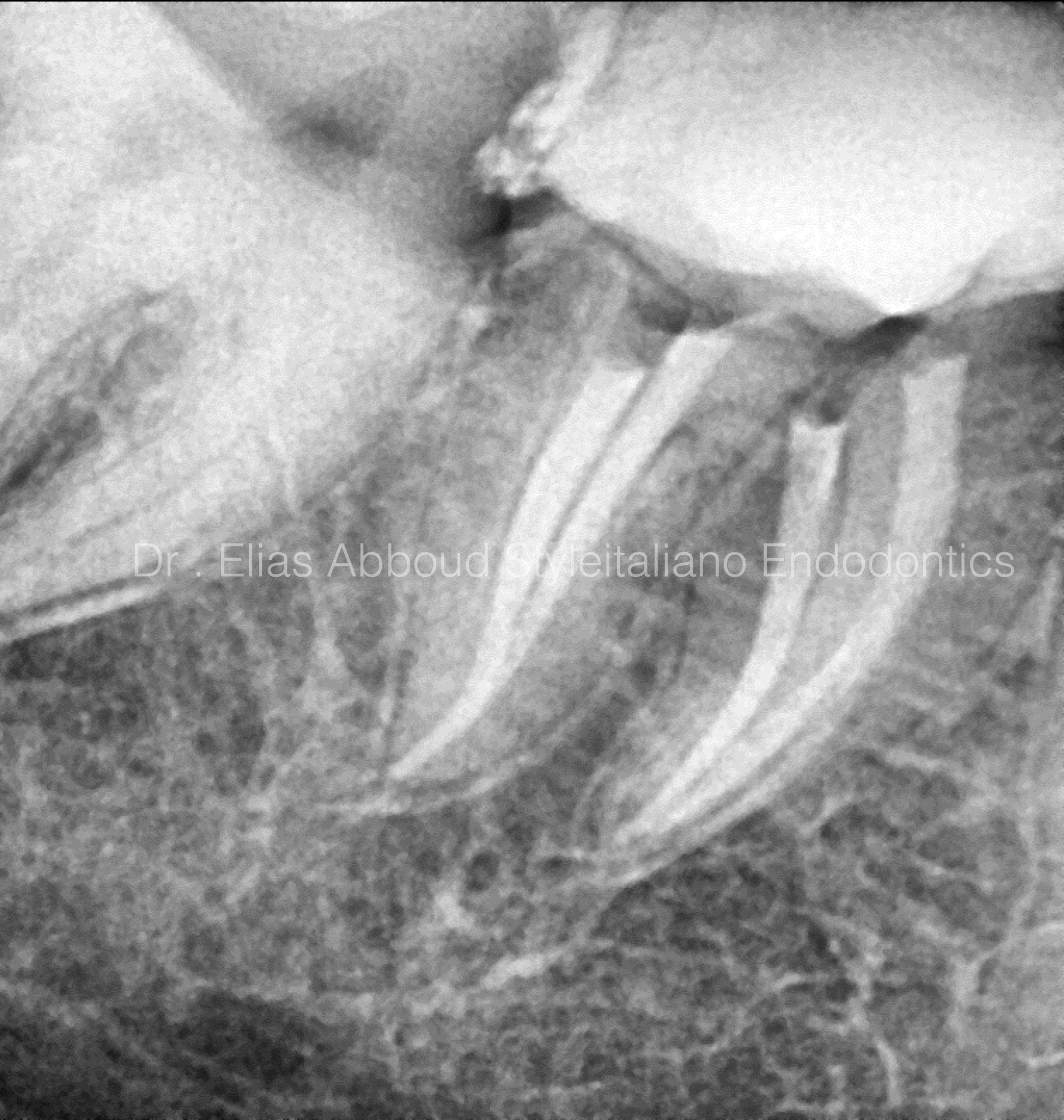
Fig. 10
Post op radiograph

Fig. 11
About the Author:
Dr . Elias Abboud Damascus Syria
2018 : B.D.S ( Damascus university )
2019 : ENDODONTICS AND OPERATIVE DENTISTRY
AT THE NATIONAL DENTAL CENTER
Conclusions
During the retreatment procedure, regaining the anatomy and cleaning the entire endodontic system is necessary for a better long term prognosis of the tooth. This will suppose the removal of the entire material. A long and sharp explorer such as Explora will give us the possibility to detect the orifices
Bibliography
Cohen's Pathways of the Pulp Ha JH, Park SS. Influence of glide path on the screw-in effect and torque of nickel-titanium rotary files in simulated resin root canals, Restor Dent Endod 2012;37:4.
Jamileh Ghoddusi , Maryam Javidi , Mohammad Hasan Zarrabi , Hossein Bagheri Flare-ups Incidence and Severity after Using Calcium Hydroxide as an Intra Canal DressingIranian Endodontic Journal, Vol. 1 No. 1 (2006), 1 April 2006 , Page 7-13
