Direct Vs Indirect restoration of endodontically treated teeth
21/10/2024
Fellow
Warning: Undefined variable $post in /home/styleendo/htdocs/styleitaliano-endodontics.org/wp-content/plugins/oxygen/component-framework/components/classes/code-block.class.php(133) : eval()'d code on line 2
Warning: Attempt to read property "ID" on null in /home/styleendo/htdocs/styleitaliano-endodontics.org/wp-content/plugins/oxygen/component-framework/components/classes/code-block.class.php(133) : eval()'d code on line 2
The restoration of endodontically treated teeth must be planned before initiating root canal treatment. Because Microbial contamination of the root canal system and peri-apical tissues is the most common reason of endodontics failure (Saunders and Saunders, 1994; Torabinejad et al., 1990). Therefore, the root canal system should be sealed both apically and laterally with appropriate root filling material in order to prevent microorganisms from reaching the root canal system. Leakage of microorganisms and tissue fluids into the root canal system can occur both apically and coronally.
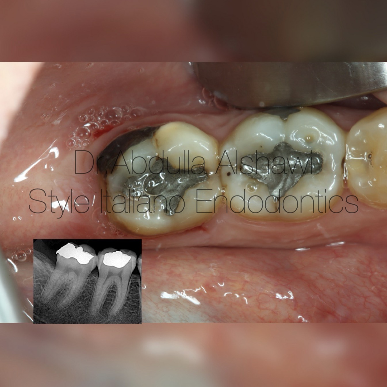
Fig. 1
A 19-year-old female patient came to the clinic with pain from cold stimulation and percussion .
pre operative x-ray shows teeth No 36 ,37 restored with amalgam
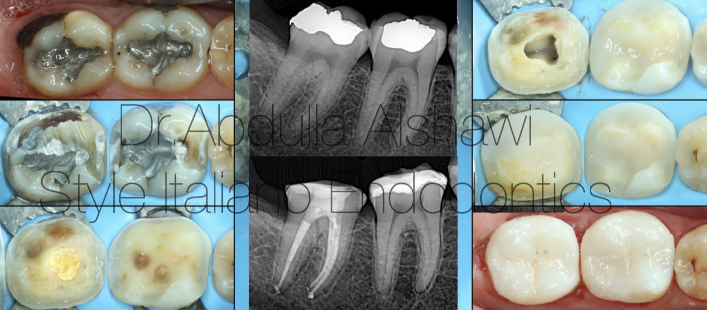
Fig. 2
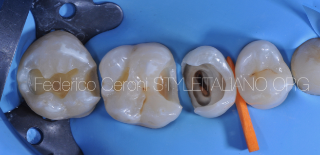
Lower first and second molars treated with amalgam restoration 2 years prior, pat suffering from pain due to recurrent caires and CL 5 caries ,after rubber dam placement and removal of all caires and unsupported enamel , all of cusps are removed because the remaining thickness is less than 3mm .
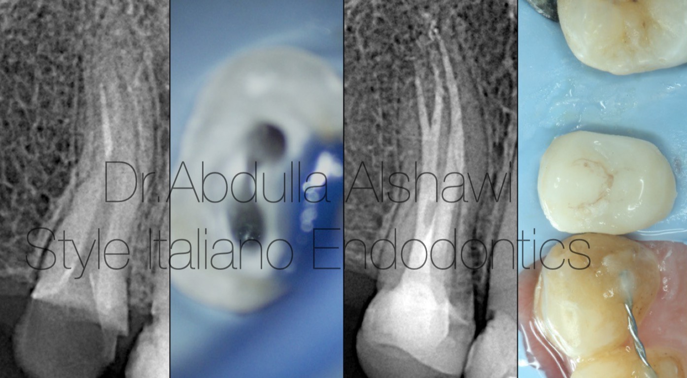
Fig. 3
Destrictive upper first premolar after ET deep margin elevation and prepared biobase restored with overlay (lithium disilicate).
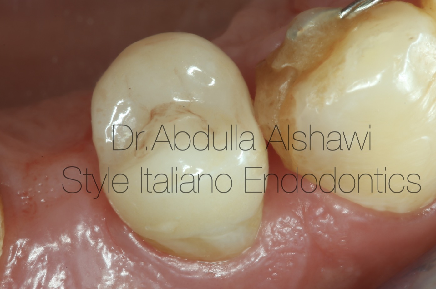
Fig. 4
Upper first premolar recall after 2 months.
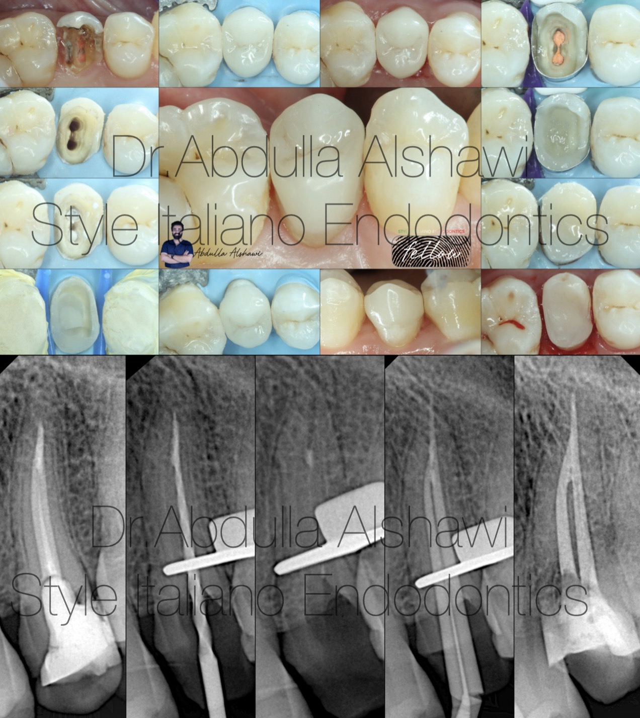
Fig. 5
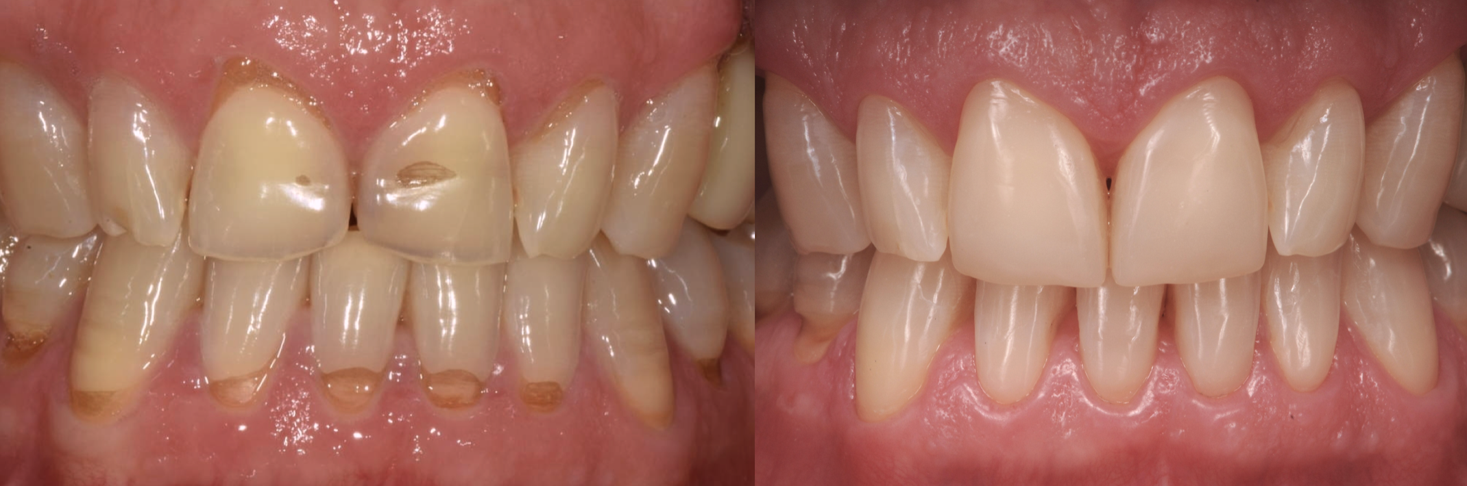
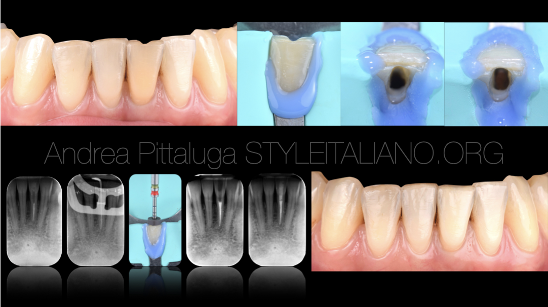
Fractured upper second premolar under level of soft tissue ,doing crown lengthening to achieve enough amount from tooth structure for RD placement and starting re-treatment, final restoration overlay.
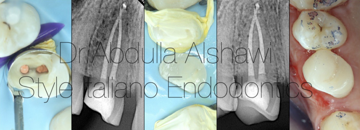
Fig. 6
Upper first premolar prepared biobase and restored with indirect overlay.
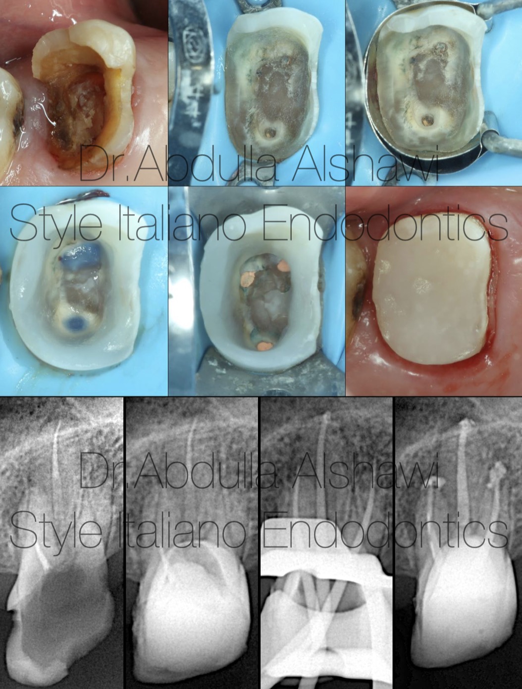
Fig. 7
Destructive upper first molar with failed root canal treatment, patient lost a lot of teeth and have no opposing tooth, so the patient refused extraction.
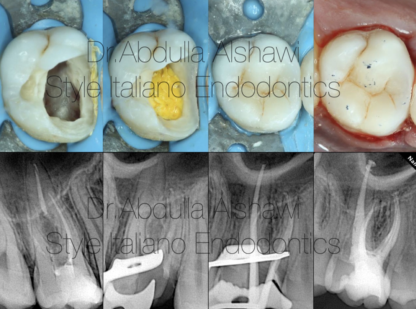
Fig. 8
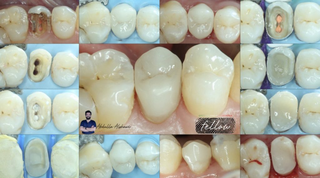
Upper first molar with failed restoration and root canal treatment, starting re-treatment with refine the cavity design, after that rubber dam placement and DME then complete RCT, finish this case with direct way
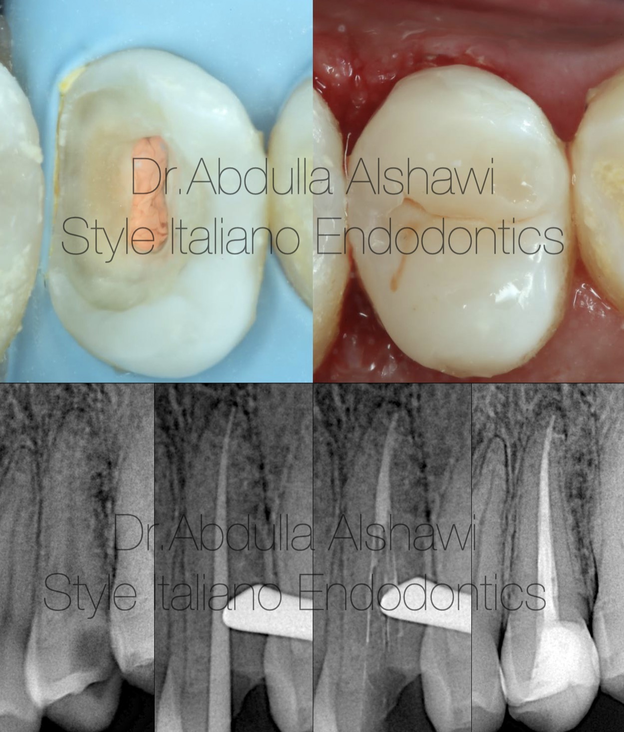
Fig. 9
Upper second premolar with distal cavity ,endo treatment and direct restoration
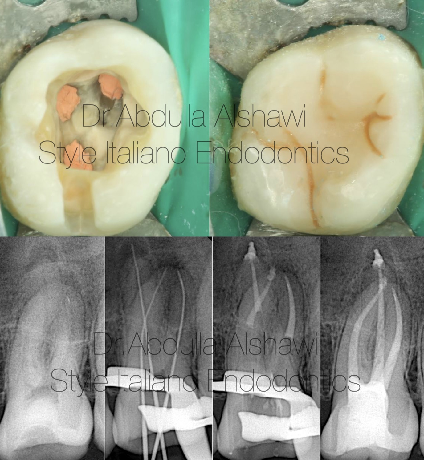
Fig. 10
Regarding all studies and research about the thickness of remaining cusps ,restoration of upper first molar is with direct way because the thickness is more than 3mm .

Fig. 11
Abuot the author :
B.D.S
Graduated from Uruk dental collage-Iraq 2018-2019
Fellow member in style Italiano endodontics.
Key opinion leader of MDclus
Conclusions
Before starting your endo treatment you must to Knowing whether the condition of the tooth is restorable or not is the first part to success treating the tooth ,also this point help you to know which type of restoration can use for finishing your case with best result .
Bibliography
1.Aquilino, S. A. and Caplan, D. J. (2002). Relationship between crown placement and the survival of endodontically treated teeth. Journal of Prosthetic Dentistry, 87(3), 256–263.
2.Assif, D., Bitenski, A., Pilo, R., Oren, E. (1993). Eect of post design on resistance to fracture of endodontically treated teeth with complete crowns. The Journal of Prosthetic Dentistry, 69(1), 36–40
3. Edelhoff D, Sorensen JA. Tooth structure removal associated with various preparation designs for posterior teeth. Int J Periodontics Restorative Dent. 2002;22:241–9.
4. Gluskin AH, Radke RA, Frost SL, Watanabe LG. The mandibular incisor: rethinking guide-lines for post and core design. J Endod. 1995;21:33–7.
5. Hunter AJ, Feiglin B, Williams JF. Effects of post placement on endodontically treated teeth. J Prosthet Dent. 1989;62:166–72.