Clinical Application of R One Mini that Emphasises on Dentin Preservation
28/06/2025
Marino Sutedjo
Warning: Undefined variable $post in /home/styleendo/htdocs/styleitaliano-endodontics.org/wp-content/plugins/oxygen/component-framework/components/classes/code-block.class.php(133) : eval()'d code on line 2
Warning: Attempt to read property "ID" on null in /home/styleendo/htdocs/styleitaliano-endodontics.org/wp-content/plugins/oxygen/component-framework/components/classes/code-block.class.php(133) : eval()'d code on line 2
In the evolving landscape of endodontics, the demand for instruments that balance effectiveness with conservation of tooth structure has never been greater. While we must shape our root canals to create volume for our irrigants to kill and solve tissue, at the same time we need to maintain as much as practical safe and sound dentin especially in the Pericervical area. Enter the R-One Mini—a reciprocating file system that designed specifically with the minimally invasive shaping philosophy in mind.
The R-One Mini was developed in collaboration with the Sapienza University of Rome, to meet the needs of clinicians seeking safer, more conservative root canal shaping, particularly in anatomically complex or narrow canals.
The system has 3 sequence of files but it require only two files. All of the files are work in reciprocating movement to enhanced fatigue resistance. First file is 19/.05 that designed to work for the coronal part of the canal as an opener. Then the first shaper is 17/.04 and followed by final shaping file of 25/.06. The system utilizes AF-R and AF-L a heat-treated nickel-titanium alloys.
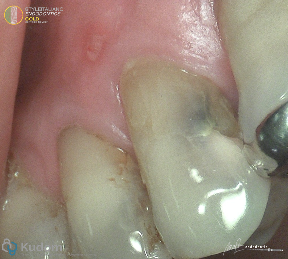
Fig. 1
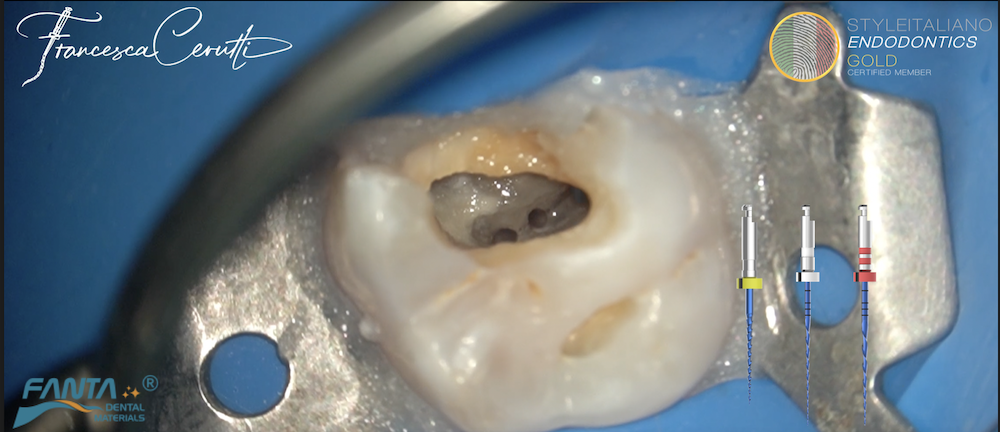
Patient came with chief complaint of pain when he used the tooth for eating on his upper left premolar. Sinus tract were seen and cavity also seen in distal area. Percussion test was +
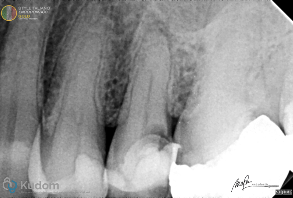
Fig. 2
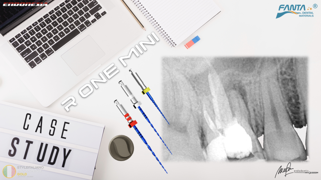
From the Pre op Xray there was an apical lesion on tooth 25 and leakage from restoration on distal area.
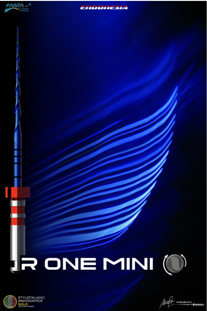
Fig. 3
Root canal treatment was performed using R One Mini
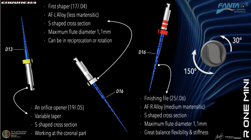
Fig. 4
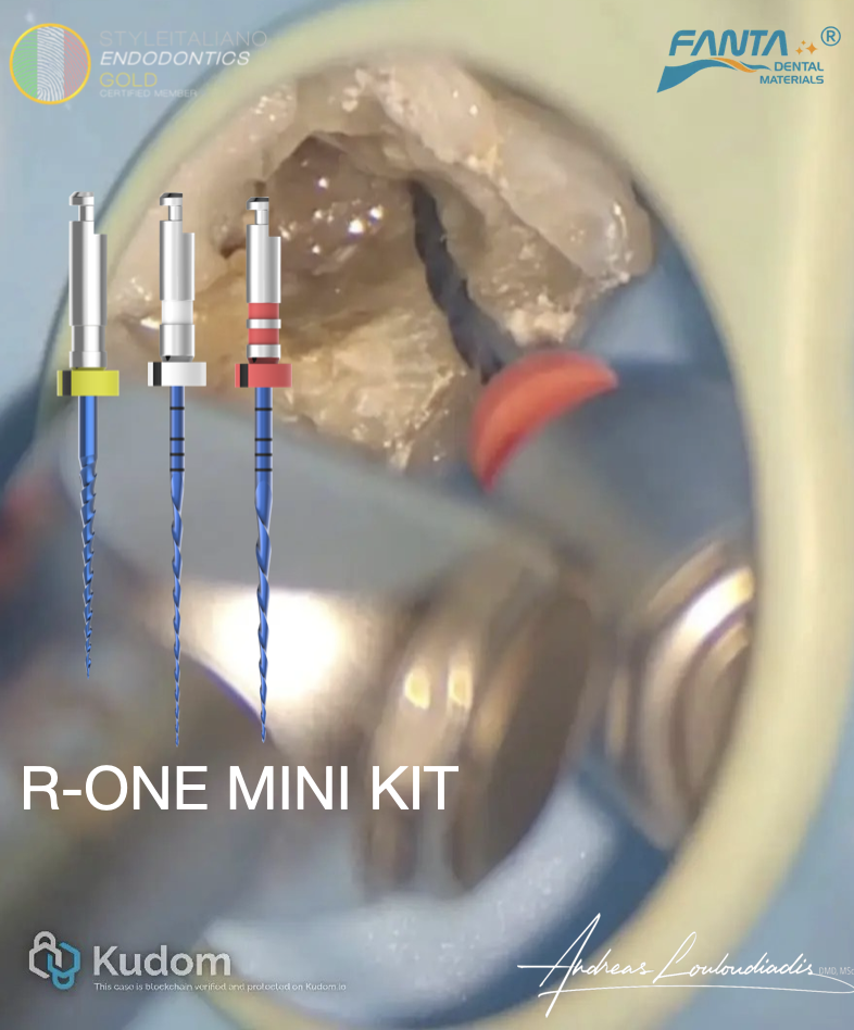
The short details about R One Mini System
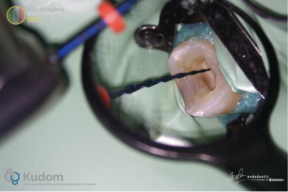
Fig. 5
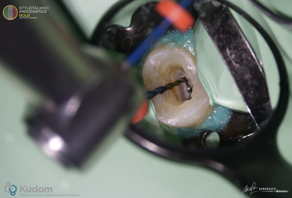
Both canals were shaped using 17/.04 and final shaping size of 25/.06
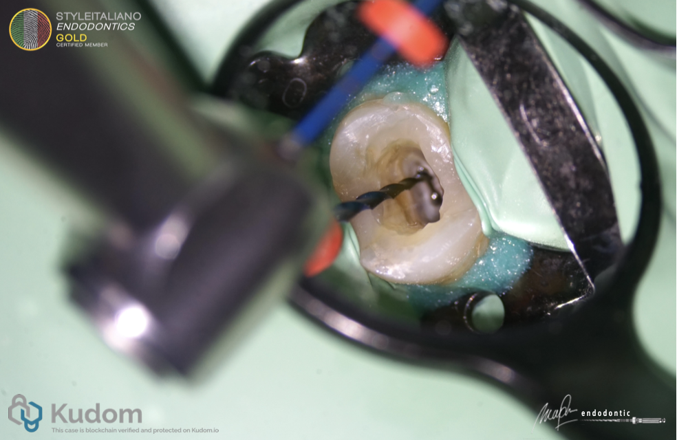
Fig. 6
Both canals were shaped using 17/.04 and final shaping size of 25/.06
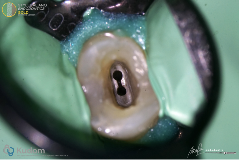
Fig. 7
After root canal preparation. Irrigation using 6% NaOCl and Sonic activation
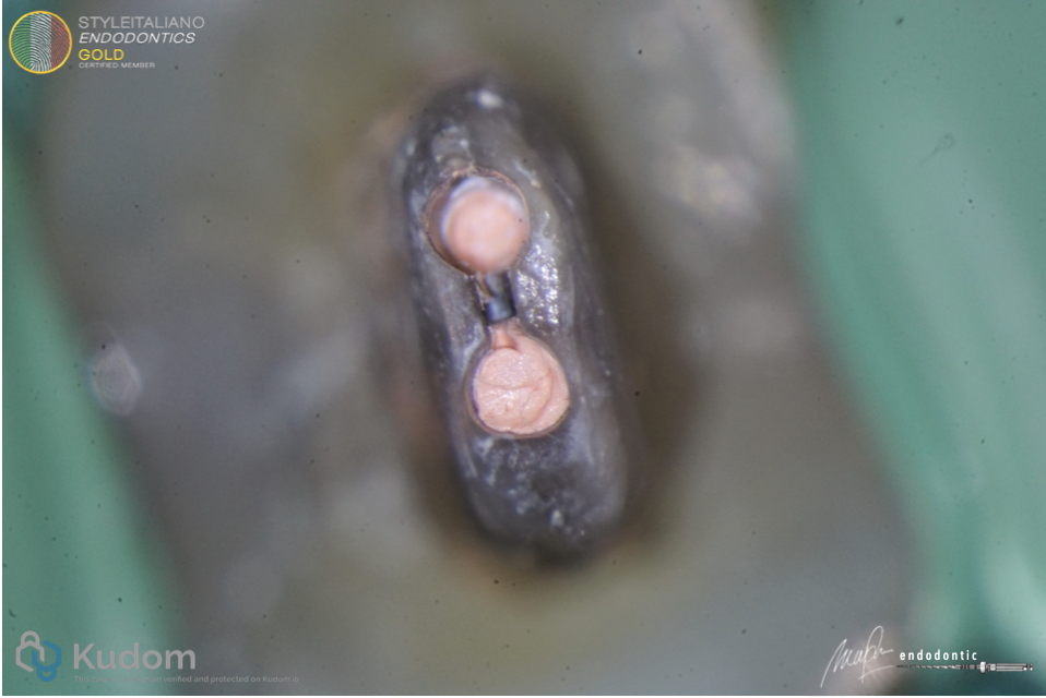
Fig. 8
After root canal obturation. Obturation using BC sealer and single cone technique
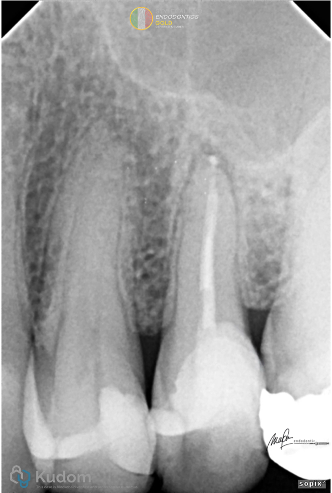
Fig. 9
Post Op Xray. Maintaining PCD area in mesial wall
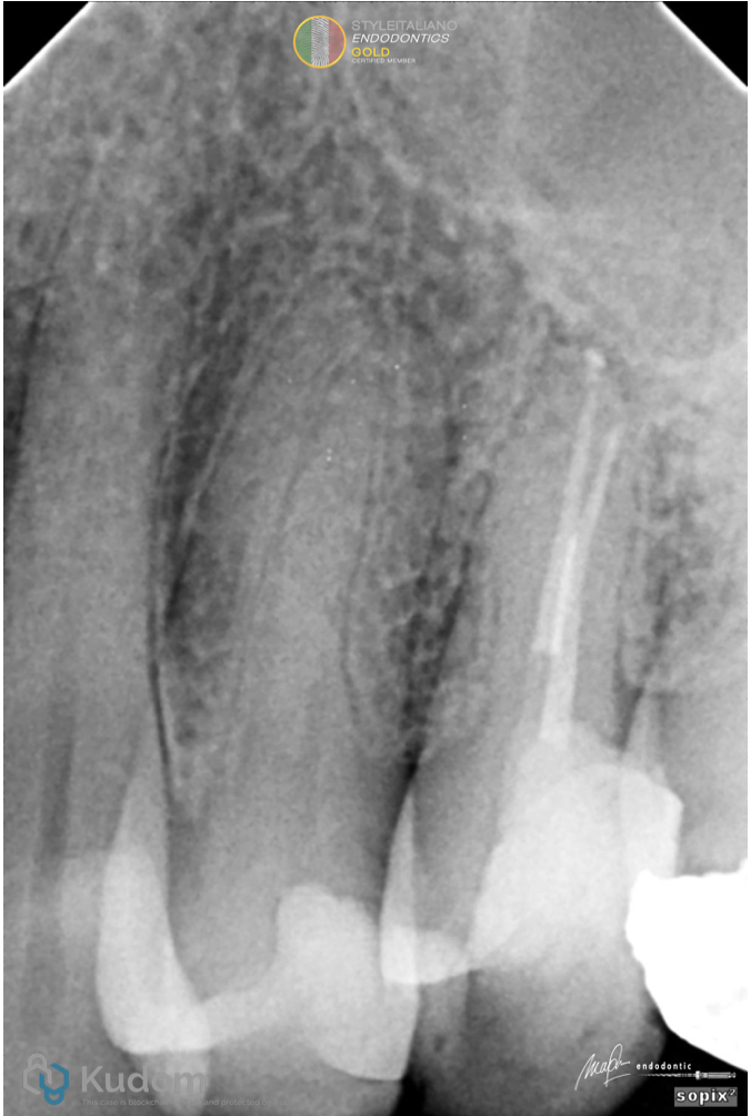
Fig. 10
Post Op Xray
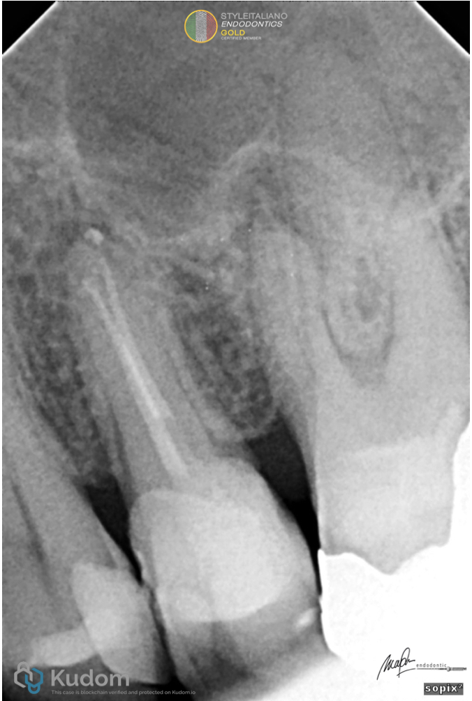
Fig. 11
Post Op Xray. One of the canal portal of exit is in position with the apical lesion
Conclusions
This R One Mini systems can minimizes the risk of procedural errors, making it a valuable addition to modern endodontic practice. Based on this clinical experience, the R-One Mini demonstrates promise for safer and more efficient root canal treatments, particularly in anatomically challenging cases.