Apical plug with Bioceramic putty
24/08/2024
Fellow
Warning: Undefined variable $post in /home/styleendo/htdocs/styleitaliano-endodontics.org/wp-content/plugins/oxygen/component-framework/components/classes/code-block.class.php(133) : eval()'d code on line 2
Warning: Attempt to read property "ID" on null in /home/styleendo/htdocs/styleitaliano-endodontics.org/wp-content/plugins/oxygen/component-framework/components/classes/code-block.class.php(133) : eval()'d code on line 2
The purpose of this article is to explain all the phases to do in a correct way an apical plug. Today on the market there was a lot of material that perform wery well like MTA or Bioceramic material in putty consistency. It’s important to know not only the technique but also when it is necessary do an apical plug. My personal advice is that is not important the apical diameter (or better is not the fundamental factor) but if the apical walls are parallel (not containing) or convergent (containing). If we are able to have with our guttaperca cone a right tug-back also with high apical diameter we are able to obturate the canal in this way, If not we have to do an apical plug.
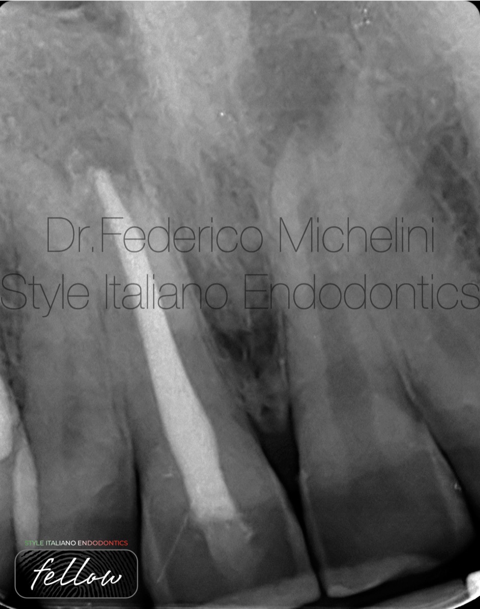
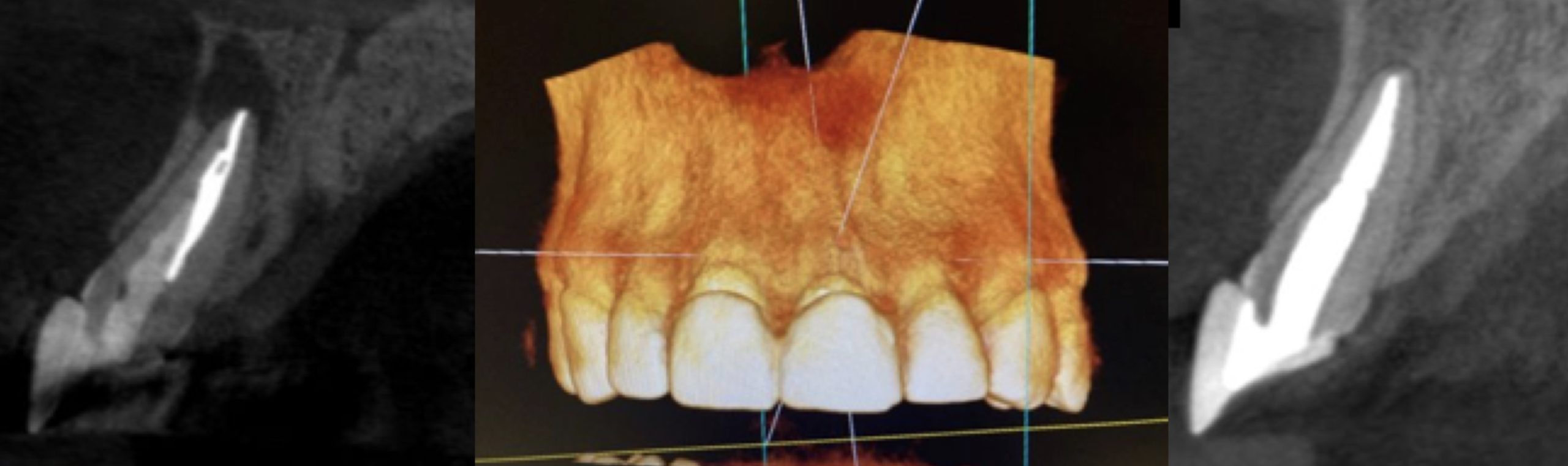
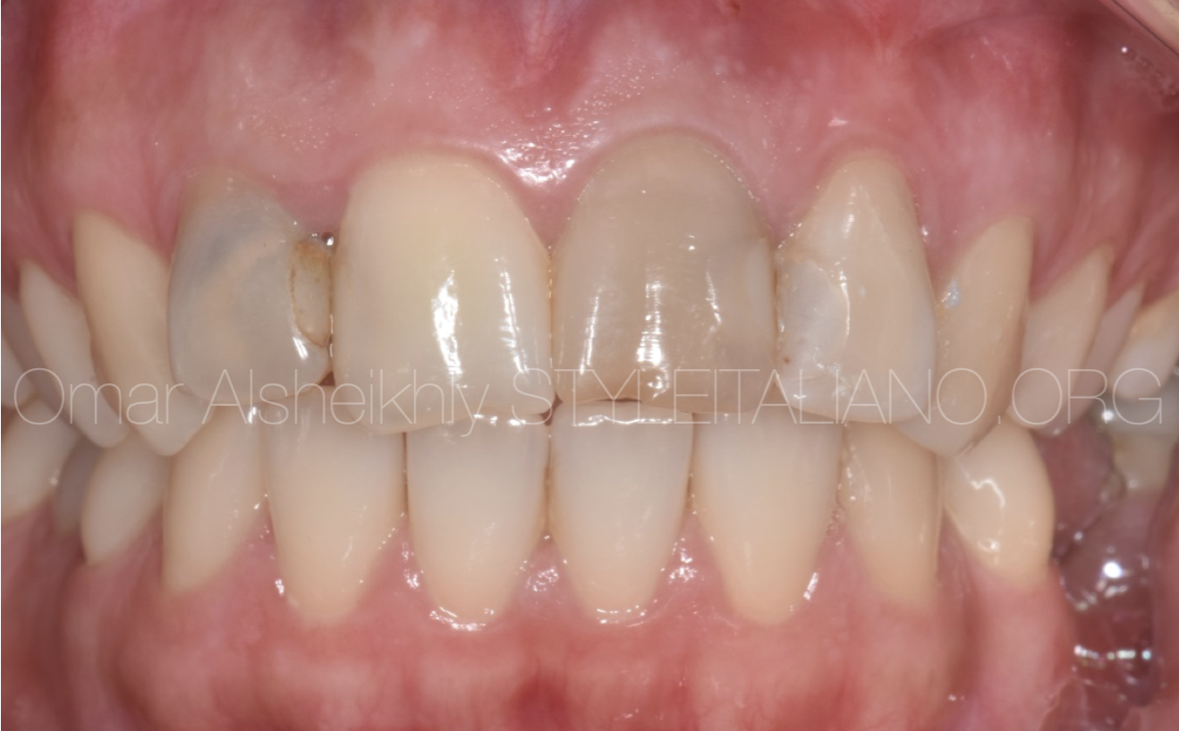
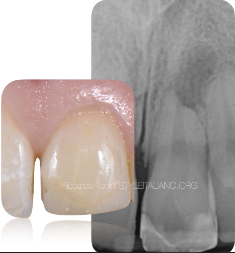
Fig. 1
Pre-operative X-ray.
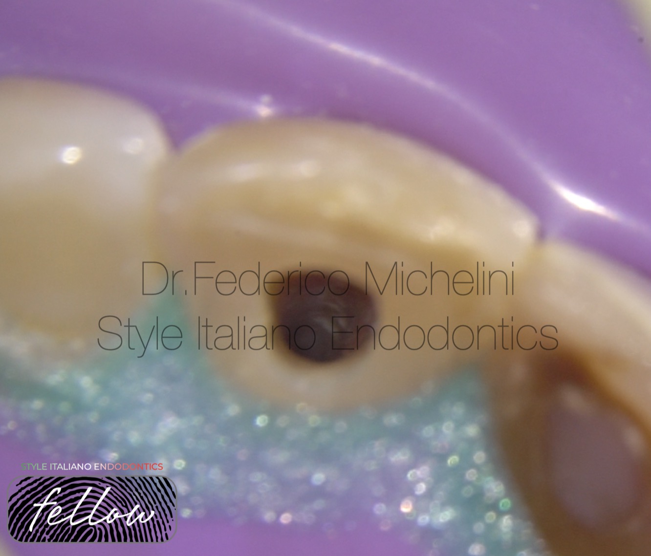
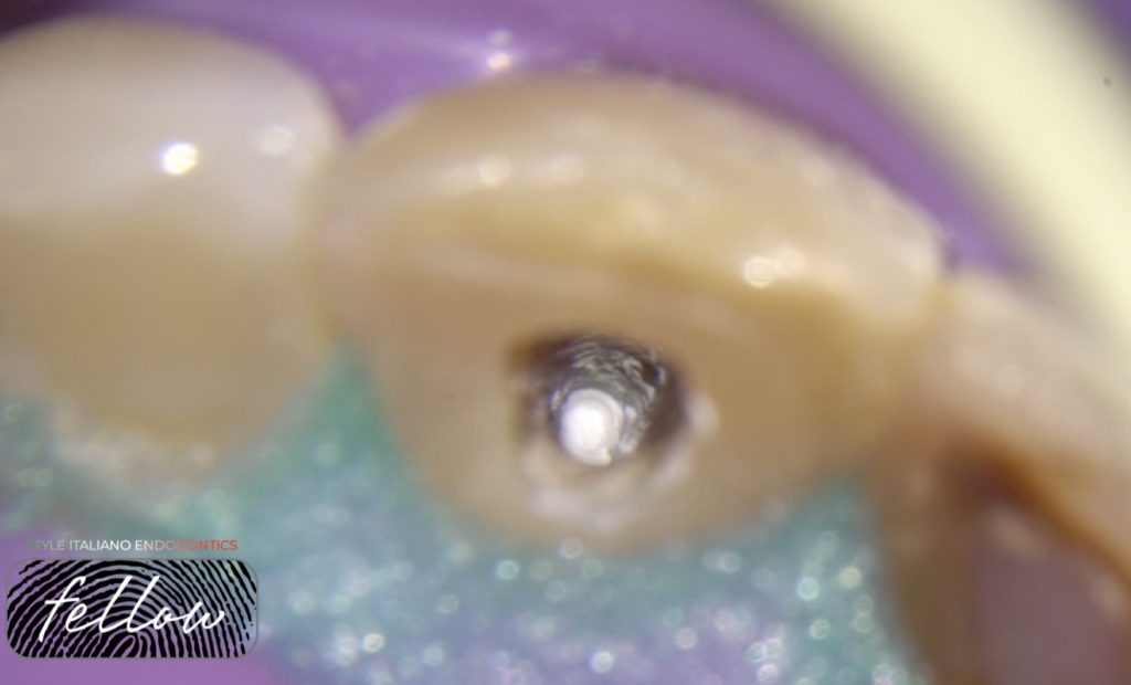
Fig. 2
Image of the canal after final irrigation protocol ready for obturation.
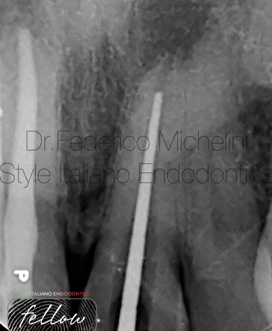
Fig. 3
X-ray with a plugger 2mm shorter of the working length.
It’s important do this type of X-ray to have the control after the compaction of the material.
When the dimension of the apex it’s very high we have the possibility to put a collagen sponge into the apical area like as stop to control the extrusion of the material.
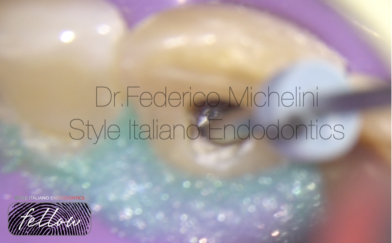
Fig. 4
First placement with dedicated tool.
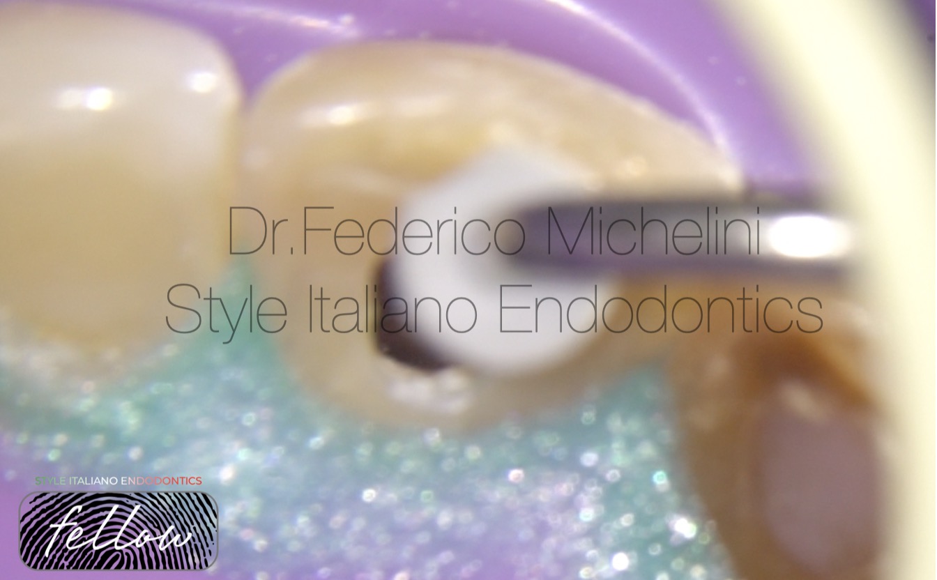
Fig. 5
Compaction with plugger. The rubber stop was positioned 2mm shorter than the working length.
We have more control over the extrusion beyond the apex.
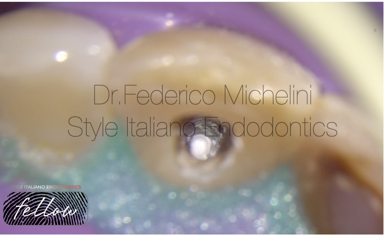
Fig. 6
Check with microscope of the first placement.
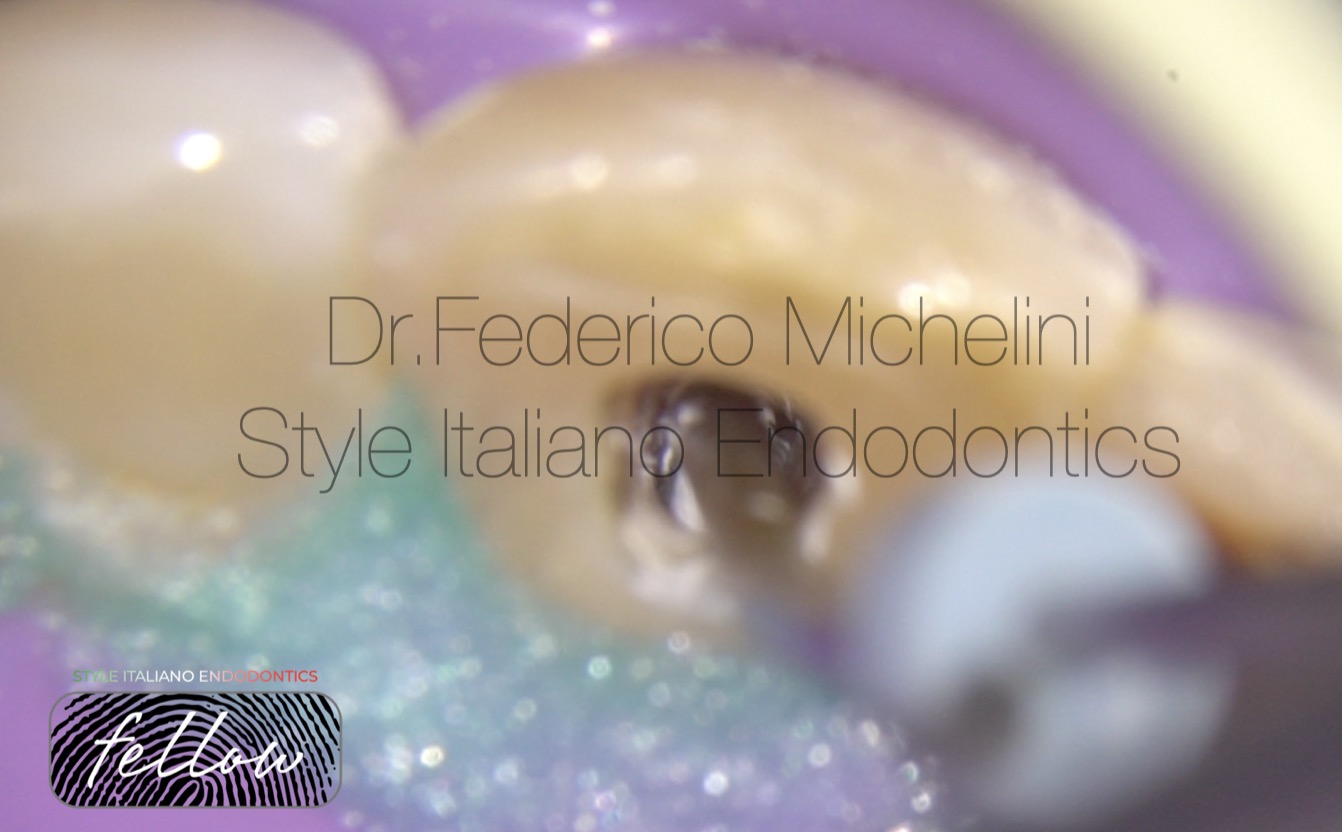
Fig. 7
Second placement of bioceramic putty.
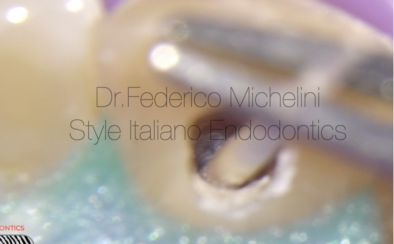
Fig. 8
Compaction with paper cone.
At the end of the 2 placement with the dedicated tool and with the plugger I finished my compaction with a paper point for more control and final adaptation of the material.
Fig. 9
Final check of the material.
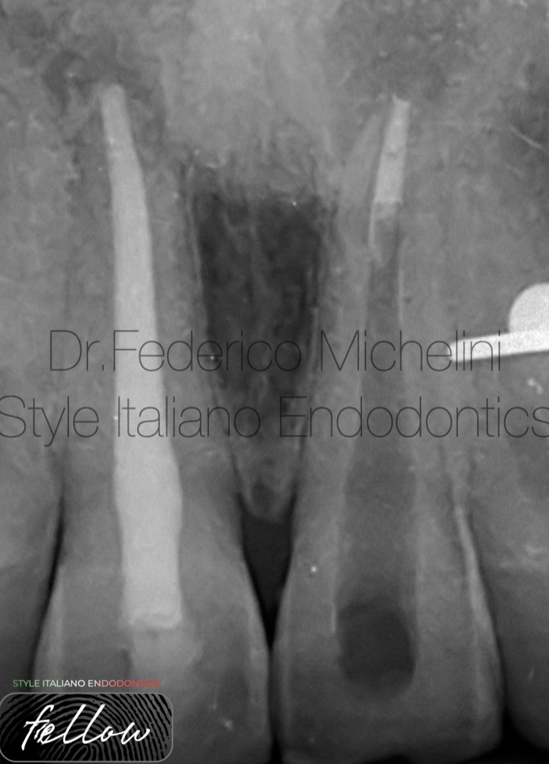
Fig. 10
X-ray to check the apical plug. This X-ray it’s very important to do because if we need to correct something we still have time ( the material doesn’t arrived to the apex, empty spaces between the placement..).
A suggestion: do this type of x-ray after 1 or 2 placement of the material because if we put too material and we need to correct something will become too difficult or impossible.
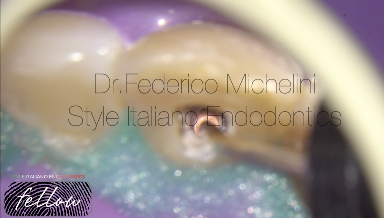
Fig. 11
In the same session after the positioning of 4-5 mm of bioceramic putty into the apical area the canal was fill with guttaperca.
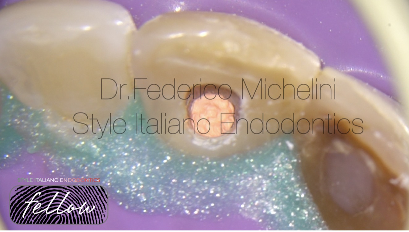
Fig. 12
Final result.
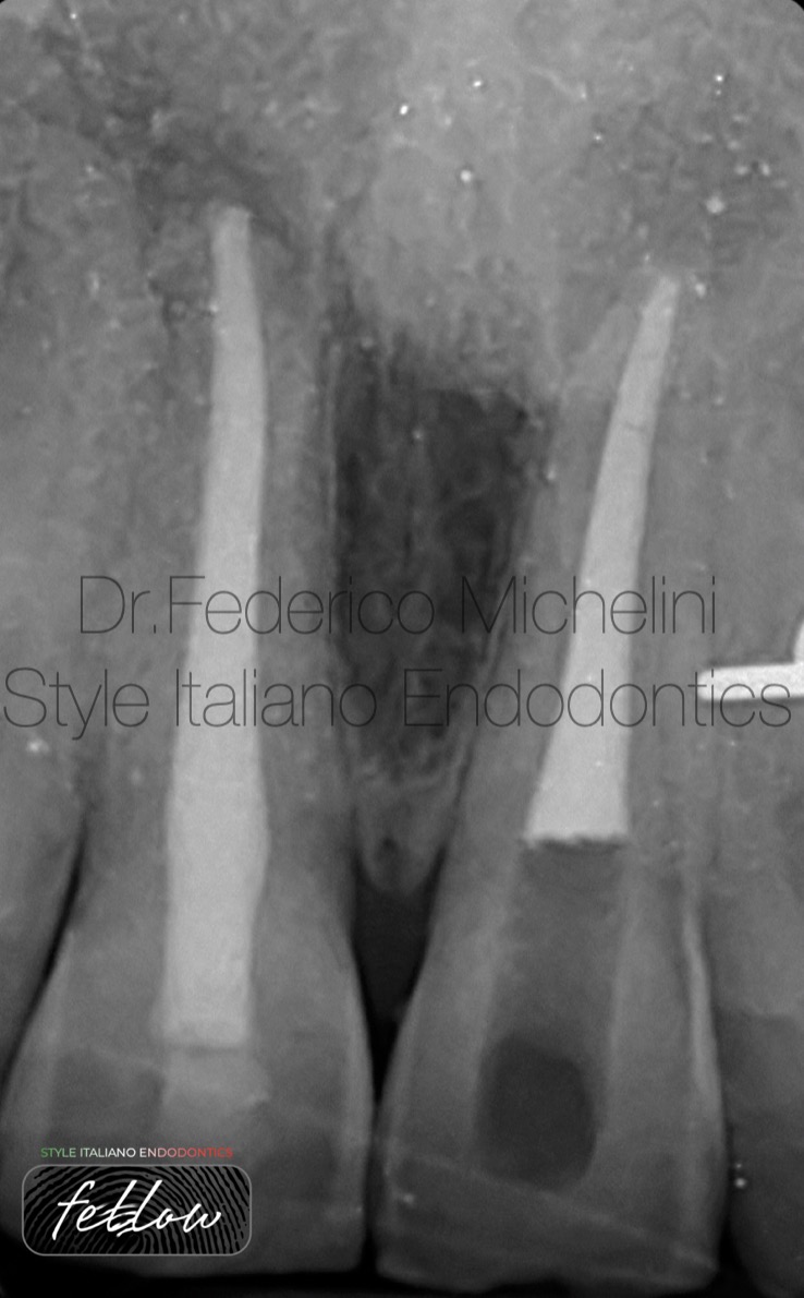
Fig. 13
X-ray with the Back-filling with guttaperca.
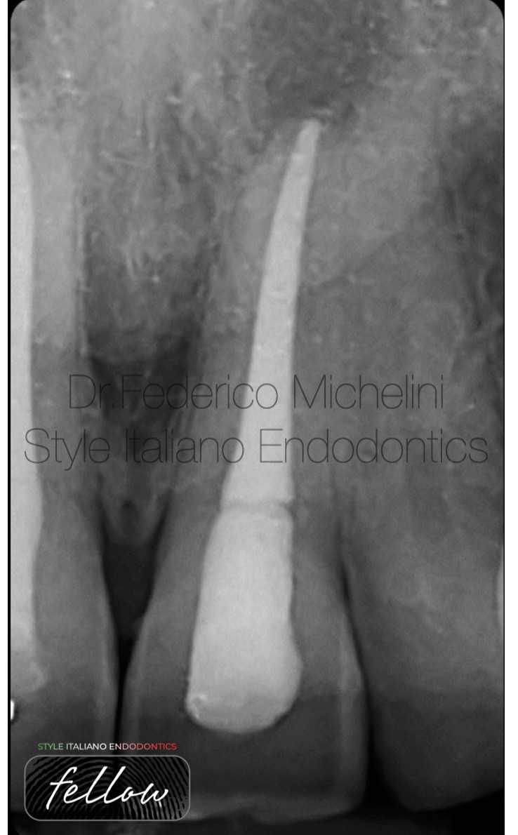
Fig. 14
Final X-ray with the composite obturation and control.
The video with all phases described above.

Fig. 15
About Dr. Michelini
2015: Graduated in dentistry from the University of Valencia.
2017: Master in Endodontics from the University of Bologna.
Speaker in national and international congresses regarding endodontics.
2017-2023: Tutor of the Master of Endodontics and of the Degree Course of Dentistry and Dental Prosthesis for the University of Bologna.
He won the Closed Meeting of Italian Society of Endodontics in 2019.
He attended the advanced course of retreatments and endodontics surgery of Italian Academy of Endodontics.
He attended the course of microscopy by Dott. Fabio Gorni.
Member of the Italian Society of Endodontics
Member Fellow of Style italiano Endodontics.
Opinion Leader and Speaker for Morita, Coltene and Labomed.
He works in Parma and Piacenza dedicating to microscopic endodontics.
Conclusions
Today we have on the market a lot of tools and materials that help us in our work. Also this, it’s important have clearly all the phases to realize an apical plug even if it is not part of a daily routine.
Bibliography
- Mineral Trioxide Aggregate and other bioactive endodontic cements: an updated overview-part II: other clinical applications and complications. Torabinejad M, et al. Int Endod J. 2018.
- Use of Mineral Trioxide Aggregate with or without a collagen sponge as an apical plug in teeth with immature apices. Tek GB, et al. J coin pediatr dent. 2021.