Cracked tooth syndrome
24/02/2024
Fellow
Warning: Undefined variable $post in /var/www/vhosts/styleitaliano-endodontics.org/endodontics.styleitaliano.org/wp-content/plugins/oxygen/component-framework/components/classes/code-block.class.php(133) : eval()'d code on line 2
Warning: Attempt to read property "ID" on null in /var/www/vhosts/styleitaliano-endodontics.org/endodontics.styleitaliano.org/wp-content/plugins/oxygen/component-framework/components/classes/code-block.class.php(133) : eval()'d code on line 2
Within the intricate landscape of dental health, the phenomenon of cracked teeth poses a challenging conundrum for both patients and dental professionals. Cracked teeth, an umbrella term encompassing various fissures and fractures within tooth structures, present a complex array of symptoms and diagnostic intricacies, often defying conventional diagnostic methodologies. This exclusive article aims to delve into the multifaceted nature of cracked teeth, exploring their diverse manifestations, diagnostic challenges, and tailored approaches to comprehensive management.
Navigating the Diagnostic Labyrinth
Diagnosing cracked teeth poses a formidable challenge due to their varied presentations and elusive symptoms. Traditional diagnostic methods, including visual inspection, tactile examination, and radiographic imaging, often fall short in identifying subtle cracks, especially in cases with intermittent symptoms. Advanced techniques, such as high-resolution intraoral cameras, spectroscopy, and thermal imaging, show promise in enhancing diagnostic accuracy by capturing detailed images and identifying microfractures within tooth structures.
The signs and symptoms of a cracked tooth can vary depending on the type and severity of the crack. Here are some common indicators that may suggest the presence of a cracked tooth:
1. Intermittent Pain: One of the primary symptoms of a cracked tooth is sporadic or intermittent pain. This pain might occur while chewing or biting down on food and can be triggered by specific types of foods or temperature changes (hot or cold).
2. Sensitivity: Increased sensitivity to hot, cold, or sweet foods and beverages, especially when the stimuli are removed, is another common sign. This sensitivity can occur in the affected tooth or even in nearby teeth.
3. Discomfort Upon Release: Patients might experience discomfort or pain when releasing biting pressure from the affected tooth. This pain might not necessarily be present while actively biting down but appears upon release.
4. Pain while Chewing: Cracked teeth can cause pain or discomfort while chewing, particularly when the pressure is exerted on the affected tooth during the chewing process.
5. Visible Damage: In some cases, a cracked tooth might be visible to the naked eye, presenting as a visible fracture line on the tooth surface. However, not all cracks are visible without magnification or specialized dental instruments.
6. Gum Inflammation: Persistent inflammation or irritation in the gums surrounding the affected tooth can sometimes be an accompanying symptom of a cracked tooth.
It’s important to note that not all cracked teeth present with noticeable symptoms, especially in the case of smaller or superficial cracks. In these instances, diagnosis might require a thorough dental examination, possibly using specialized tools or imaging techniques to detect the presence of a crack.
Detecting a cracked tooth on an X-ray
It can be challenging, especially if the crack is small or extends in a way that’s not clearly visible on the radiograph. However, certain types of cracks or associated complications might be observable through X-rays, depending on their nature and extent. Here’s how cracks may appear on dental X-rays:
1. Vertical Root Fractures (VRFs): A vertical root fracture, which occurs in the root of the tooth, can sometimes be detected through X-rays. However, VRFs might not always be easily visible, especially in the early stages. They may appear as a thin, vertical line extending from the root of the tooth.
2. Craze Lines or Superficial Cracks: These minute cracks within the enamel are often not visible on X-rays because they are limited to the outer surface of the tooth and may not extend deep enough to be captured by traditional dental radiographs..
3. Cracks Extending Into the Pulp: In some cases, a larger crack that extends deeper into the tooth, potentially affecting the pulp or nerve tissue, might exhibit signs on X-rays. This could appear as a discontinuity or shadow within the tooth structure, but smaller cracks might not be easily discernible.
4. Secondary Signs: While the crack itself might not always be visible, X-rays can reveal secondary signs that suggest the presence of a crack, such as changes in the surrounding bone, localized inflammation, or subtle alterations in the periodontal ligament space adjacent to the affected tooth.
It’s important to note that not all cracks are detectable through routine X-rays. Sometimes, additional imaging techniques, such as cone-beam computed tomography (CBCT) or specialized imaging with higher resolution, might be necessary to visualize intricate or smaller cracks more effectively.
Managing a dental crack involves a tailored approach based on the type, severity, and individual circumstances of the patient. Here’s various strategies for managing a dental crack:
1. Observation and Monitoring: In cases where the crack is superficial and doesn’t cause discomfort, a dentist may recommend regular monitoring without immediate intervention. Consistent dental check-ups allow for tracking any changes in the crack’s progression.
2. Protective Measures: For minor cracks that cause minimal symptoms, dental professionals might apply dental sealants or bonding to shield the cracked area, reducing sensitivity and preventing further damage.
3. Custom Restorations: Larger or more noticeable cracks, particularly those impacting the cusps or chewing surfaces, might require custom restorations such as onlays or inlays. These restorative treatments provide stability and protection to the affected tooth..
4. Crown Placement: Extensive cracks compromising the tooth’s structural integrity often necessitate the placement of dental crowns. Crowns cover the entire visible portion of the tooth, restoring strength and safeguarding against additional cracking.
5. Root Canal Therapy: When a crack extends into the inner pulp of the tooth, causing pain or infection, root canal therapy becomes necessary. This procedure involves cleaning the infected area and sealing the root canal to alleviate discomfort and prevent further complications.
6. Consideration for Extraction: In scenarios where the crack is severe, irreparable, or extends deeply below the gum line, extraction might be the only viable solution. Following extraction, replacement options such as dental implants or bridges can restore functionality and aesthetics.
7. Individualized Plans: Every patient’s situation is unique, and treatment plans must be personalized. Factors like overall oral health, the extent of the crack, and patient preferences are considered while determining the most suitable course of action.
8. Preventive Practices: Post-treatment, maintaining good oral hygiene through regular brushing, flossing, and dental check-ups is crucial. Additionally, avoiding habits that stress the teeth, such as chewing hard objects or clenching/grinding, helps prevent future cracks or damage.
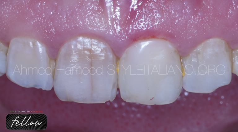
Fig. 1
Surface Craze Lines:
Minute cracks within the enamel, often imperceptible and largely benign, occasionally causing slight sensitivity but not requiring active intervention.
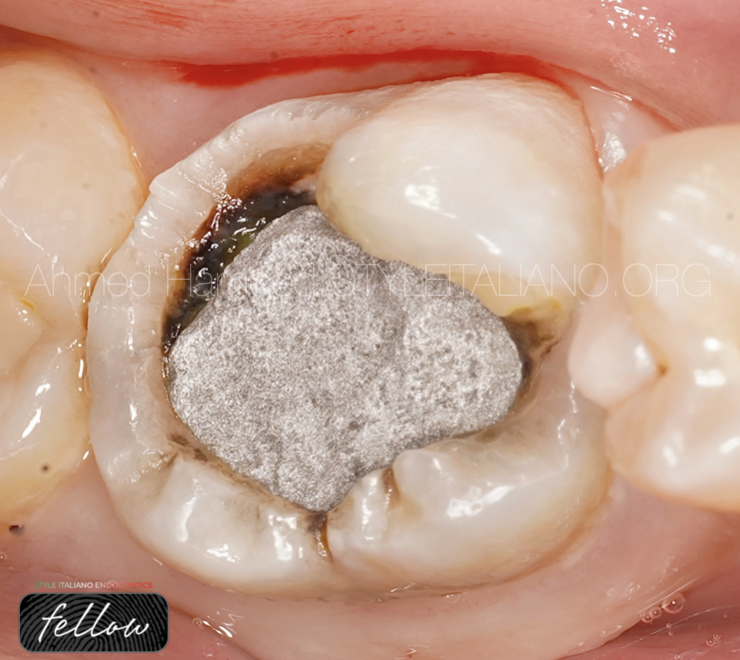
Fig. 2
Fractured Cusps:
Cracks affecting the tooth’s chewing surface, commonly arising from existing large fillings or structural weaknesses, leading to discomfort during biting or chewing..
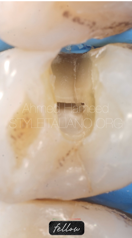
Fig. 3
Cracked Tooth Syndrome:
Characterized by deeper cracks originating from the chewing surface and extending towards the root, presenting with intermittent sharp pain or sensitivity upon release of biting pressure
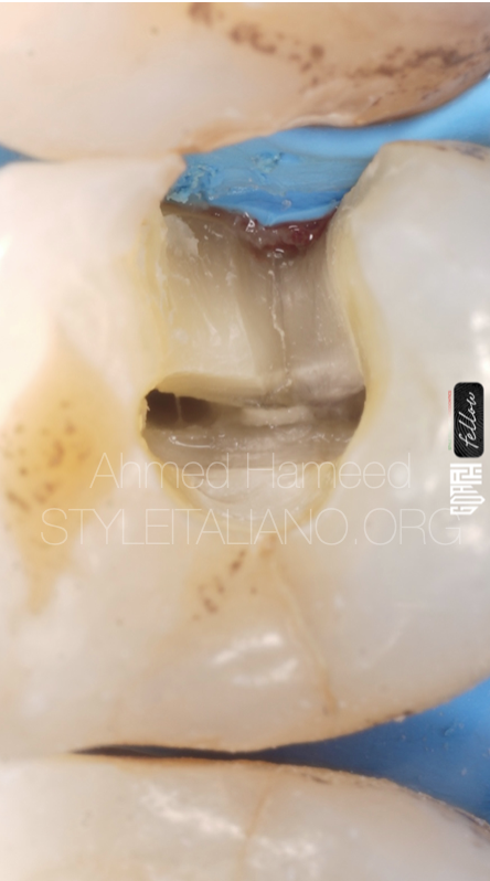
Fig. 4
Cracked Tooth Syndrome:
Characterized by deeper cracks originating from the chewing surface and extending towards the root, presenting with intermittent sharp pain or sensitivity upon release of biting pressure
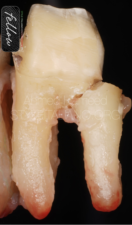
Fig. 5
Split Teeth:
An advanced stage where the tooth separates into distinct segments due to an extensive crack, necessitating complex treatments, including possible extraction and replacement
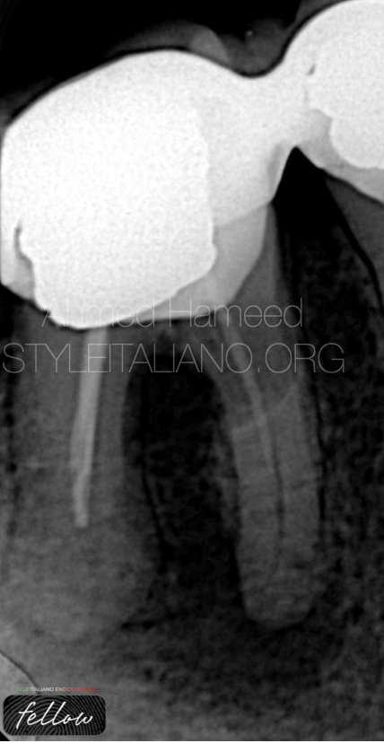
Fig. 6
Figure 6:
One of the landmarks that may refer to an extensive root fracture or crack is the J shape lesion extinding from the apical part of the root to the coronal part which may be with root resorbsion as obvious in this x-ray.
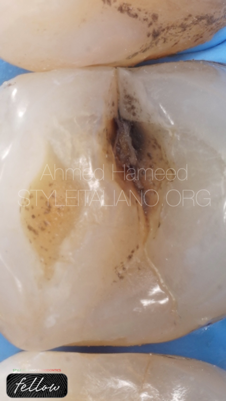
Fig. 7
This clinical case shows a typical crack line on enamel extending in mesio distal direction,pt is 49 y female came seeking for dental treatment for tooth 24,after taking history and chief complaint the pt suffering from pain on hot,cold,and biting on this tooth,it was slightly tender on percussion,the patient had no molars to bite on so all the chewing forces were mainly on premolars which gives the impression of crack extending to the pulp specially the tooth was not resbonding on thermal test.
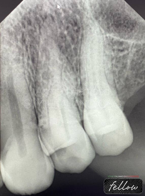
Fig. 8
Peri apical radiograph do not tell that much about crack condition however it shows some periapical changes and small lesion on mesial aspect that need a very good reading of radiograph.
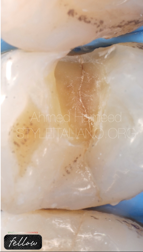
Fig. 9
After caries removal we can see that the crack line extends into dentin that means we need mor drilling chasing the crack down.
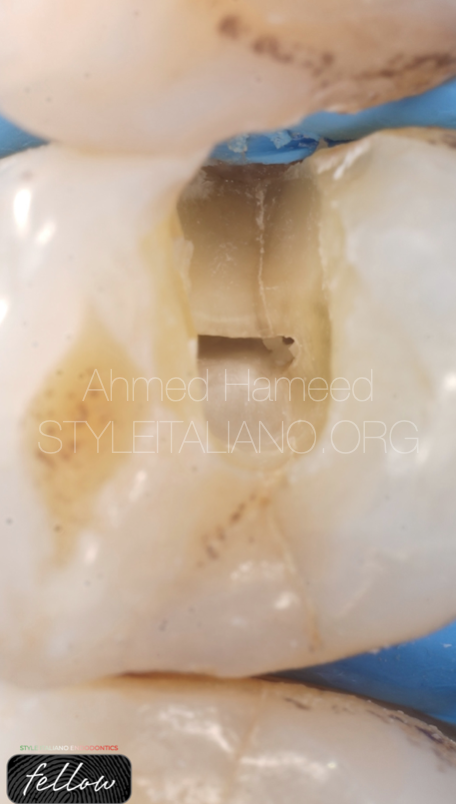
Fig. 10
This picture shows how the crack extends towards the pulp causing pul necrosis as it obvious from the empty chamber and the crack still going down .
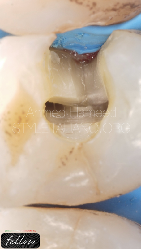
Fig. 11
This picture shows how the crack extends down towards the bone that means this my be a vertical tooth fracture and it needs extraction.
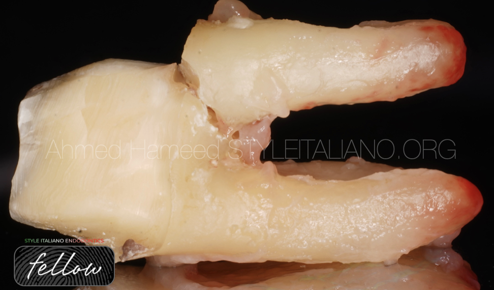
We can see here clearly that the type of fracture here is split that means there is nothing to do here.
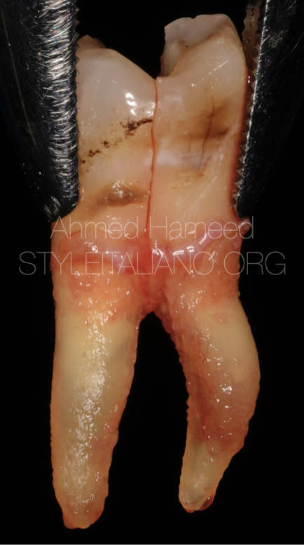
Fig. 12
Extraction is the only management here.

Fig. 13
Conclusions
Cracked teeth stand as a perplexing realm within dentistry, encompassing various types with diverse clinical presentations and diagnostic challenges. An individualized approach, integrating advanced diagnostic tools and tailored treatment modalities, remains paramount in addressing the complexities associated with each form of crack. As dental science continues to evolve, ongoing research and technological innovations promise to unravel the mysteries of cracked teeth, striving for improved patient care and outcomes in the realm of dental health.
Bibliography
1. Lynch, C. D., & McConnell, R. J. (2002). The cracked tooth syndrome: an elusive diagnosis. Journal of the Irish Dental Association, 48(2), 57-63.
2. Cameron, C. E. (2015). Cracked tooth syndrome: Diagnosis, treatment, and correlation with symptoms. A report of four cases. Journal of Endodontics, 41(1), 78-85. doi:10.1016/j.joen.2014.09.020
3. Banerji, S., Mehta, S. B., & Millar, B. J. (2010). Cracked tooth syndrome. Part 1: Aetiology and diagnosis. British Dental Journal, 208(10), 459-463. doi:10.1038/sj.bdj.2010.372
4. Lynch, C. D., & McConnell, R. J. (2007). The cracked tooth syndrome. Journal of the Canadian Dental Association, 73(8), 721-725.
5. Homewood, C. I., & Teplitsky, P. E. (2017). Cracked tooth syndrome: Overview of literature. Journal of the Michigan Dental Association, 99(8), 30-33.
6. American Association of Endodontists. (2020). Cracked Teeth. Retrieved from https://www.aae.org/patients/symptoms/cracked-teeth/
7. Chai, H., Lee, J. J., & Manton, D. J. (2015). The cracked tooth syndrome: An elusive diagnosis. Journal of the American Dental Association, 146(6), 398-406. doi:10.1016/j.adaj.2015.01.010
8. Yaman, P., Dennison, J. B., & Cheung, G. S. (2016). Conservative approaches for the treatment of cracked teeth. Dental Clinics of North America, 60(2), 285-297. doi:10.1016/j.cden.2015.11.004
9. Moule, A. J., & Moule, C. A. (2014). Cracked tooth syndrome: Aetiology, diagnosis and treatment. Australian Dental Journal, 59(2), 133-134. doi:10.1111/adj.12166
10. Hargreaves, K. M., & Berman, L. H. (2016). Cohen’s Pathways of the Pulp (11th ed.). St. Louis, MO: Elsevier.

