Five Canals, One Tooth: A Microscope-Assisted Endo-Resto Case
28/09/2026
Warning: Undefined variable $post in /home/styleendo/htdocs/styleitaliano-endodontics.org/wp-content/plugins/oxygen/component-framework/components/classes/code-block.class.php(133) : eval()'d code on line 2
Warning: Attempt to read property "ID" on null in /home/styleendo/htdocs/styleitaliano-endodontics.org/wp-content/plugins/oxygen/component-framework/components/classes/code-block.class.php(133) : eval()'d code on line 2
Successful endodontic treatment depends on the integration of accurate diagnosis, three-dimensional knowledge of root canal anatomy, effective chemomechanical preparation, and a restorative strategy that preserves the structural integrity of the treated tooth.
Mandibular molars commonly present with complex root canal configurations. Although the presence of two distal canals is a recognized anatomical variation, the identification of a middle distal canal introduces an additional challenge during access preparation, canal negotiation, shaping, and obturation.
The detection and management of additional canals require a combination of anatomical knowledge, adequate visualization, careful exploration, and a systematic treatment protocol. Magnification can facilitate the identification of anatomical landmarks and canal orifices, while effective irrigation and activation contribute to the cleaning and disinfection of the root canal system.
The restorative phase is equally important. Following endodontic treatment, the remaining tooth structure must be evaluated and managed with the aim of providing a durable coronal seal and restoring function while respecting the biological and mechanical requirements of the tooth.
This case report presents the endodontic and restorative management of a cariously involved mandibular molar diagnosed with symptomatic irreversible pulpitis. The case highlights the identification of an additional middle distal canal, the treatment of five root canals, a comprehensive irrigation activation protocol, warm obturation, and the subsequent restoration of the tooth using a direct coronal build-up, margin elevation, a bio-base, and an adhesive overlay.
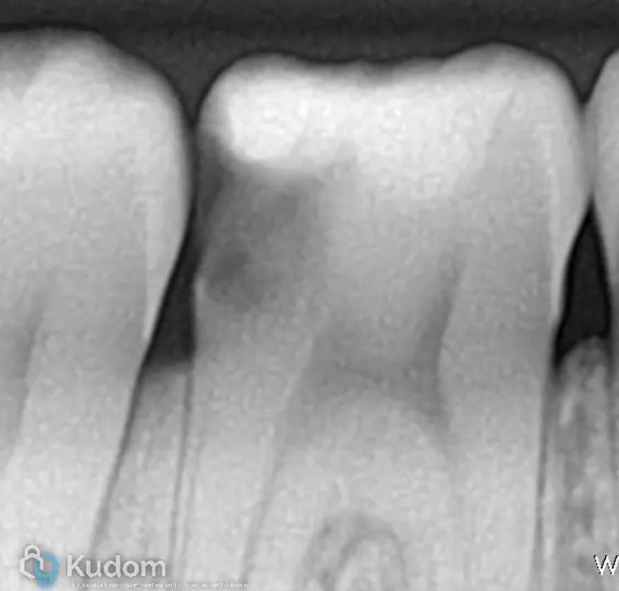
Fig. 1
A patient presented for evaluation of a mandibular molar with extensive carious involvement.
A preoperative bitewing radiograph revealed caries extending to the pulp chamber. Following clinical and radiographic examination, the tooth was diagnosed with irreversible pulpitis.
The preoperative radiographic assessment was used to evaluate the extent of the carious lesion and the condition of the tooth before treatment.
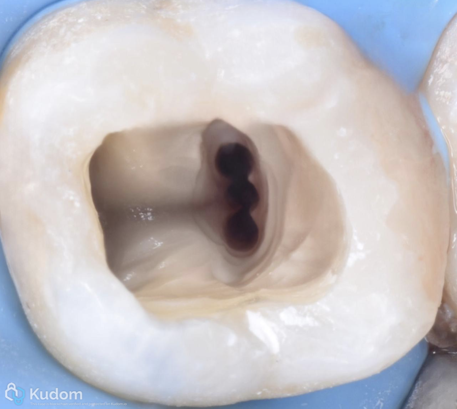
Fig. 2
Following appropriate anesthesia and rubber dam isolation, access cavity preparation was performed under high magnification.
The access cavity was refined to provide adequate visualization and access to the pulpal floor while maintaining a conservative approach to the remaining tooth structure.
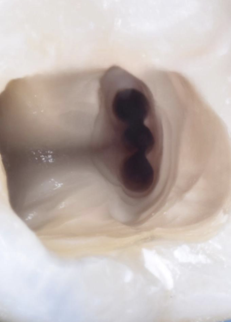
During exploration of the pulpal floor, an additional canal orifice was identified in the distal aspect of the tooth.
In addition to the expected canal anatomy, a middle distal canal was located. This finding increased the total number of canals to five.
The identification of the middle distal canal was a key anatomical finding in this case. Its successful negotiation and subsequent treatment emphasized the importance of carefully examining the pulpal floor and not relying exclusively on the most common anatomical configuration.
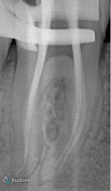
Fig. 3
Canal negotiation and shaping
Following canal identification, all five canals were negotiated and prepared.
The canals were shaped to a final preparation size of 25 taper 4%
The shaping protocol was performed with the aim of creating a reproducible canal geometry while facilitating irrigation, disinfection, and subsequent obturation.
The final preparation was standardized across the five canals at size 25 with a 4% taper.
The successful shaping of the complete canal system, including the middle distal canal, allowed for a consistent approach to the subsequent irrigation and obturation procedures.
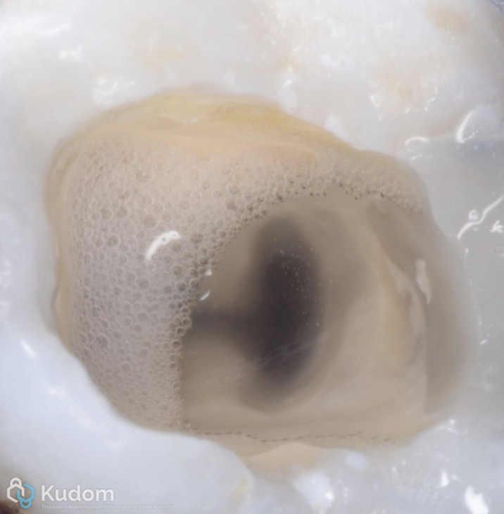
Fig. 4
Irrigation and activation
Following mechanical preparation, the canals were subjected to a comprehensive irrigation and activation protocol.
Multiple cycles of sonic and ultrasonic activation were used to enhance the distribution and agitation of the irrigating solutions within the prepared canal system.
The objective of the activation protocol was to facilitate the removal of debris and promote effective chemical cleaning of the root canal system, including areas that may be difficult to access through instrumentation alone.
The use of repeated activation cycles was particularly relevant in this case because of the presence of five canals and the additional middle distal anatomy.
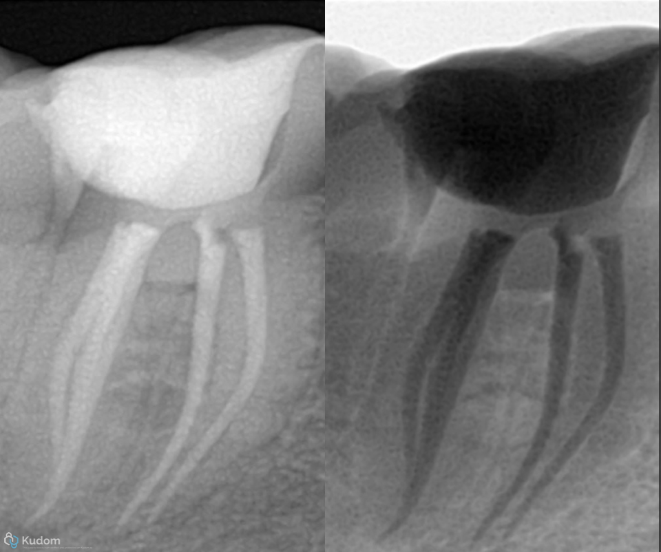
Fig. 5
Root canal obturation
Following completion of shaping and irrigation, the canals were obturated using a warm modified obturation technique.
The objective of obturation was to achieve a dense and continuous filling of the prepared root canal spaces while maintaining the established working lengths and canal anatomy.
The obturation procedure was performed for all five canals.
The selected technique allowed for the adaptation of the obturation material to the prepared canal spaces and the three-dimensional anatomy of the root canal system.
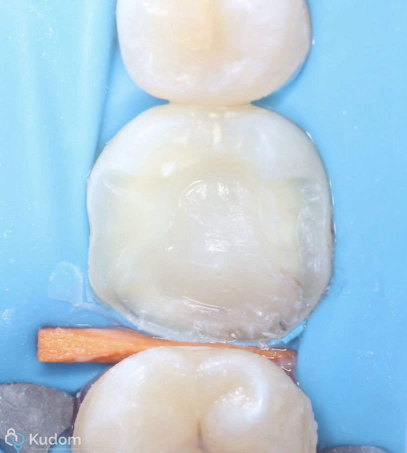
Fig. 6
Coronal build-up and margin management
Following completion of the endodontic treatment, the restorative phase was initiated.
The coronal tooth structure was evaluated, and the existing defect was managed to facilitate a predictable definitive restoration.
A coronal build-up was performed to reconstruct the missing tooth structure and establish a suitable foundation for the restorative procedure.
The restorative margin was subsequently elevated to improve the accessibility and management of the deep margin.
The purpose of this step was to create a more accessible and manageable restorative margin while facilitating the subsequent adhesive restorative procedures.
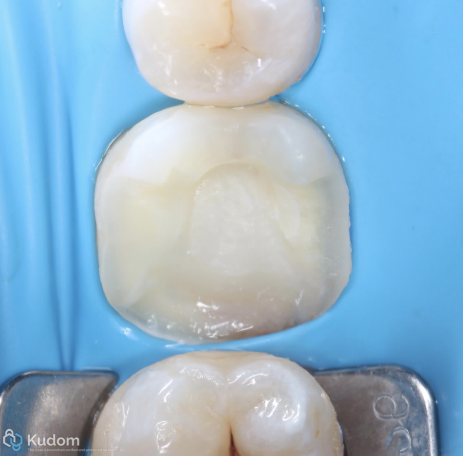
Fig. 7
Bio-base and adhesive overlay
Following the build-up and margin elevation, a bio-base was performed to provide a suitable foundation for the definitive restoration.
The tooth was then prepared for an adhesive overlay.
The definitive restoration was designed to restore the anatomy and function of the treated molar while maintaining a conservative approach to the remaining tooth structure.
The overlay was fabricated using lithium disilicate
The restorative design was selected according to the remaining tooth structure, occlusal requirements, and the need to provide a durable coronal seal.
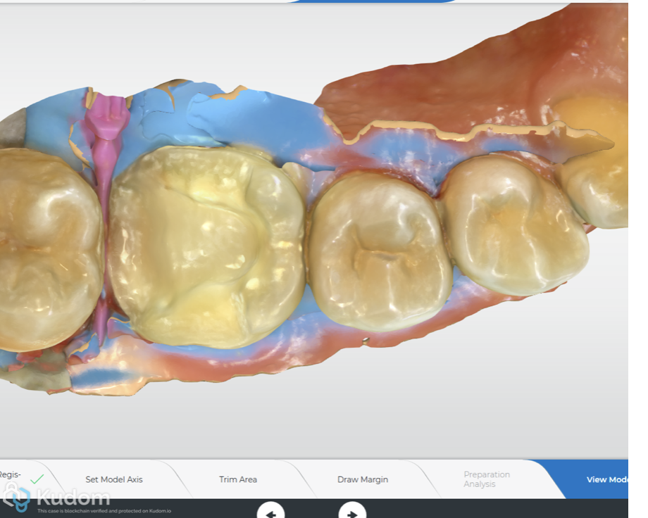
Fig. 8
Intraoral scanning under rubber dam
An intraoral scan was performed under rubber dam isolation following completion of the coronal build-up and preparation.
Scanning under isolation allowed the restorative workflow to be completed while maintaining the working field under controlled conditions.
The digital impression was used for the fabrication of the definitive overlay.
This approach integrated endodontic isolation and restorative digital workflow into a single treatment sequence.
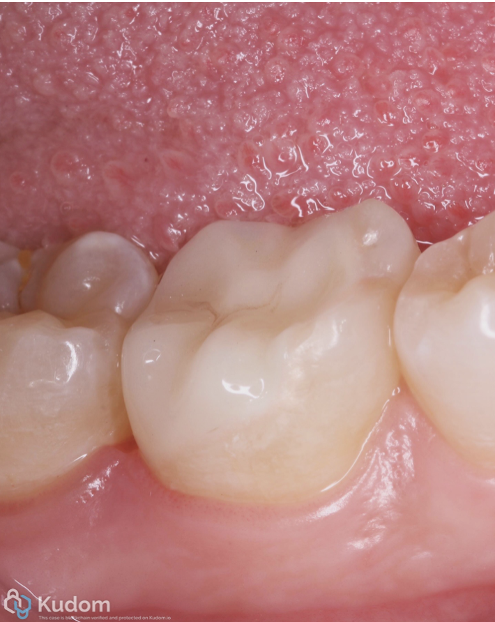
Fig. 9
Final outcome
The final radiographic and clinical assessment demonstrated the completion of endodontic treatment and definitive restoration of the tooth.
The five root canals, including the middle distal canal, were obturated, and the tooth was restored using a definitive overlay.
The final restoration provided the intended coronal anatomy and function.
Fig. 10
About the author:
Dr Mazen Ahmed
Conclusions
The management of complex endodontic anatomy begins with recognizing that the expected anatomy is not always the actual anatomy. In this case, the identification of a middle distal canal allowed complete treatment of a five-canal mandibular molar through a systematic approach combining magnification, controlled instrumentation, active irrigation, and warm obturation.
Equally important, the restorative phase was considered from the outset as part of the overall treatment strategy. Margin elevation, bio-base reconstruction, digital scanning under rubber dam isolation, and definitive overlay restoration provided a structured transition from endodontic treatment to definitive coronal rehabilitation.
This case reinforces the importance of treating the tooth as a whole: finding the anatomy, controlling the biology, and preserving the remaining structure.
Bibliography
- Ahmed HMA, Neelakantan P, Arias A, et al. Morphology and prevalence of middle canals in the mandibular molars: a systematic review. Clinical Oral Investigations. 2018;22:1679–1691.
- Căpută PE, Retsas A, Kuijk L, Chávez de Paz LE, Boutsioukis C. Ultrasonic irrigant activation during root canal treatment: a systematic review. Journal of Endodontics. 2019;45(1):31–44.e13. doi:10.1016/j.joen.2018.09.010.
- Taylor A, Burns L. Deep margin elevation in restorative dentistry: a scoping review. Journal of Dentistry. 2024;146:105066. doi:10.1016/j.jdent.2024.105066.
- Lane J, Sadeghzadeh-Araghi A, Jackson G, Bonsor S. Survival and success rates of endodontically treated teeth restored with full veneer crowns or full cuspal coverage onlays: a systematic review. European Journal of Prosthodontics and Restorative Dentistry. 2024;32(1):45–55. doi:10.1922/EJPRD_2547Lane11.
- Al-Meshal SA, et al. Remaining tooth structure and prognosis of restored endodontically treated teeth: a systematic review. International Endodontic Journal. 2025.