Nonsurgical Root Canal Treatment with an Apical Plug in a Patient on Active Oral Bisphosphonate Therapy with Root Resorption
14/09/2026
Warning: Undefined variable $post in /home/styleendo/htdocs/styleitaliano-endodontics.org/wp-content/plugins/oxygen/component-framework/components/classes/code-block.class.php(133) : eval()'d code on line 2
Warning: Attempt to read property "ID" on null in /home/styleendo/htdocs/styleitaliano-endodontics.org/wp-content/plugins/oxygen/component-framework/components/classes/code-block.class.php(133) : eval()'d code on line 2
Deciding whether to extract a tooth in a patient on bisphosphonate therapy is not a matter of "extraction is always contraindicated." It requires assessing the patient's actual risk of medication-related osteonecrosis of the jaw (MRONJ), which depends on a few key factors. Route of administration is the first: oral bisphosphonates carry a much lower risk than intravenous or subcutaneous antiresorptives (e.g., zoledronate, denosumab). Duration matters too: for oral bisphosphonates, risk stays low for the first three to four years and increases afterward; for high-dose IV therapy in oncologic patients, risk tends to rise after twelve to twenty-four months. The indication also affects risk, since oncologic protocols use much higher cumulative doses than osteoporosis treatment. Corticosteroid or antiangiogenic therapy, diabetes, and poor periodontal/oral hygiene independently raise risk further.
Whenever a tooth can realistically be saved, extraction should be avoided regardless of risk category, since it is the most consistently reported MRONJ trigger. Root canal treatment, including surgery-avoiding techniques such as an apical plug, should be first choice whenever enough tooth structure and periodontal support remain for a functional restoration, an approach supported by recent clinical evidence in patients under antiresorptive therapy.
In the case presented, a 68-year-old woman had been on active oral bisphosphonate therapy for approximately 5 years, placing her above the lowest risk bracket, though still well below IV/oncologic regimens. She presented with pain, percussion tenderness, and mobility of a mandibular second molar with distal root resorption from an adjacent impacted third molar. Removing the third molar would have addressed the cause mechanically, but extraction itself carries MRONJ risk; both teeth were therefore managed conservatively: root canal treatment with an apical plug on the second molar, and clinical/radiographic monitoring of the impacted third molar rather than extraction, which would ordinarily be the indicated management for this type of external pressure resorption.
Fig. 1
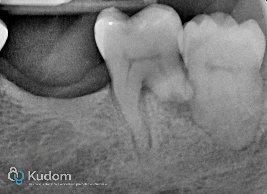
The preoperative periapical radiograph showed the mandibular second molar rotated mesially into the space left by the missing first molar, positioned close to the mesial surface of the impacted third molar. The distal root exhibited a radiolucent defect consistent with external root resorption, caused by chronic mechanical pressure from the adjacent impacted third molar.
External root resorption adjacent to an impacted third molar is thought to follow a two-phase mechanism: an initial mechanical or follicular injury to the protective cementum and periodontal ligament at the point of contact, followed by odontoclastic activity driven by local inflammatory mediators, a process considered biologically comparable to the physiological resorption of primary tooth roots by an erupting permanent successor.
The patient's history of active oral bisphosphonate therapy, ongoing for approximately five years, required specific considerations before treatment. Informed consent addressed the rationale for a conservative, non-surgical approach given her medication history, and her prescribing physician's records confirmed the therapy was uncomplicated and still active. Additional pre-treatment considerations included confirming the tooth was restorable and suitable for conservative management rather than extraction, planning strict asepsis throughout instrumentation, and anticipating that the distal resorption would likely preclude conventional apical obturation, favouring an apical plug technique.
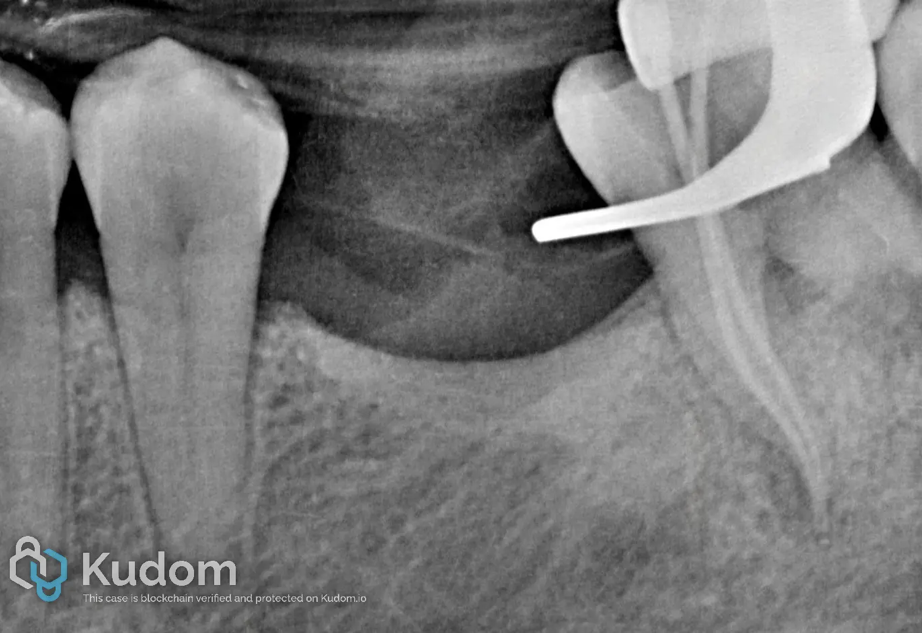
Fig. 2
Following local anesthesia, access cavity preparation and rubber dam isolation, the mesial canals, presenting a regular and fully formed root morphology, were instrumented mechanically with SlimShaper rotary files (Zarc, Gijón, Spain), using the E-Connect S+ endodontic motor (Eighteeth, Changzhou, China), up to a 25 apical diameter. Irrigation was performed with 5.25% sodium hypochlorite (NaOCl) delivered via a side-vented irrigation needle (IrriFlex, Produits Dentaires SA, Vevey, Switzerland). Gutta-percha cone fit was verified radiographically, and confirmed adequate apical control and a reproducible working length.
A final irrigation sequence with 17% EDTA followed by 5.25% NaOCl preceded drying with matched sterile paper points (Zarc). Irrigant was activated ultrasonically with the Actor I Pro Ultrasonic Endo Activation Device (Bomedent, Shanghai, China).
The distal canal presented a fundamentally different scenario: external root resorption at the apical third had created an irregular, wider-than-normal apical foramen, making rotary shaping unsuitable and unable to establish a reliable apical stop. This canal was managed exclusively with manual stainless-steel K-files (Dentsply Sirona, Charlotte, NC, USA), instrumented progressively up to a size 80 to clean the walls without further weakening the resorption-affected dentin, given the already wide apical diameter created by the resorptive defect. The same irrigation and ultrasonic activation protocol was followed, and the canal was dried before proceeding to the apical plug technique, described in the following section.
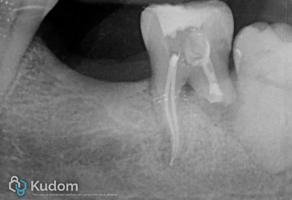
Fig. 3
The two mesial canals were obturated using a single-cone technique, in accordance with the shaping achieved with SlimShaper rotary instrumentation. NeoSealer Flo bioceramic sealer (Avalon Biomed, Houston, TX, USA) was applied to the canal walls, and matched gutta-percha cones were seated to full working length, providing a predictable, bioactive seal along the length of both canals.
The distal canal required a fundamentally different obturation strategy due to the apical resorptive defect, which precluded conventional cone-based filling. A fibrin matrix was first introduced and condensed to working length using a hand plugger, acting as an internal scaffold to contain the subsequently placed bioceramic material and prevent its extrusion beyond the compromised apical anatomy. Against this matrix, an apical plug of NeoPutty (Avalon Biomed, Houston, TX, USA) was condensed with a hand plugger, establishing an artificial apical stop in the absence of a natural constriction. Once the plug was placed and radiographically verified, the remainder of the canal was backfilled entirely with NeoPutty, providing a continuous bioceramic fill from the apical barrier to the canal orifice.
Following obturation of both canals, the access cavity was temporarily restored with Cavit (3M, Saint Paul, MN, USA), allowing adequate coronal sealing while the bioceramic materials completed their setting reaction prior to definitive restoration.
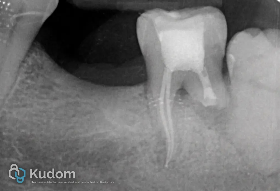
Fig. 4
After 7 days, following completion of the endodontic phase and temporary sealing, the tooth was restored with a direct composite restoration to allow long-term monitoring of both the endodontically treated distal root and the immediately adjacent impacted third molar, prior to any decision regarding definitive coronal coverage. G-ænial Posterior composite (GC Corporation, Tokyo, Japan), shade A3, was selected for its handling characteristics and adequate radiopacity, allowing future radiographic differentiation between restorative material and the underlying bioceramic obturation.
The restoration was built up incrementally following standard adhesive protocol, restoring the access cavity and re-establishing anatomical form. Particular attention was given to occlusal contacts: the restoration was deliberately taken out of full occlusal contact, relieving the tooth of functional loading in centric and eccentric movements. This choice reflects the tooth's compromised periodontal support secondary to the distal root resorption and its history of mobility at presentation, and was intended to reduce mechanical stress on the healing apical region during the monitoring period, rather than to provide a definitive functional restoration at this stage.
The overall treatment plan therefore prioritized a conservative, monitoring-oriented approach: preserving the tooth and the adjacent impacted third molar without immediate further intervention, while periodically reassessing both the resorptive defect and the periapical status radiographically and clinically over time.
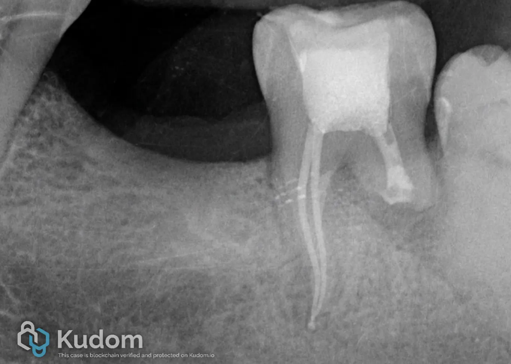
Fig. 5
The patient was reviewed at 6 months following completion of treatment. Clinically, she reported a marked reduction in tenderness to percussion and resolution of the discomfort present at initial presentation, with no spontaneous pain, swelling, or sinus tract formation. Tooth mobility, present at baseline due to the compromised periodontal support from the distal resorptive defect, was also noted to have improved, consistent with resolution of the local inflammatory process.
The postoperative periapical radiograph confirmed adequate three-dimensional obturation of both canals, with the mesial canals showing a well-condensed single-cone fill and the distal canal showing satisfactory adaptation of the apical plug and bioceramic backfill against the resorbed root surface, without radiographic evidence of extrusion beyond the apex. Furthermore, the radiolucent area previously noted at the furcation level showed a visible reduction in size compared with the preoperative image, suggestive of early bone healing in this region.
Given the patient's medication history and the conservative, monitoring-oriented treatment strategy adopted, follow-up is planned to continue at regular intervals, combining clinical assessment with periodic radiographic control, to evaluate the longer-term stability of the resorptive defect, the continued reduction of the furcation lesion, and the condition of the adjacent impacted third molar, which remains untreated and under observation as part of the same conservative management rationale.

Fig. 6
About the author: Dr Giulia Malvicini
Conclusions
Few practical takeaways emerge from this case. First, oral bisphosphonate therapy is not a fixed risk category: risk increases with duration, rising above the lowest-risk bracket after about four years, even without changing the drug or route. A patient on oral bisphosphonates for five years, as in this case, carries a different risk than one in their first year, a distinction that is easy to miss if only the drug is recorded in the patient's history, without noting how long they have been taking it. In practice, this means duration of therapy should be re-verified at every relevant visit, not assumed to be static once documented.
Second, an open or resorbed apex is not, by itself, a reason to extract or operate. A fibrin matrix combined with a bioceramic apical plug can create an artificial apical stop, allowing complete canal obturation without apical surgery. This is particularly relevant in cases of external pressure resorption from adjacent impacted teeth, where the resorptive defect can otherwise be mistaken for an untreatable apex. For this specific type of resorption, standard management as outlined in the current classification calls for extracting the impacted tooth generating the pressure, but extraction itself carries MRONJ risk. Both teeth were therefore managed conservatively: root canal treatment with an apical plug on the second molar, and clinical and radiographic monitoring of the impacted third molar rather than extraction.
Importantly, should the patient's bisphosphonate therapy be discontinued in the future, her MRONJ risk profile would change accordingly, and extraction of the impacted third molar could then be reconsidered as the definitive treatment for the underlying cause of the resorption, in line with the standard management this case has, for now, deliberately set aside. Continued monitoring over time therefore serves a dual purpose: tracking the stability of the resorptive defect and the periapical status, while also reassessing, at each review, whether the systemic circumstances that justified this conservative deviation still apply.
Bibliography
- Ruggiero SL, Dodson TB, Aghaloo T, Carlson ER, Ward BB, Kademani D. American Association of Oral and Maxillofacial Surgeons' Position Paper on Medication-Related Osteonecrosis of the Jaws—2022 Update. J Oral Maxillofac Surg. 2022;80(5):920-943.
- Bedogni A, Mauceri R, Fusco V, et al. Italian position paper (SIPMO-SICMF) on medication-related osteonecrosis of the jaw (MRONJ). Oral Dis. 2024. doi:10.1111/odi.14887.
- American Association of Endodontists. Guidelines for Medication-Related Osteonecrosis of the Jaw: An Update. AAE Clinical Practice Guidelines. Available at: aae.org.
- Zavalloni G, Spinelli A, Coppini M, Mauceri R, Campisi G, Lenzi J, Gandolfi MG, Prati C, Zamparini F. Endodontic treatment and management of patients under antiresorptive treatment: a scoping review on MRONJ risk. Clin Oral Investig. 2025.
- Gürses G, Akçakaya A, Aktı A, Aydin O. The effect of impacted third molars on second molar external root resorption, a cross-sectional cone beam computed tomography study. Med Oral Patol Oral Cir Bucal. 2023;28(6):e504-511.
- Abbott PV, Lin S. Tooth resorption—Part 2: A clinical classification. Dent Traumatol. 2022;38:267-285.
- Hamad SA. External root resorption of second molars due to impacted third molars. J Clin Exp Dent. 2024;16(12):e1489-e1494.
- Bhandare RS, Mattigatti S. Management of an Open Apex Using a Platelet-Rich Fibrin Membrane as an Internal Matrix and Mineral Trioxide Aggregate as an Apical Barrier: A Case Report. Cureus. 2024;16(12):e75316.