

Surgical Endodontics on Teeth 2.4 and 2.5 with Symptomatic Apical Periodontitis Following Failed Previous Surgery
17/04/2026
Warning: Undefined variable $post in /home/styleendo/htdocs/styleitaliano-endodontics.org/wp-content/plugins/oxygen/component-framework/components/classes/code-block.class.php(133) : eval()'d code on line 2
Warning: Attempt to read property "ID" on null in /home/styleendo/htdocs/styleitaliano-endodontics.org/wp-content/plugins/oxygen/component-framework/components/classes/code-block.class.php(133) : eval()'d code on line 2
Surgical endodontics represents a predictable treatment option in cases of persistent apical periodontitis when orthograde retreatment is not feasible or has previously failed. The introduction of microsurgical techniques, including magnification, ultrasonic retro-preparation, and the use of bioceramic materials, has significantly improved clinical outcomes and long-term success rates.
However, failure of initial surgical endodontic procedures may occur due to inadequate apical sealing, incomplete removal of pathological tissue, or persistent intraradicular infection. In such cases, secondary surgical intervention can be considered a valid approach to achieve resolution of the pathology and proper healing.
The present case describes the management of symptomatic apical periodontitis affecting teeth 2.4 and 2.5 following failed previous surgical endodontic treatment, highlighting the role of modern microsurgical techniques in achieving a predictable outcome.
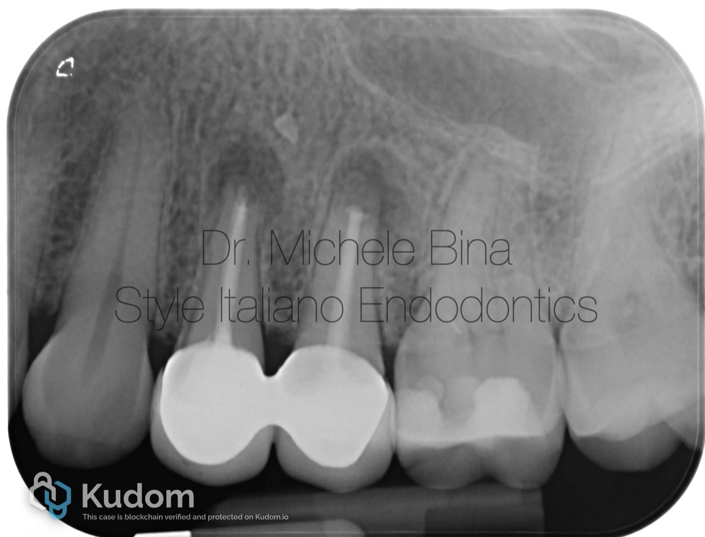
Fig. 1
Patient M.V., in good systemic health, presented with symptomatic apical periodontitis affecting teeth 2.4 and 2.5 after failed previous surgical endodontic treatment. Tooth 2.4 showed a sinus tract, indicating persistent infection.
Secondary surgical endodontic treatment was performed. Following flap elevation and lesion removal, apicectomy and ultrasonic retro-preparation were carried out. Retrograde filling was completed using a premixed bioceramic material delivered with a MAP System. The site was irrigated and sutured with e-PTFE to achieve primary closure.
This case demonstrates the effectiveness of surgical retreatment in managing persistent periapical pathology.
Full case video
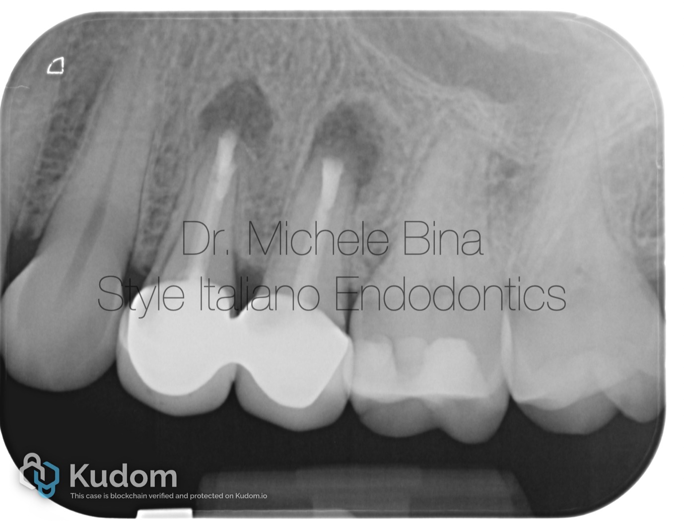
Fig. 2
Post operative rx
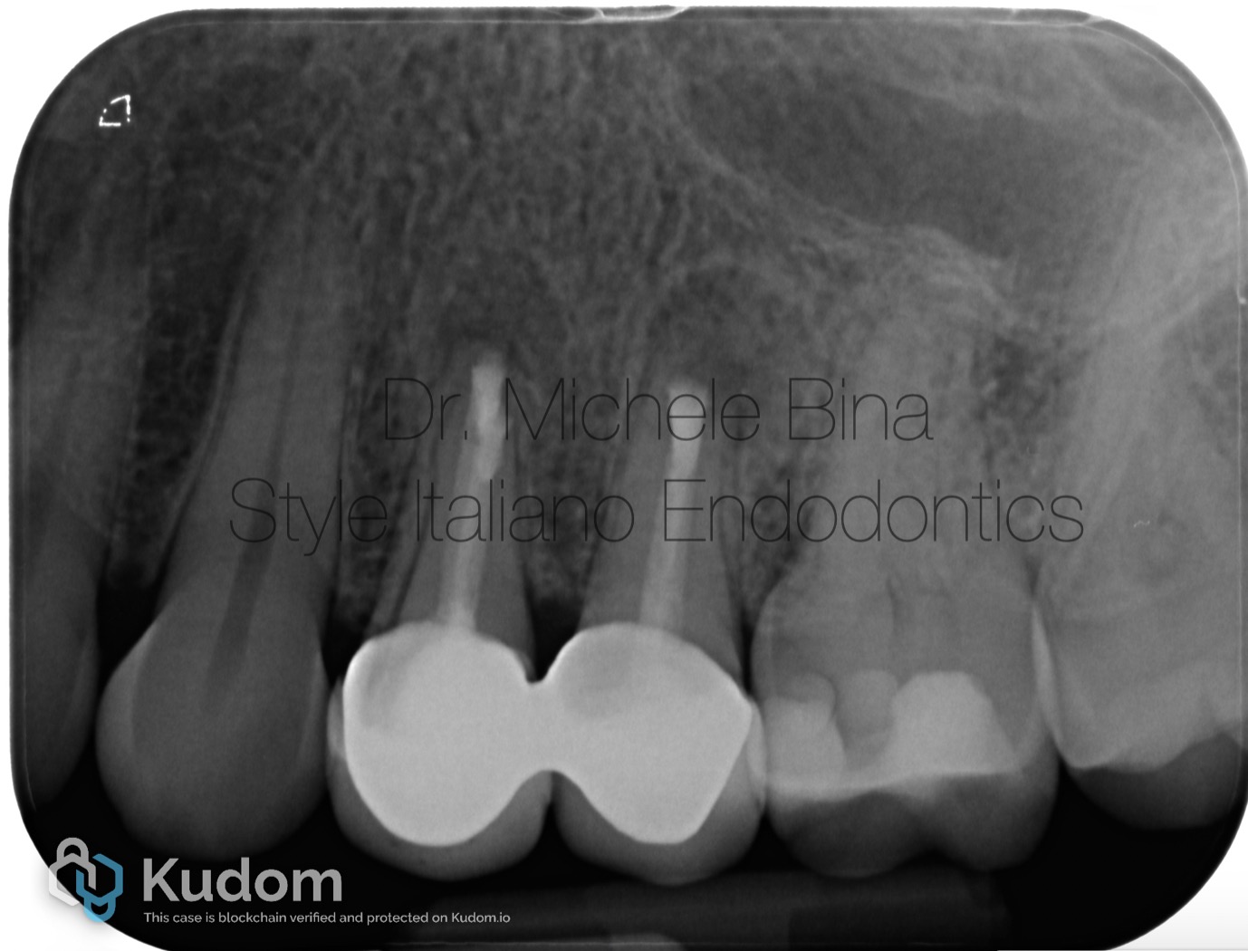
Fig. 3
Rx with follow up at 6 months
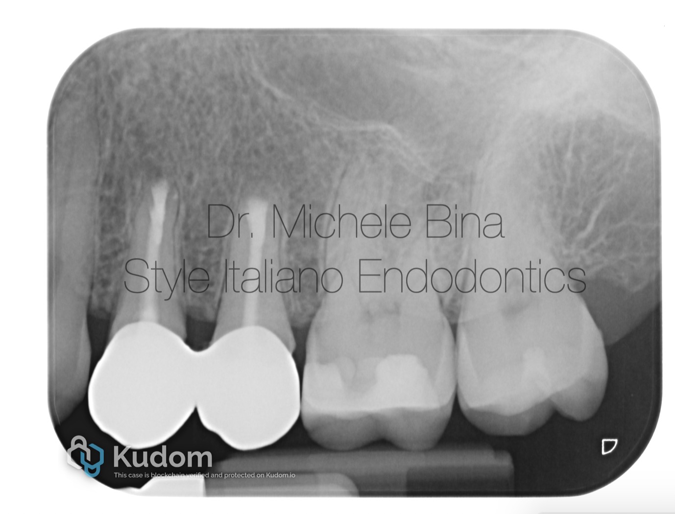
Fig. 4
Rx with follow up at 20 months with complete healing
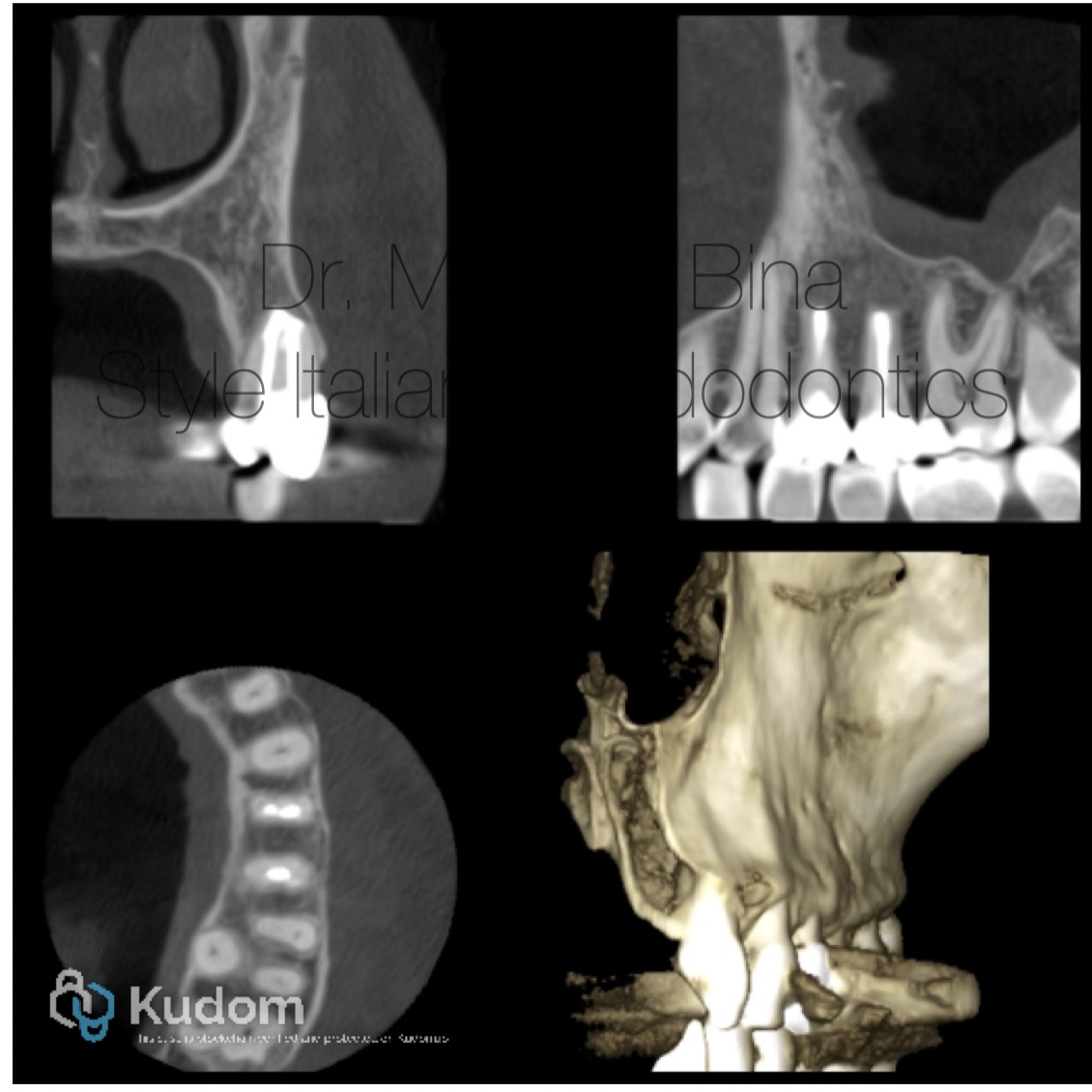
Fig. 5
Post operative CBCT with complete healing

Fig. 6
About the Author:
Michele Bina
Graduated with honors from the University of Cagliari in 2013, Dr. Michele Bina completed a Master’s degree in Clinical Endodontics at the same university in 2015. Since 2016, he has served as a tutor for the aforementioned program. In 2024, he earned a Master’s degree in Oral Surgery from the University of Padua.
Dr. Bina is a member of the Italian Academy of Endodontics, a fellow of Style Italiano, and an opinion leader for Simit. He practices privately in Cagliari, specializing in endodontics, restorative dentistry, prosthodontics, and oral surgery.
Conclusions
Secondary surgical endodontic treatment represents a reliable option in cases of persistent apical periodontitis following failed previous surgery. The use of modern microsurgical techniques, including ultrasonic retro-preparation and bioceramic materials, allows for effective apical sealing and promotes predictable healing.
In the present case, accurate diagnosis, proper case selection, and adherence to contemporary surgical protocols resulted in successful management of the periapical pathology and resolution of the infection.
These findings support the role of surgical retreatment as a valid and predictable approach in complex endodontic cases.
Bibliography
Shinbori N, Grama AM, Patel Y, Woodmansey KF.
Clinical outcome of endodontic microsurgery using bioceramic root repair material.
J Endod. 2015;41(10):1715–1719.
Pinto D, Marques A, Pereira J, Palma PJ, Santos JM.
Long-term prognosis of endodontic microsurgery: a systematic review and meta-analysis.
Int Endod J. 2020;53(12):1601–1617.
Setzer FC, Kohli MR, Shah SB, Karabucak B, Kim S.
Outcome of endodontic surgery: a meta-analysis of the literature—Part 2: Comparison of endodontic microsurgical techniques with and without the use of higher magnification.
J Endod. 2012;38(1):1–10.
