
The importance of magnification to manage ledge/perforation
20/04/2025
Fellow
Warning: Undefined variable $post in /home/styleendo/htdocs/styleitaliano-endodontics.org/wp-content/plugins/oxygen/component-framework/components/classes/code-block.class.php(133) : eval()'d code on line 2
Warning: Attempt to read property "ID" on null in /home/styleendo/htdocs/styleitaliano-endodontics.org/wp-content/plugins/oxygen/component-framework/components/classes/code-block.class.php(133) : eval()'d code on line 2
Retreatment are always a challenge.
One of the most difficult things is to manage ledge or/and perforation into a canal.
When we have to work inside the canal the use of microscope it’s crucial to see and have the control of all procedures.
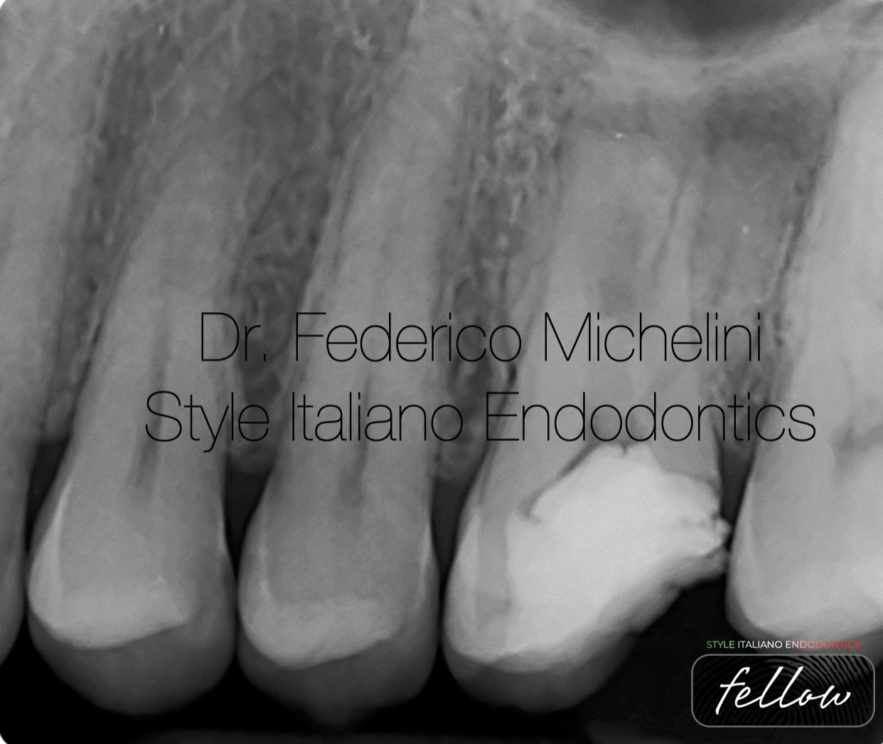
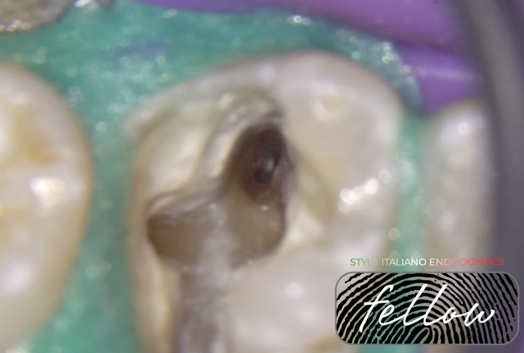
Fig. 1
Pre-operative X-ray.
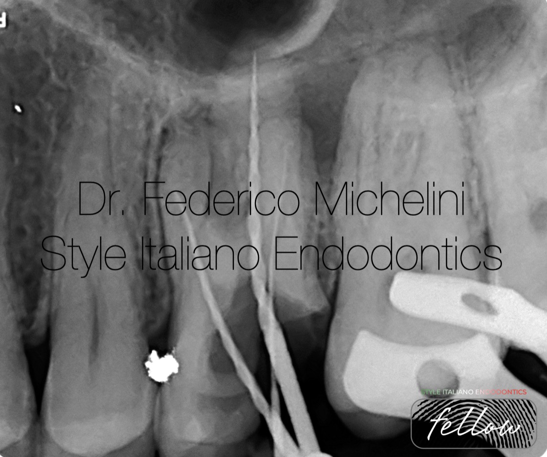
Fig. 2
Second Pre-operative X-ray.
The first tip is to do a second X-ray when there is a problem to solve it. In this case there is a ledge/perforation into a mesio-vestibular canal and disto-vestibular canal was blocked in the apical part.
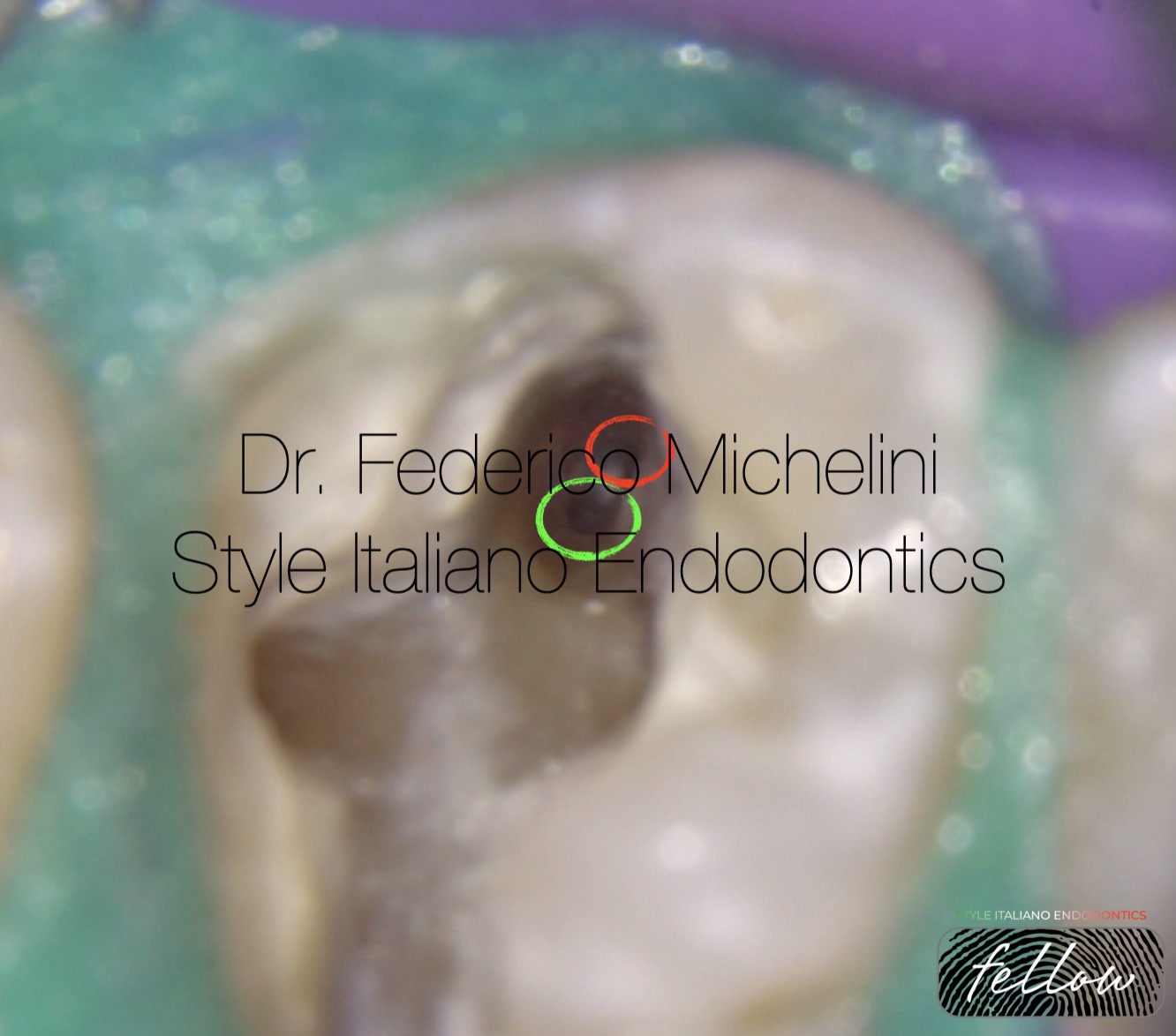
Fig. 3
Thanks magnification and ultrasonic tips I identified the ledge/peroration and after that i search, found and shape the original canal.
Green circle = Original canal
Red circle = Ledge/perforation
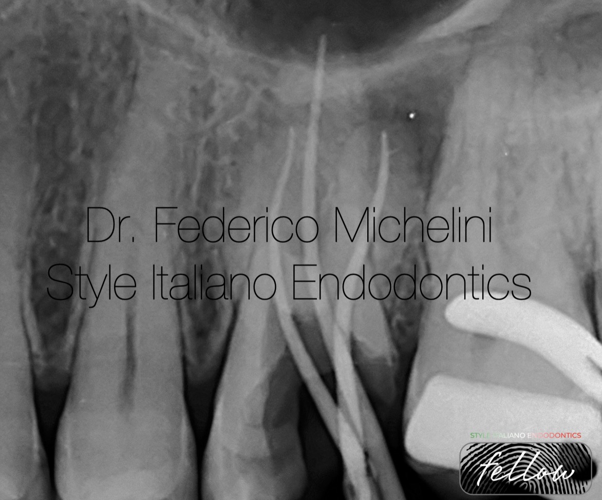
Fig. 4
X-ray with working length.
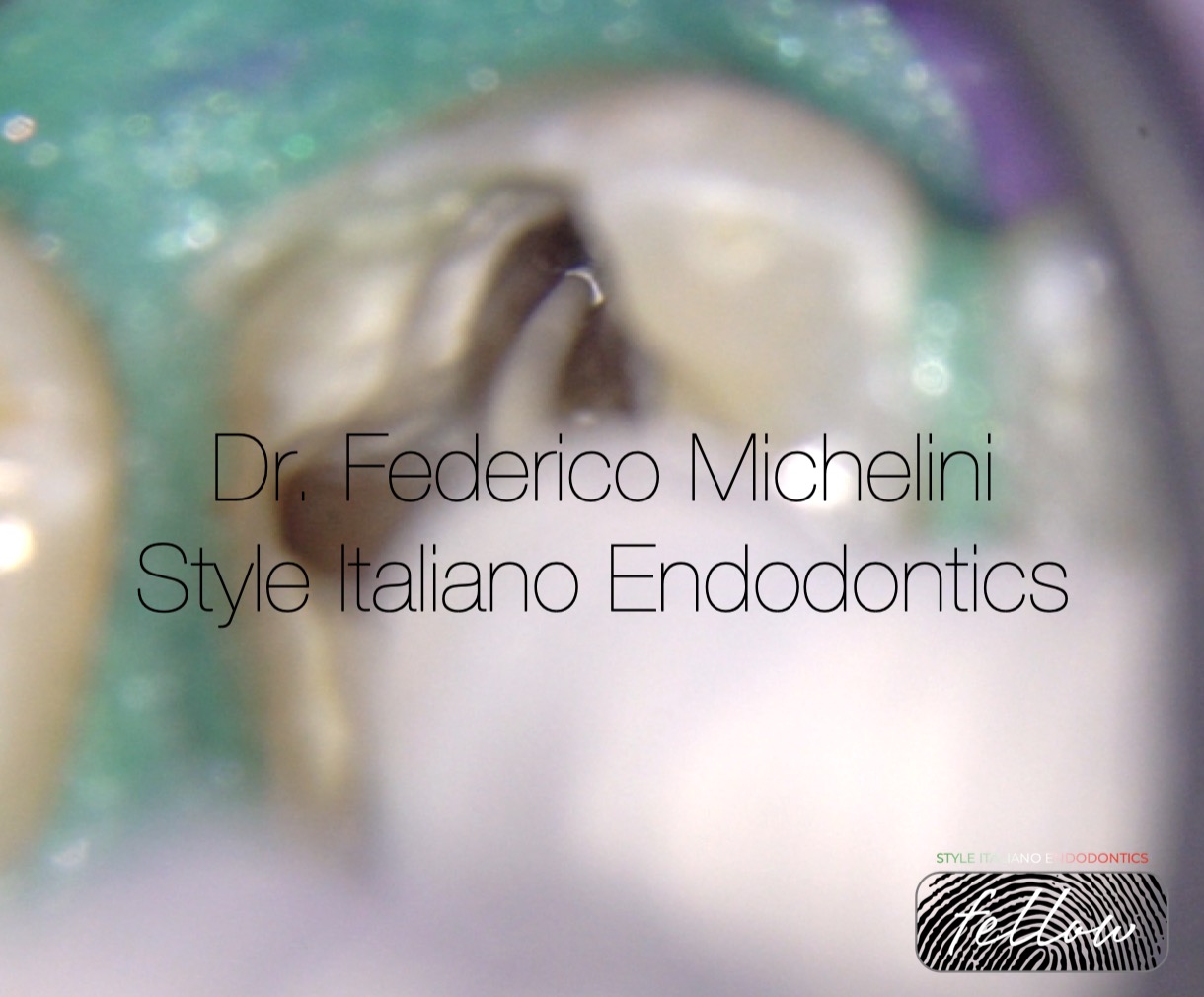
Fig. 5
Bioceramic sealer into mesio-vestibular canal.
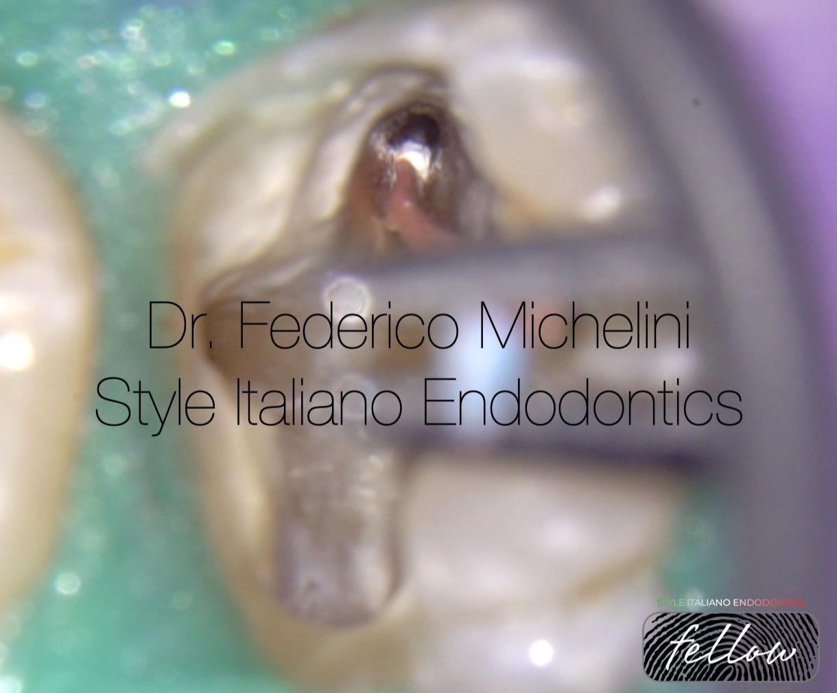
Fig. 6
Obturation of mesio-vestibular canal.
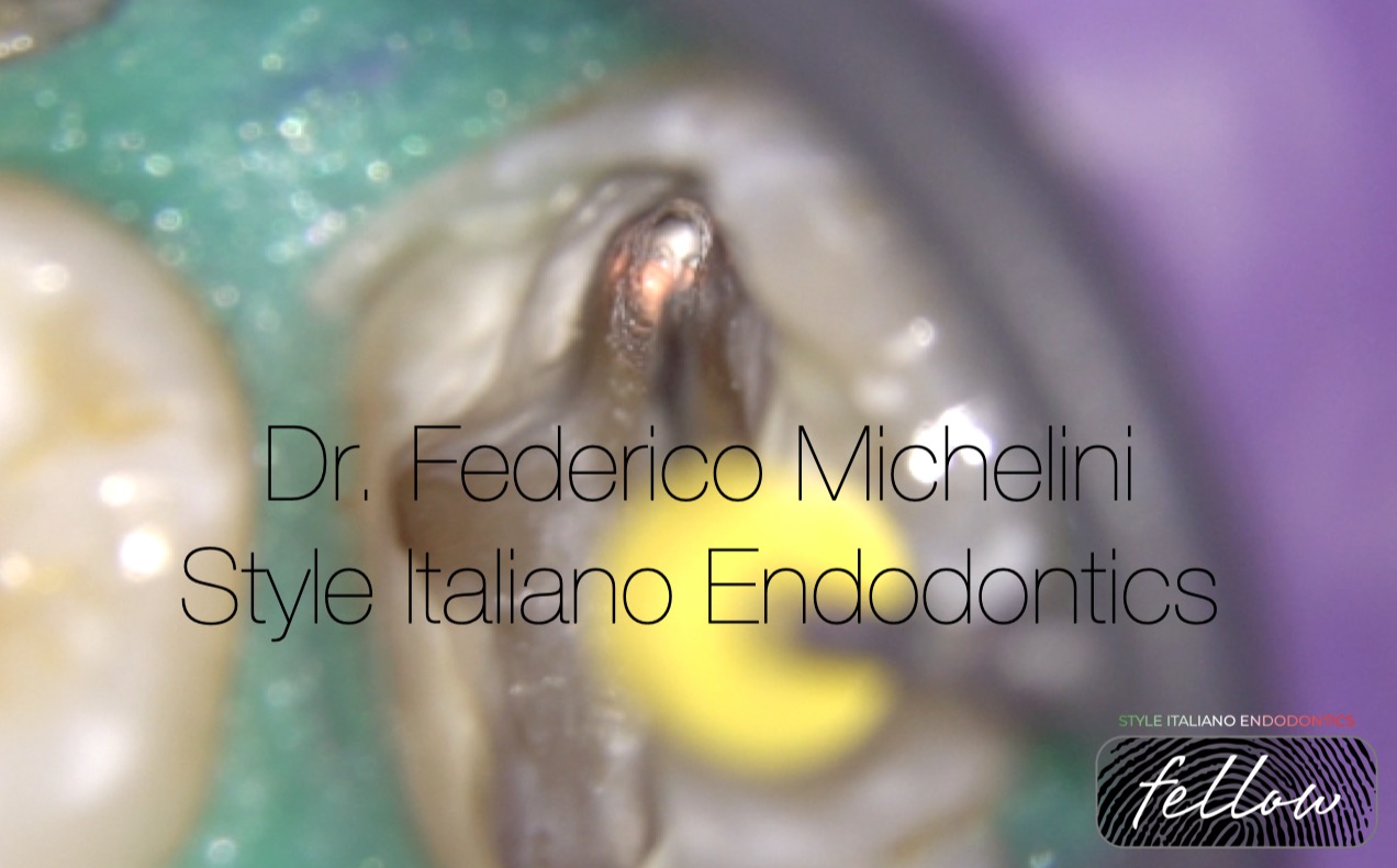
Fig. 7
Perforation repair with bioceramic material putty.
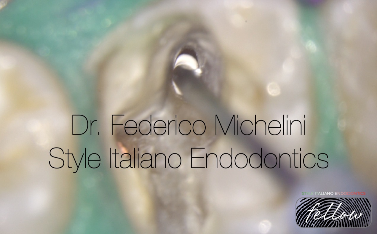
Fig. 8
Obturation of mesio-vestibular canal. Original canal and perforation. After that the canal was fill with the injection of guttaperca.
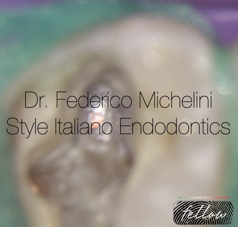
Fig. 9
X-ray with the obturation of the canals.
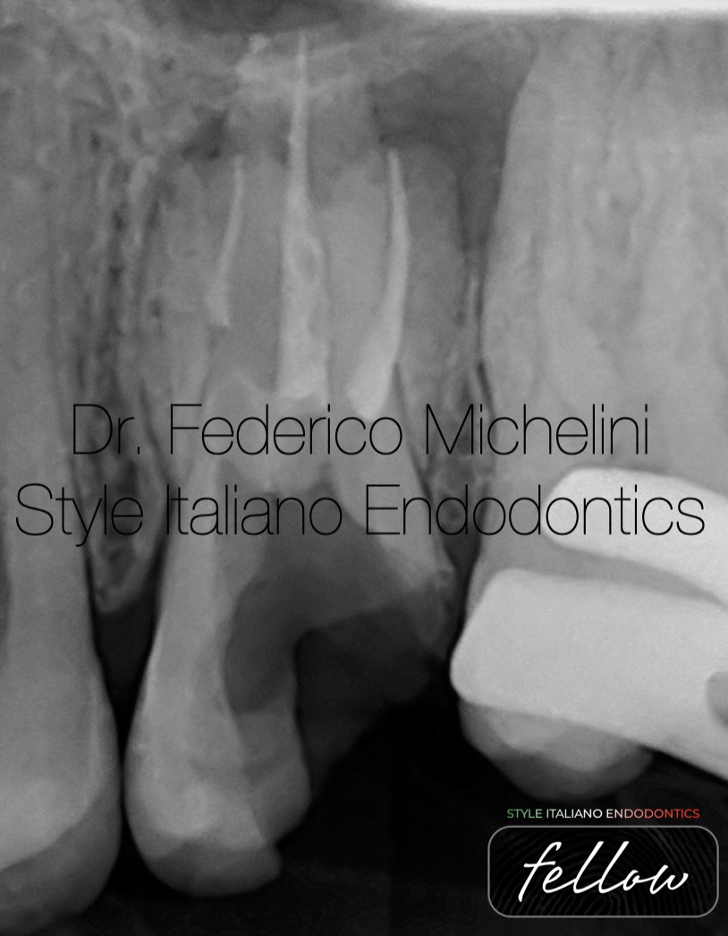
Fig. 10
Post operative X-ray.
The video show how to manage the obturation of the original canal and the ledge/perforation.

Fig. 11
About the Author
2015: Graduated in dentistry from the University of Valencia.
2017: Master in Endodontics from the University of Bologna.
Speaker in national and international congresses regarding endodontics.
2017-2024 Tutor of the Master of Endodontics and of the Degree Course of Dentistry and Dental Prosthesis for the University of Bologna.
He won the Closed Meeting of Italian Society of Endodontics in 2019.
He attended the advanced course of retreatments and endodontics surgery of Italian Academy of Endodontics.
He attended the course of microscopy by Dott. Fabio Gorni.
Active Member of the Italian Society of Endodontics (SIE)
Member Fellow of Style italiano Endodontics.
Opinion Leader and Speaker for Morita and Coltene.
He works in Parma and Piacenza dedicating to microscopic endodontics
Conclusions
The possibility thanks magnification to see into the canal it’s a huge plus to be able to resolve this type of problem.
It’s very important also know various technique and material to choose them in a right way.
Bibliography
- Estrela C, Decurcio DA, Rossi-Fedele G, Silva JA, Guedes OA, Borges ÁH. Root perforations: a review of diagnosis, prognosis and materials. Braz Oral Res. 2018 Oct 18;32.
- Kabtoleh A, Aljabban O, Alsayed Tolibah Y. Fracture Resistance of Molars With Simulated Strip Perforation Repaired With Different Calcium Silicate-Based Cements. Cureus. 2023 Jan 31;15.
- Zamparini F, Prati C, Taddei P, Spinelli A, Di Foggia M, Gandolfi MG. Chemical-Physical Properties and Bioactivity of New Premixed Calcium Silicate-Bioceramic Root Canal Sealers.
- Del Fabbro M, Taschieri S, Lodi G, Banfi G, Weinstein RL. Magnification devices for endodontic therapy. Cochrane Database Syst Rev. 2015 Dec 9;2015(12).
