Unusual anatomy in upper second molar
20/09/2020
Zaher Al-taqi
Warning: Undefined variable $post in /var/www/vhosts/styleitaliano-endodontics.org/endodontics.styleitaliano.org/wp-content/plugins/oxygen/component-framework/components/classes/code-block.class.php(133) : eval()'d code on line 2
Warning: Attempt to read property "ID" on null in /var/www/vhosts/styleitaliano-endodontics.org/endodontics.styleitaliano.org/wp-content/plugins/oxygen/component-framework/components/classes/code-block.class.php(133) : eval()'d code on line 2
The hard tissue repository of the human dental pulp takes on numerous configurations and shapes. A thorough knowledge of tooth morphology, careful interpretation of angled radiographs, proper access preparation and a detailed exploration of the interior of the tooth are essential prerequisites for a successful treatment outcome. CBCT, Magnification and illumination are aids that must be utilized to achieve this goal. in this case report article we will demonstrate the importance of such tools to have successful RCT .
Fig. 1
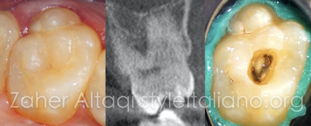
25 years old patient presented to the clinic with sever pain in the upper left area . he described it with a severe , pulsating and pressure like pain that prevents him from sleeping for 2 nights despite all the analgesics taken . Upon clinical examination we found sever tenderness on percussion related to tooth 27 . Vitality test reveals no response and negative result. So we are facing a necrotic case with symptomatic apical periodontitis .
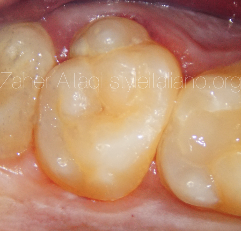
Fig. 2
Clinically we've noticed an extra cusp on the buccal aspect of the tooth which may indicate an extra root that is not visible or clear on the 2d periapical radiograph , So we did a CBCT to know more about the internal anatomy .
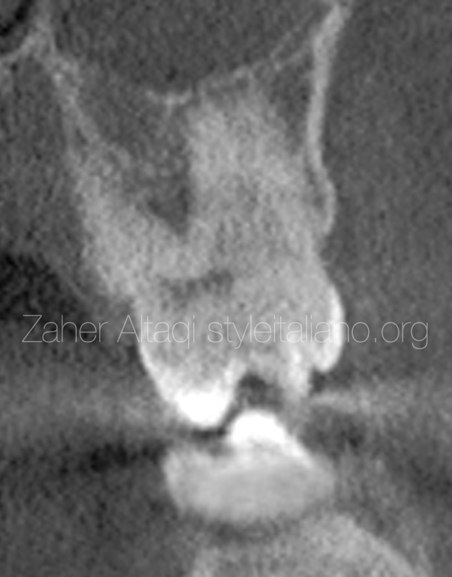
Fig. 3
CBCT coronal view shows extra root located on the buccal side which is related to the extra cusp .
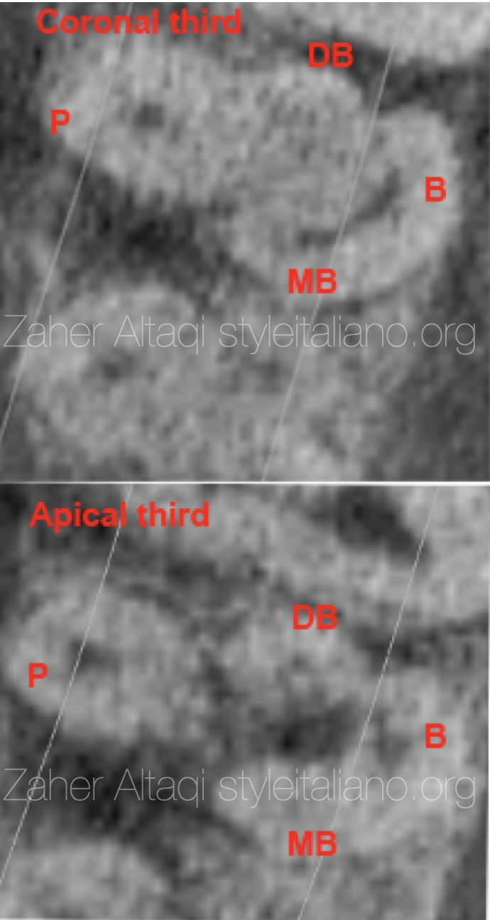
Fig. 4
CBCT axial view shows that the extra buccal root canal is connected to the MB one with an isthmus in the coronal third, then continues as a separate root canal .
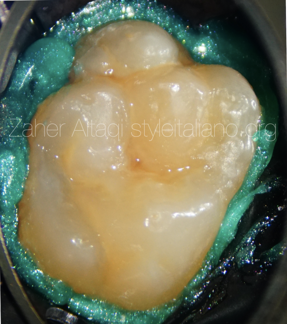
Fig. 5
Rubber dam isolation is mandatory in endodontics .
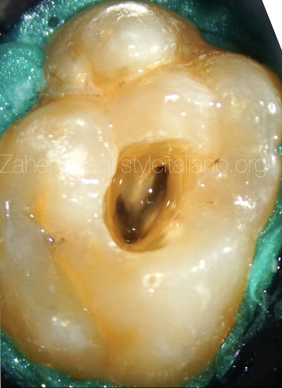
Fig. 6
Access cavity started with very well knowledge about the internal anatomy and how the access should be done in order to shape and clean the whole root canals system
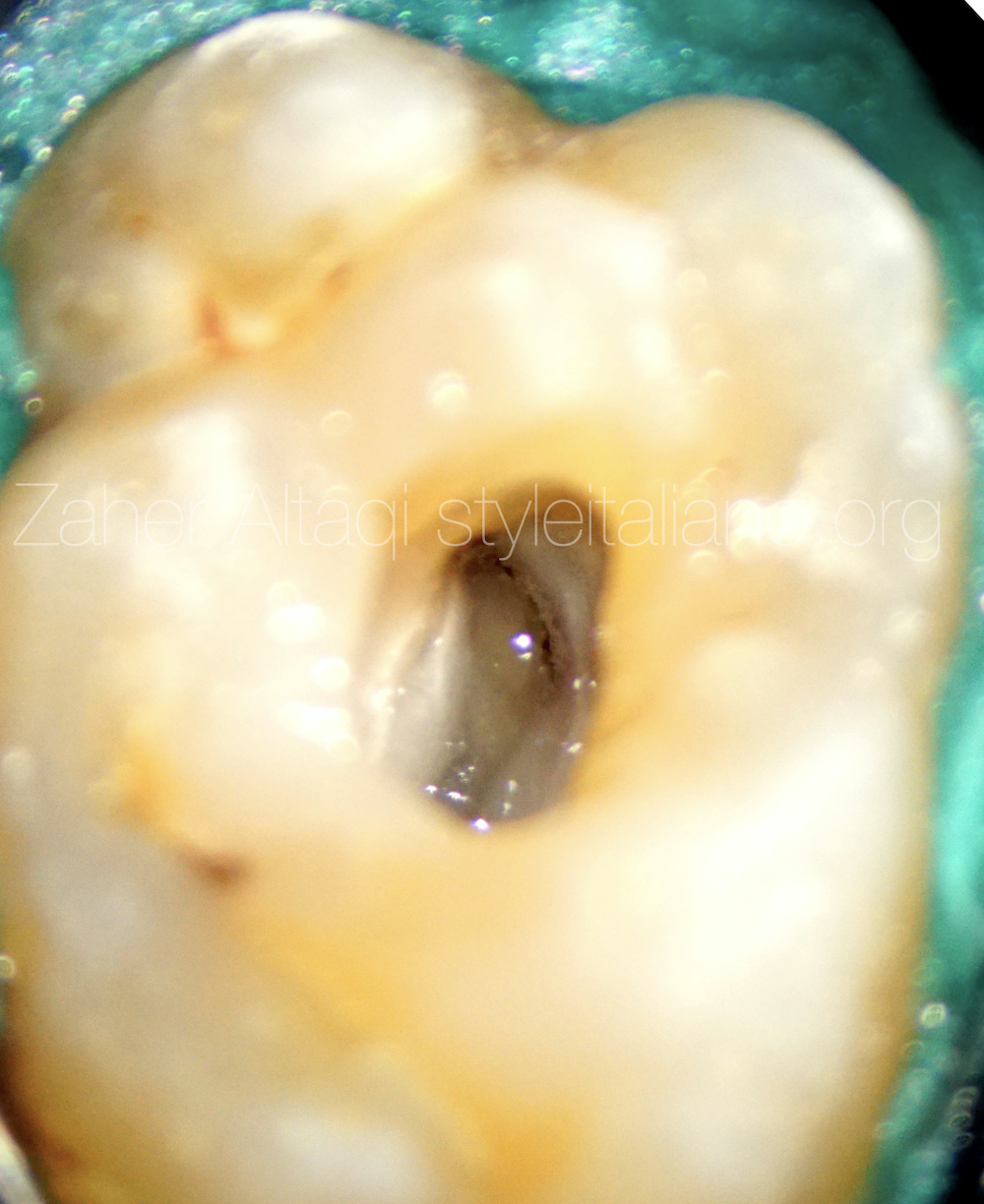
Fig. 7
MB and B roots with isthmus connection .
Routine cleaning , shaping and warm obturation was done with resin core and composite for coronal restoration .
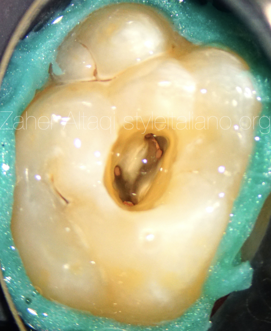
Fig. 8
Warm vertical obturation obturation was done .
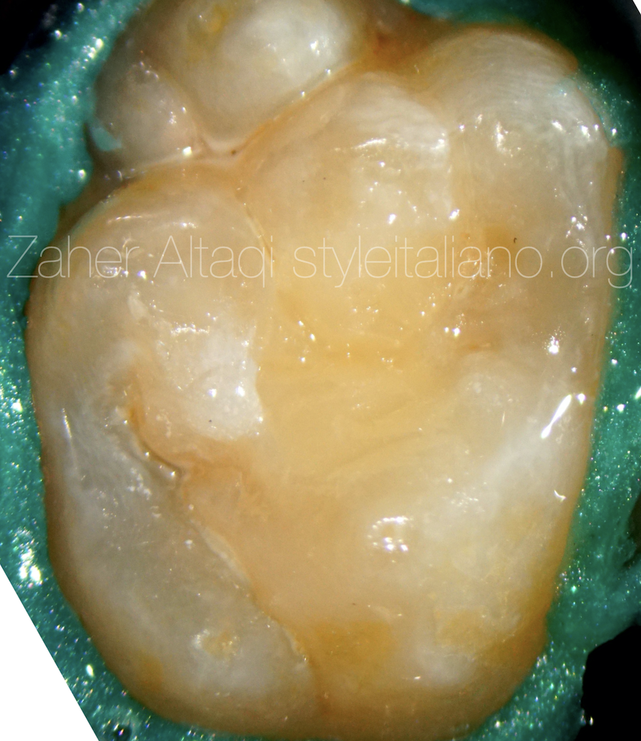
Fig. 9
Coronal seal with composite restoration.
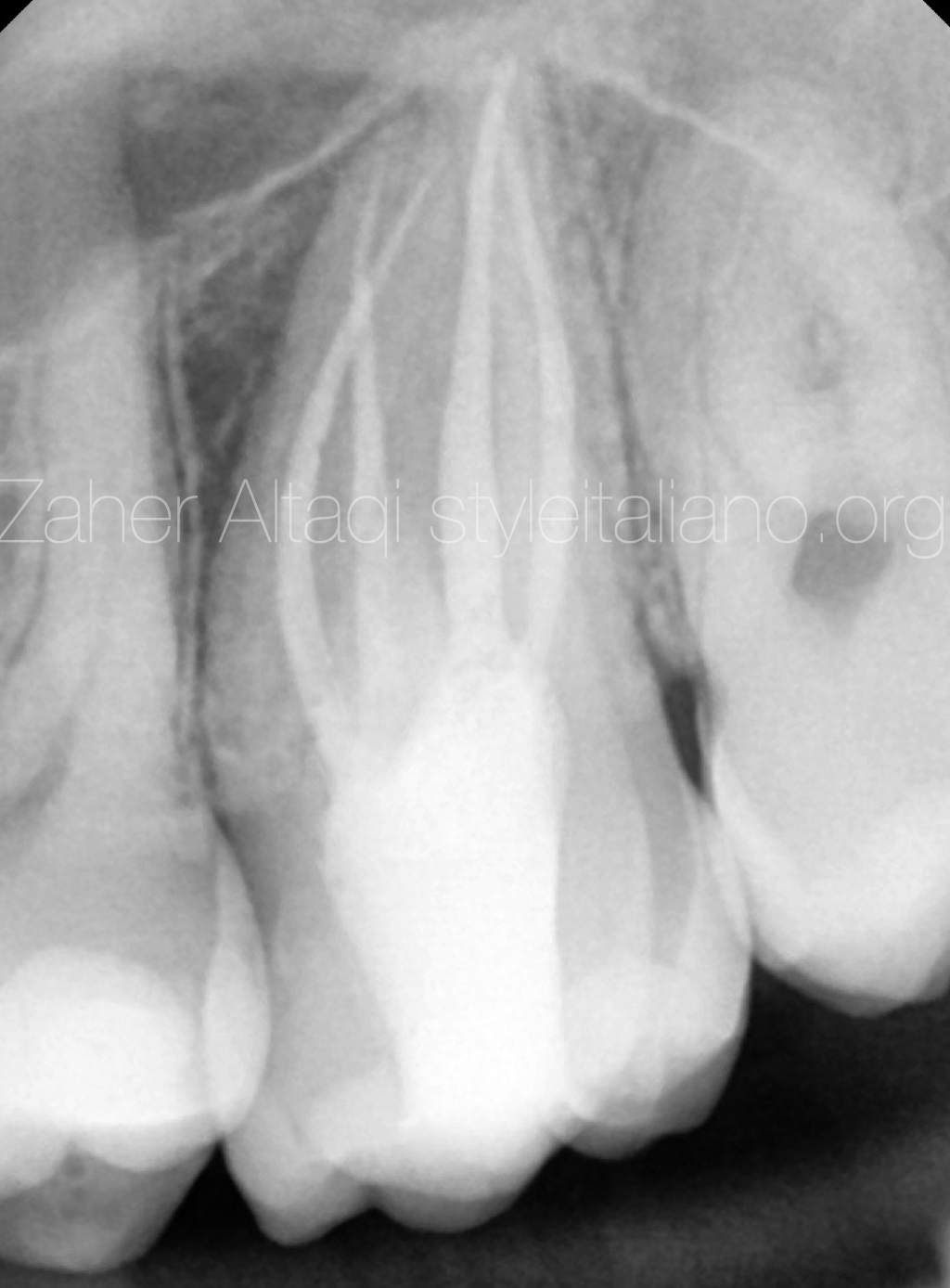
Fig. 10
Post- op radiograph with the 4 root canals treated .
Conclusions
It is important to visualize and to have knowledge of internal anatomy relationships before undertaking endodontic therapy. Unusual Clinical Tooth morphology may reveal the presence of unusual internal anatomy like extra root or canals . CBCT provide a great help for such cases because it show us the 3rd dimension which is something we really need for those cases .
Bibliography
Vertucci, FJ. Root canal morphology and its relationship to endodontic procedures. Endodontic Topics 2005, 10, 3–29
Versiani M, Basrani B, Sousa-Neto MD. The Root Canal Anatomy in Permanent Dentition. Switzerland: Springer International Publishing 2019.