Removal of broken instrument beyond the curvature
03/05/2020
Ivan Mirovic
Warning: Undefined variable $post in /var/www/vhosts/styleitaliano-endodontics.org/endodontics.styleitaliano.org/wp-content/plugins/oxygen/component-framework/components/classes/code-block.class.php(133) : eval()'d code on line 2
Warning: Attempt to read property "ID" on null in /var/www/vhosts/styleitaliano-endodontics.org/endodontics.styleitaliano.org/wp-content/plugins/oxygen/component-framework/components/classes/code-block.class.php(133) : eval()'d code on line 2
Management of broken instruments beyond the curvature always presents significant challenge, even for experienced clinicians.
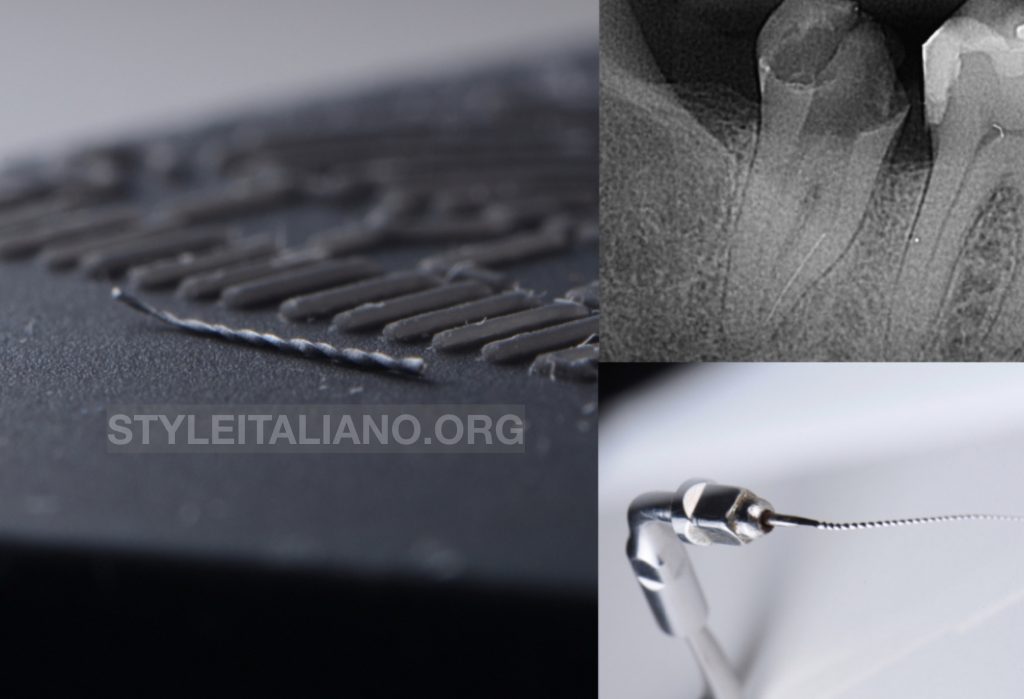
Patient was referred for retreatment of tooth #37. Tooth was diagnosed with previously initiated therapy and asymptomatic apical periodontitis. The long broken fragment of endodontic instrument in the mesial root was evident (Figure 1).
In cases of broken instruments beyond the curvature the first and the most conservative option is bypassing the broken fragment. This was done using stainless steel K file size 10. After successful bypassing canal was shaped using hand instruments in order to minimize the risk of successive breakage of rotary files. All other canals were shaped using rotary instruments. During shaping canals were copiously irrigated using full strength NaOCl.
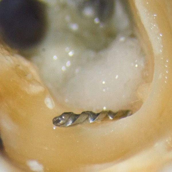
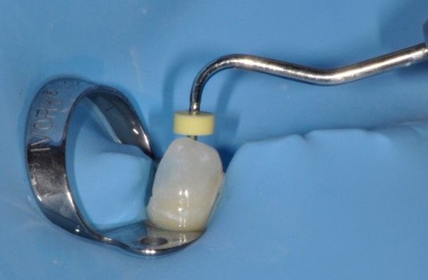
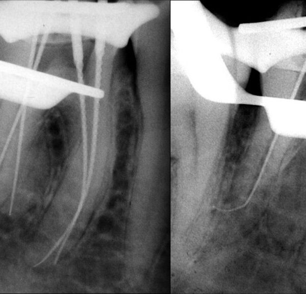
Following shaping precurved K file size 15 was placed in the endochuck (Figure 2) and activated using ultrasonics at low power. This allowed the instrument to pop up out of the canal (Figure 3,4). Obturation was completed in the same visit using warm vertical condesation technique as described by Herbert Schilder (Figure 5,6). Patient was sent back to referral dentist for final restoration. Full crown coverage was recommended.
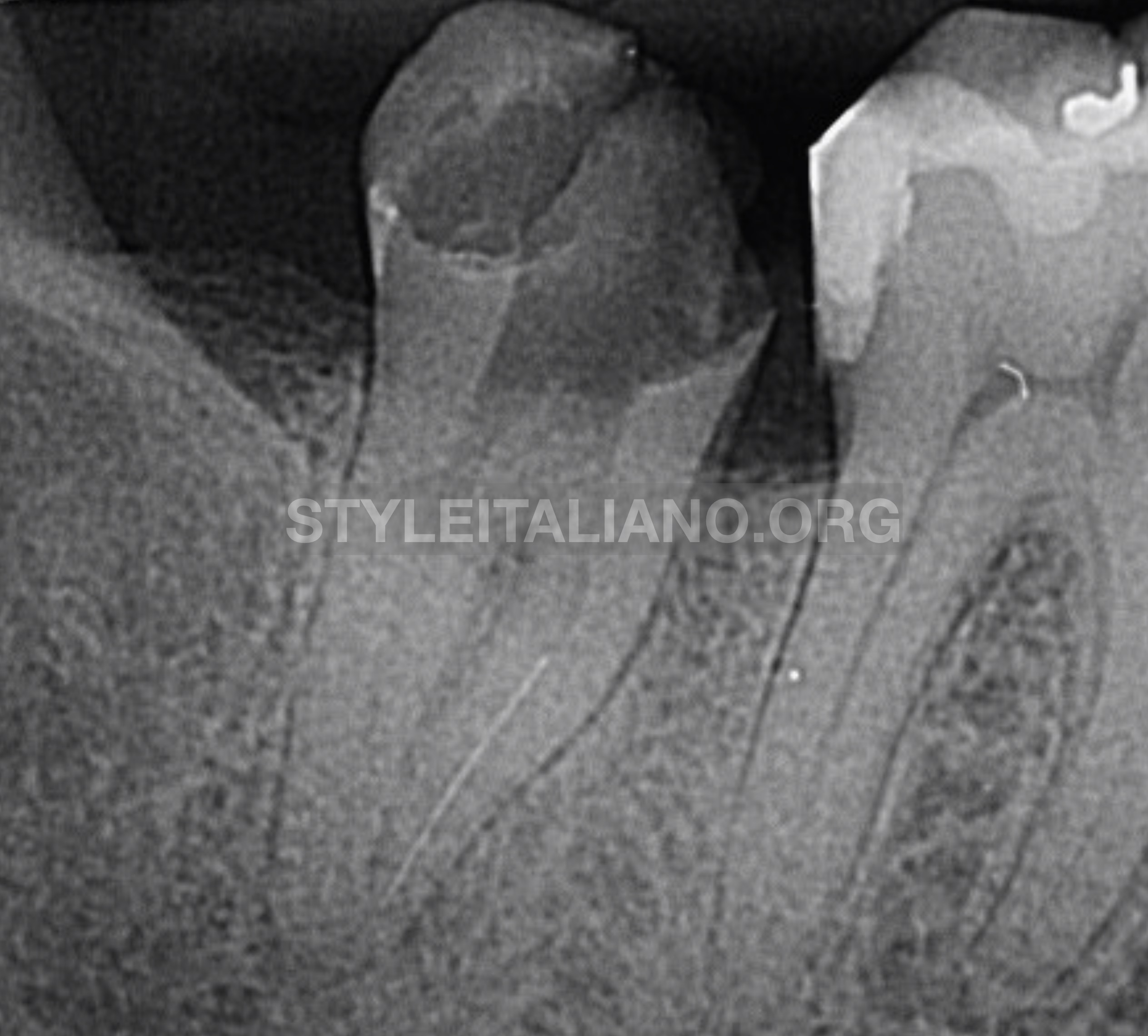
Fig. 1
Pre-operative radiograph tooth #37. Broken instrument was located in mesiobuccal canal.
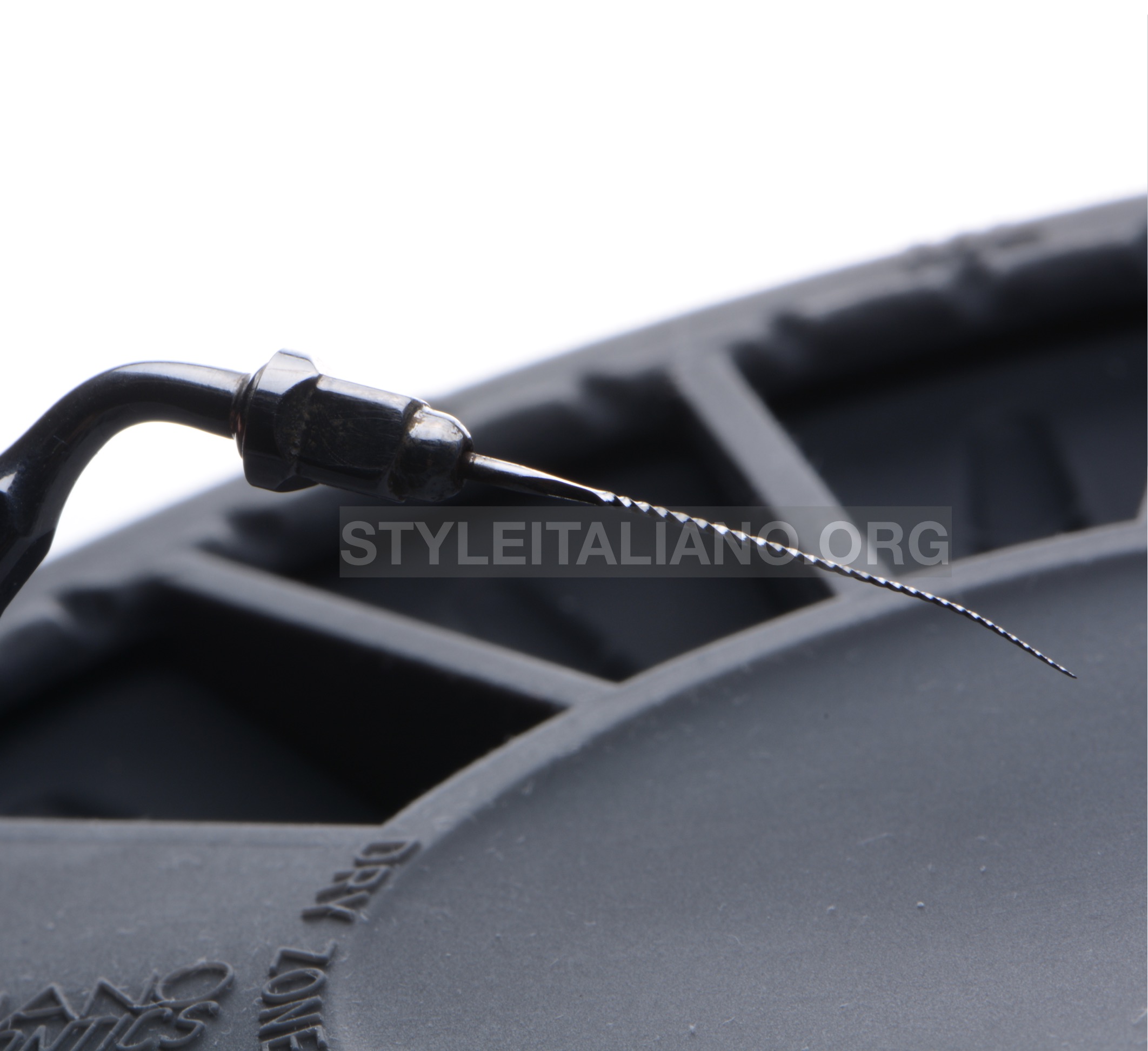
Fig. 2
Endochuck with K-file size 15
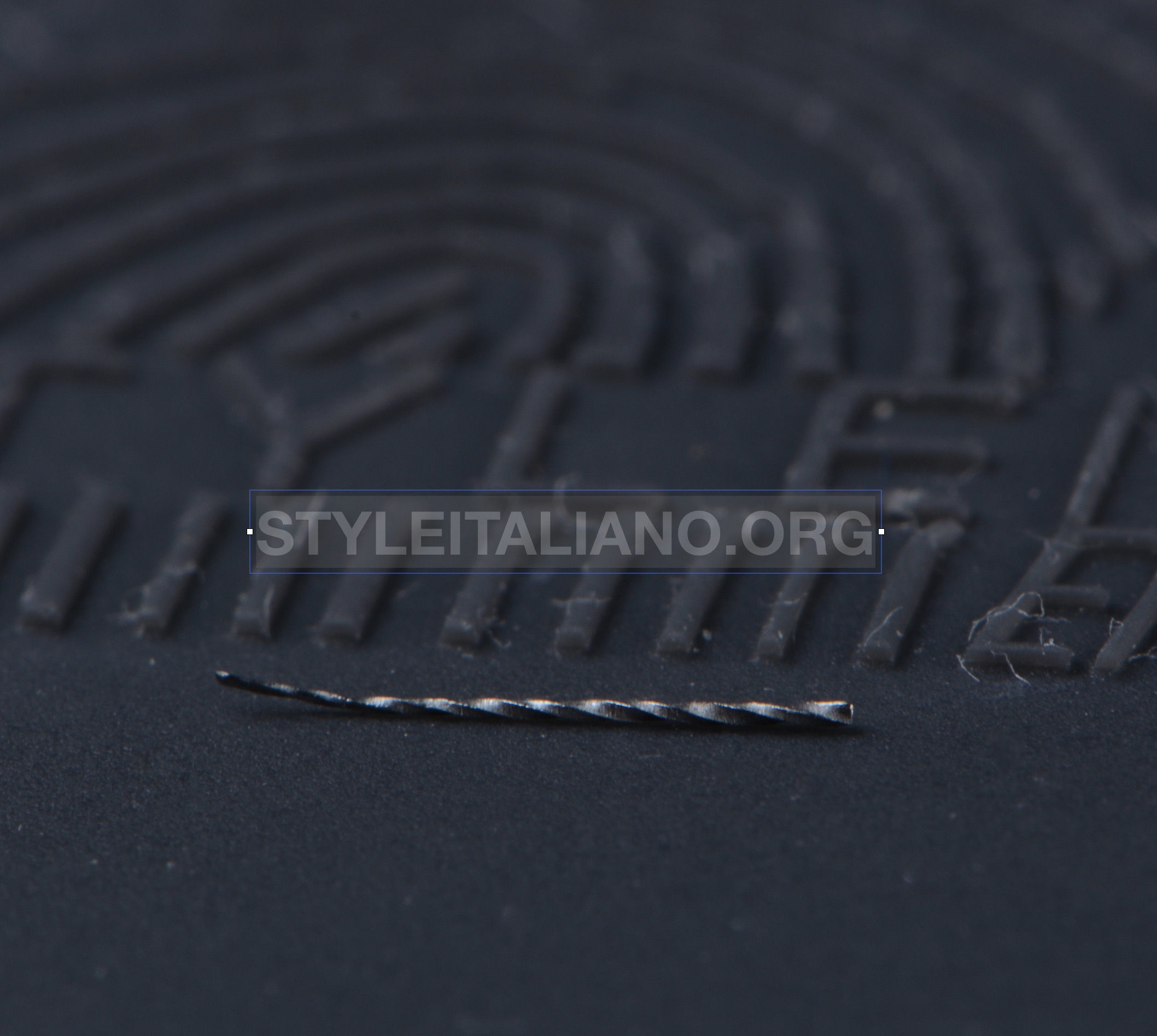
Fig. 3
Broken instrument removed
Fig. 4
Broken instrument
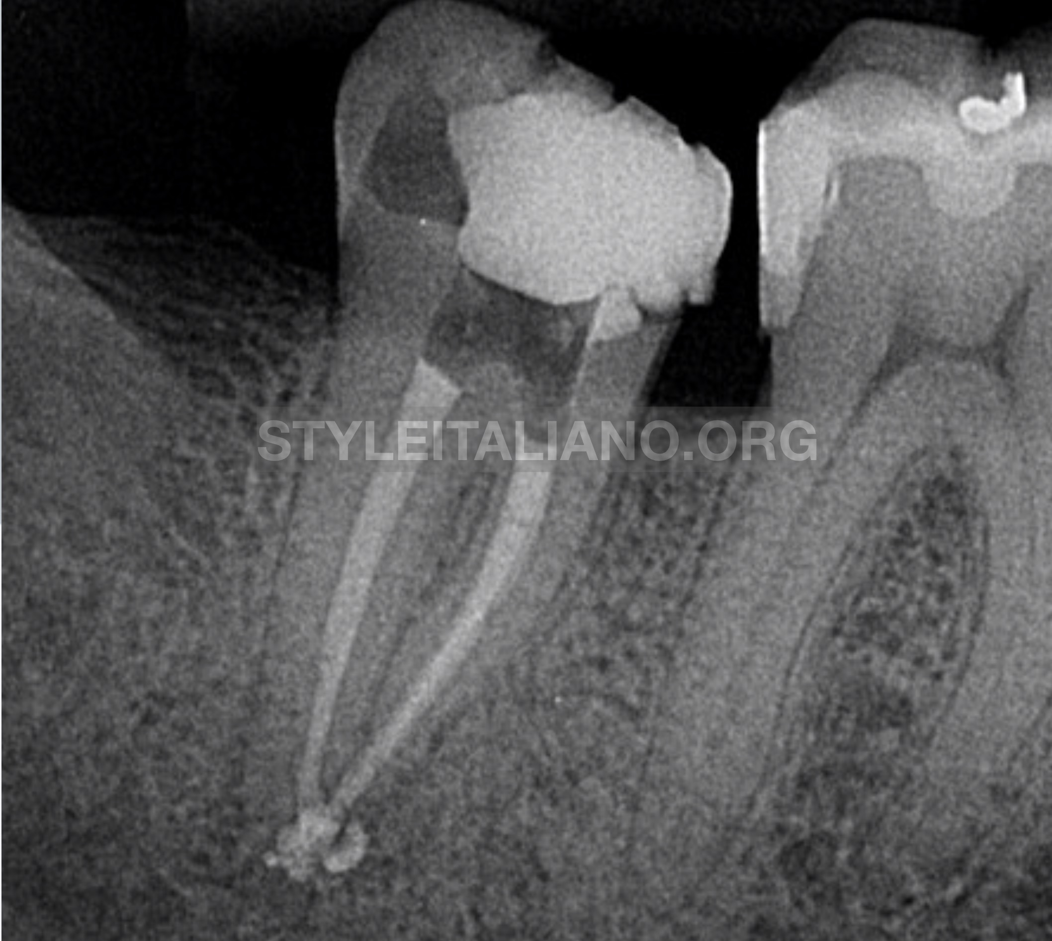
Fig. 5
Post-operative radiograph, straight-on angled film
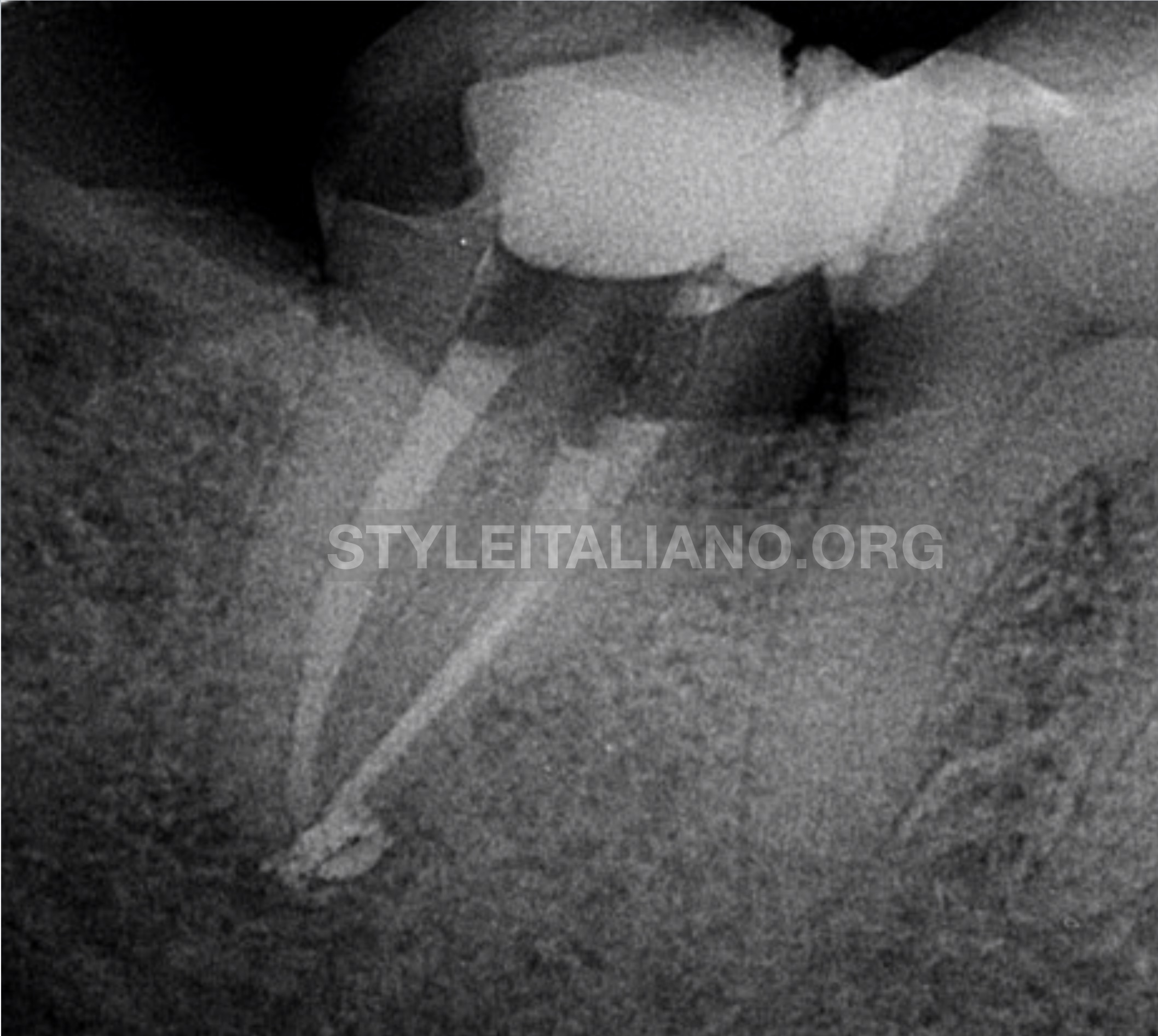
Fig. 6
Post-operative radiograph, distal oblique view. One distal canal and two mesial canals are evident.
Conclusions
When dealing with broken instruments we should search for the most conservative solution that will allow instrument removal and successive cleaning and shaping of root canal system. Described technique presents useful option in cases of broken instruments beyond curvature.
Bibliography
- Ruddle CJ: Ch. 25, Nonsurgical endodontic retreatment. In Pathways of the Pulp, 8th ed., Cohen S, Burns RC, eds., St. Louis: Mosby, pp. 875-929, 2002
- Berutti E, Castelluci A: Cleaning and shaping the root canal system. In: Castellucci A, editor. Endodontics, Vol. II. Florence: Tridente S.r.l; 2006. p. 396-437
- Schilder H. Filling root canals in three dimensions, Dent Clin North Am; 723-744, November 1967.
- Roda RS, Gettleman BH: Nonsurgical retreatment. In: Hargreaves KM, Cohen S, editors. Cohen’s pathways of the pulp, ed 10. New York: Elsevier; 2011. p. 890-952