Removal of an Anatomic Post and Core A Case Report
30/08/2020
Marc Kaloustian
Warning: Undefined variable $post in /var/www/vhosts/styleitaliano-endodontics.org/endodontics.styleitaliano.org/wp-content/plugins/oxygen/component-framework/components/classes/code-block.class.php(133) : eval()'d code on line 2
Warning: Attempt to read property "ID" on null in /var/www/vhosts/styleitaliano-endodontics.org/endodontics.styleitaliano.org/wp-content/plugins/oxygen/component-framework/components/classes/code-block.class.php(133) : eval()'d code on line 2
Different types pf posts can be placed during restorative procedures; prefabricated ones such as the screw posts, the paraposts or more recently the ceramic post and fiber posts, or the anatomical post commonly called cast post and core. In cases of non surgical retreatment The removal of a post can be challenging depending on the degree of retention of the post, the length, diameter, form and the luting cement. Furthermore, many dentists believe that the retrieval of posts can lead to iatrogenic errors such as vertical root fracture, perforation or the removal of an excessive amount of tooth Structure. Neverthless, a retrospective study by Abbot in 2004 on 1600 teeth with posts removed showed a very low number of fractures, less than 0.06 %, and the removal of the post in less than 3 minutes. Therefore, when a proper technique is applied during this procedure, the risks of harming the tooth are negligible.
Many techniques have been proposed for the removal of post, most of them are based on the traction concept with appliances like the little Giant, the Gonon kit, the Masseran kit or the Ruddle post removal system. However, when dealing with cast post and core with non precious hard metal, there are difficulties related to grabbing and the usage of conventional retrieval kits. Thus, ultrasound vibration becomes indicated and allows for a safe and a more conservative removal of the post. A study of Gariddo et al. (2009) showed in fact that even when a gripping method is used, a prior application of ultrasonic vibration facilitates tremendously the removal of the post by disrupting the cement.
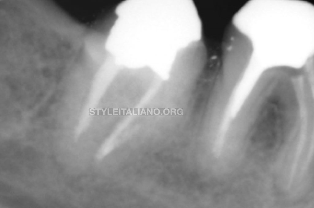
A case of a lower first right molar (46) was selected for a non surgical retreatment. The periapical preliminary Xray showed a poor endodontic primary treatment with short obturation in the distal root and in both mesial canals. Tooth was symptomatic on percussion and radiolucency was noticed on both roots. A diagnosis of symptomatic apical periodontitis was established.
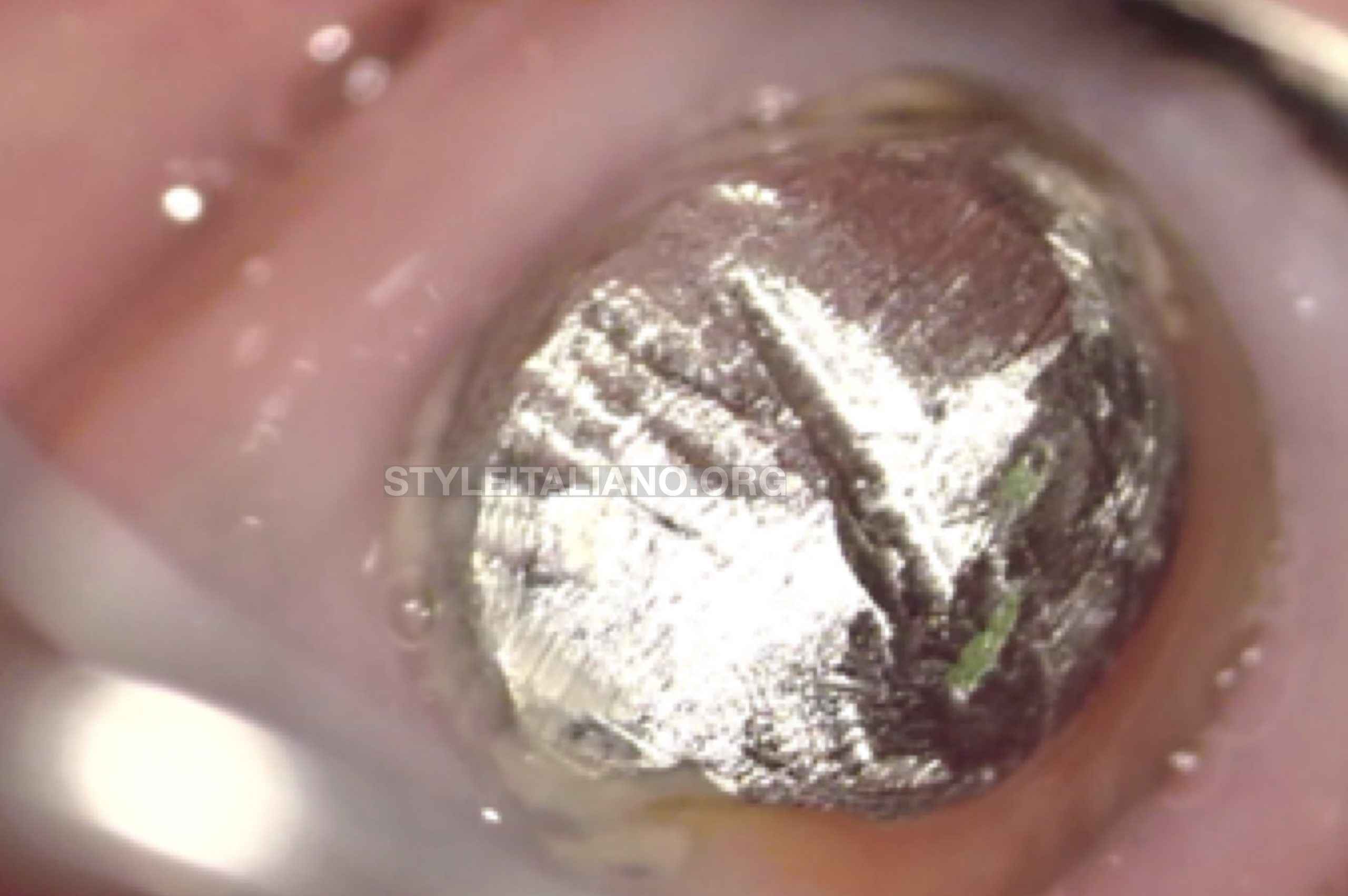
Fig. 1
Upon clinical examination, the presence of a good ferrule effect was noticed specially in the buccal aspect of the tooth. Nonetheless, a leaking cast post and core on the lingual limit of the restoration was present. Therefore a Non Surgical root canal retreatment was planned prior to a full coverage crown.

Fig. 2
After mandibular block anesthesia using Articaine 4 % with epinephrine and a buccal infiltration, the tooth was isolated with W8A wingless clamp and medium rubber dam. An additional isolation was also obtained by injecting the liquid dam around the preparation.
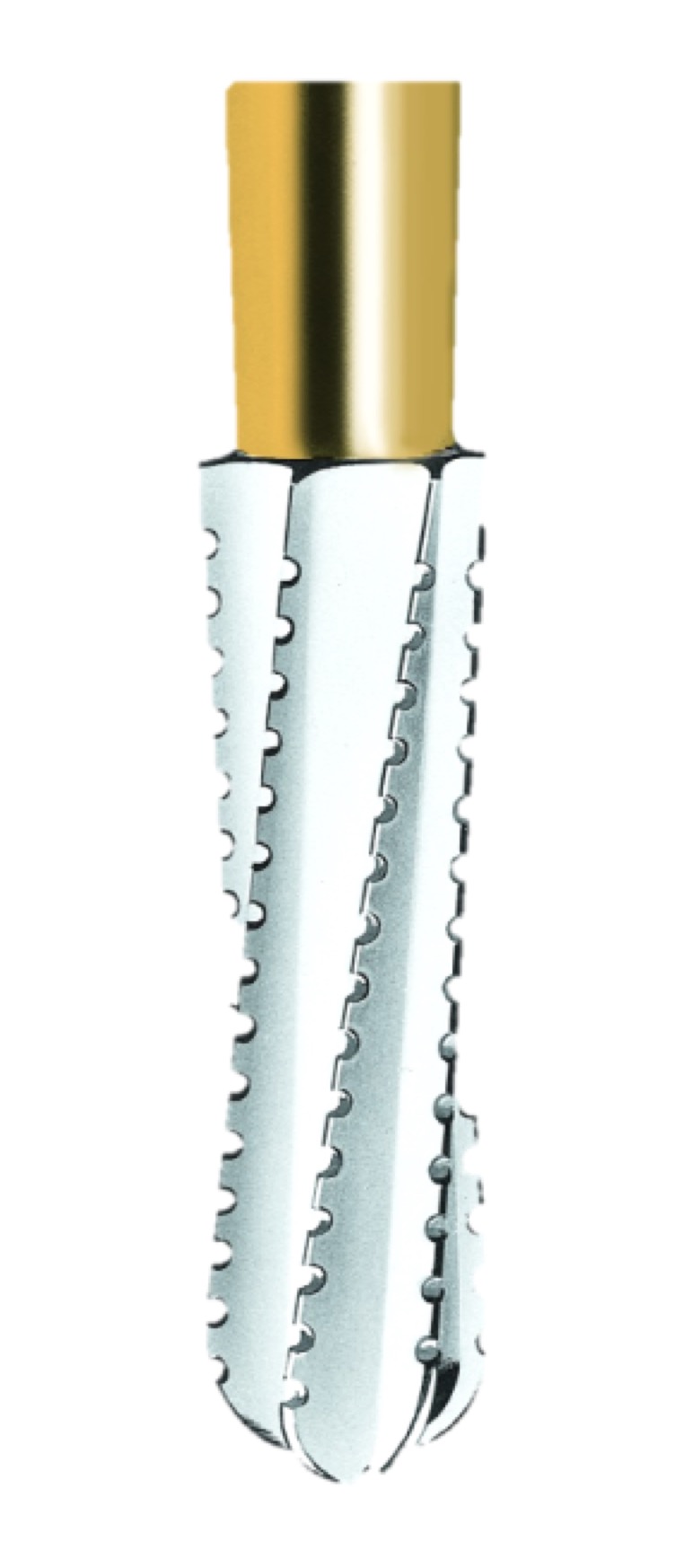
Fig. 3
The core separation was initiated under microscope magnification X10 using a transmetal bur.
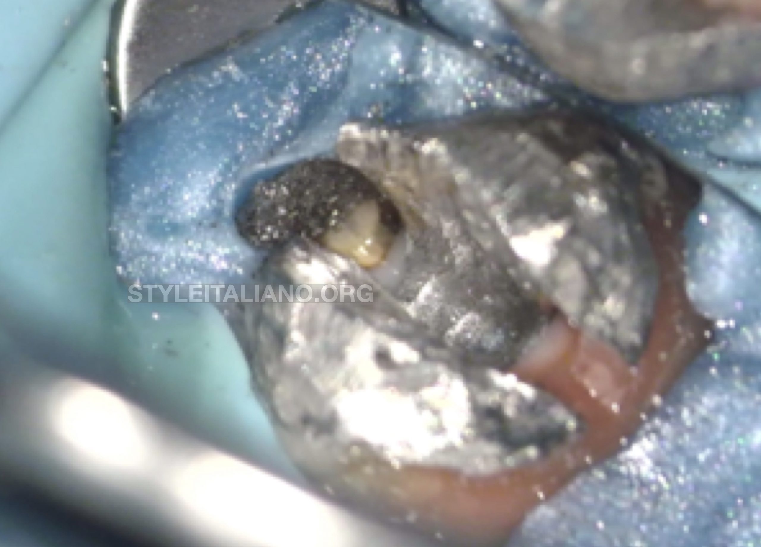
Fig. 4
The metal core aspect after the use of the transmetal bur.
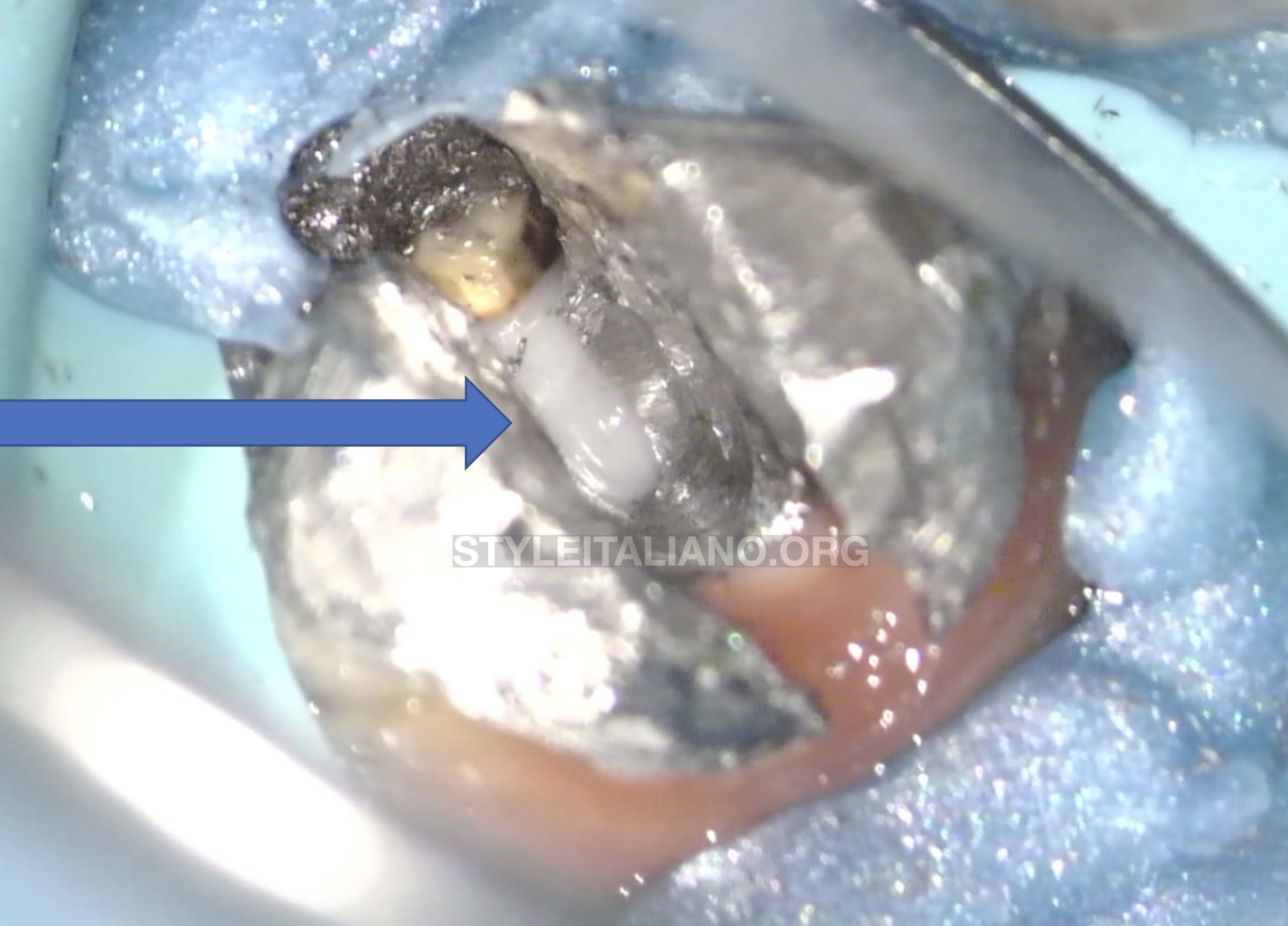
Fig. 5
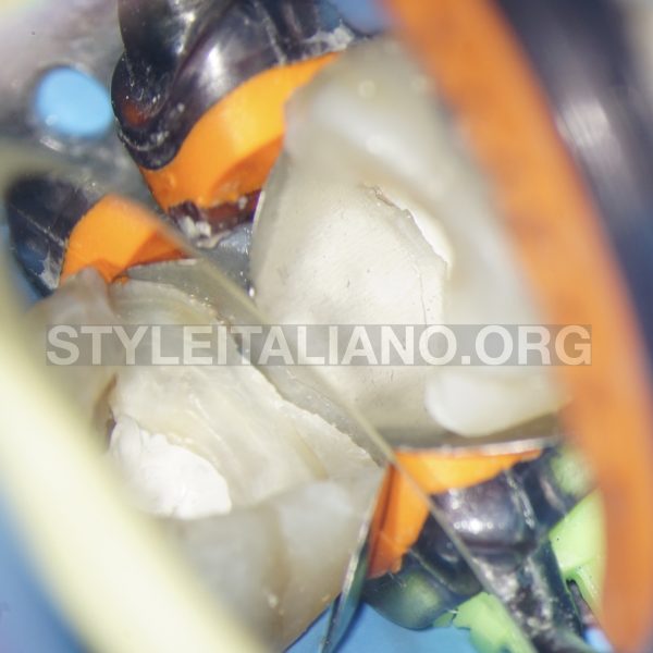
A long Surgical round carbide bur was used to separate the 2 parts precisely and completely without touching the tooth structure.
The image shows the exposure of the cement covering the pulpal floor after the 2 parts were partially separated by using the round surgical long carbide bur.
Removal of the mesial part of the metal core.
The removal of the distal part took around 3 minutes. This was done under coolant irrigation with breaks after each cycle of 15 seconds of vibration as suggested by Domenici et al. (2005) to avoid any rase of temperature over 10℃.
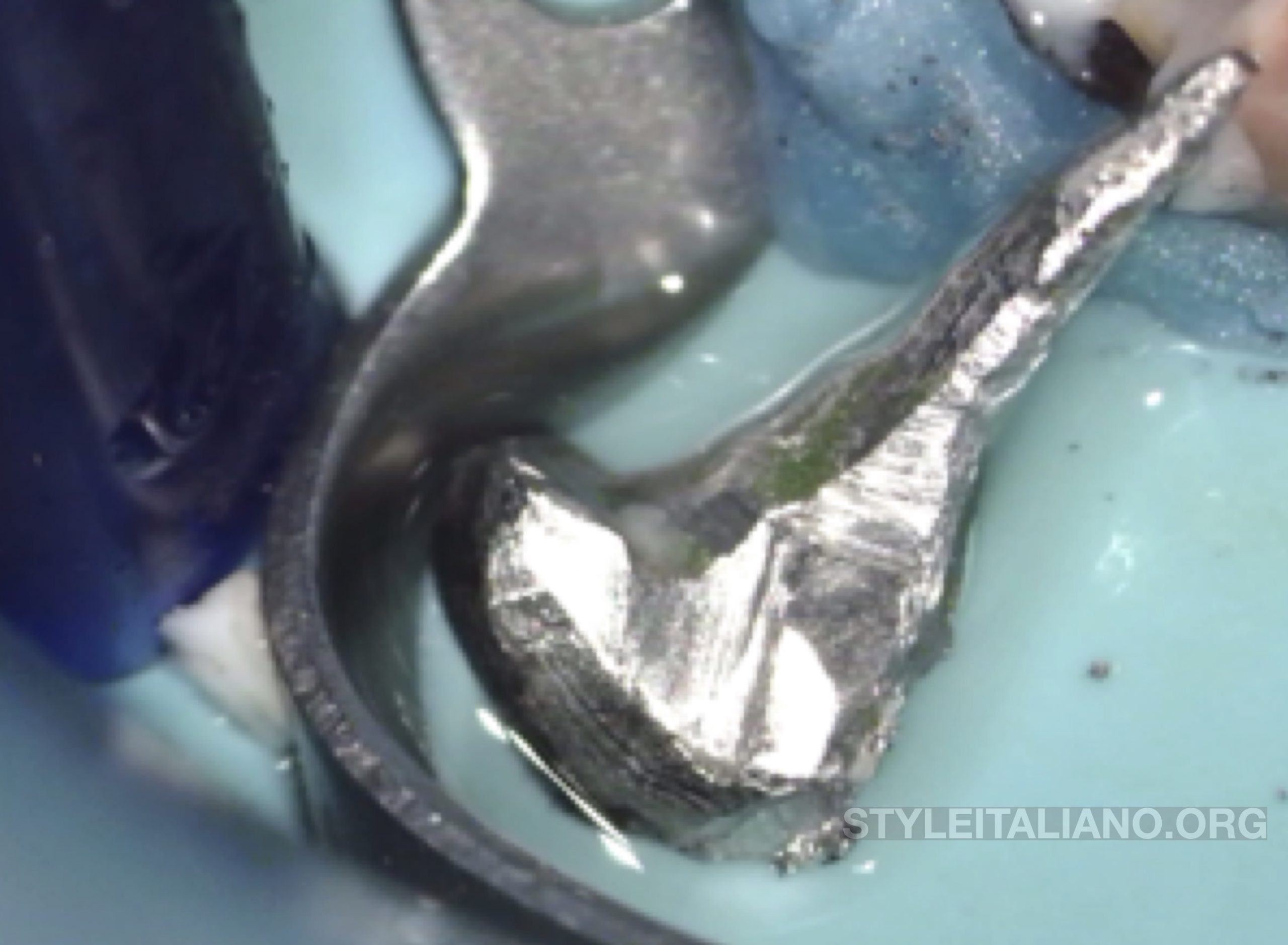
Fig. 6
Post and core removed from the distal root
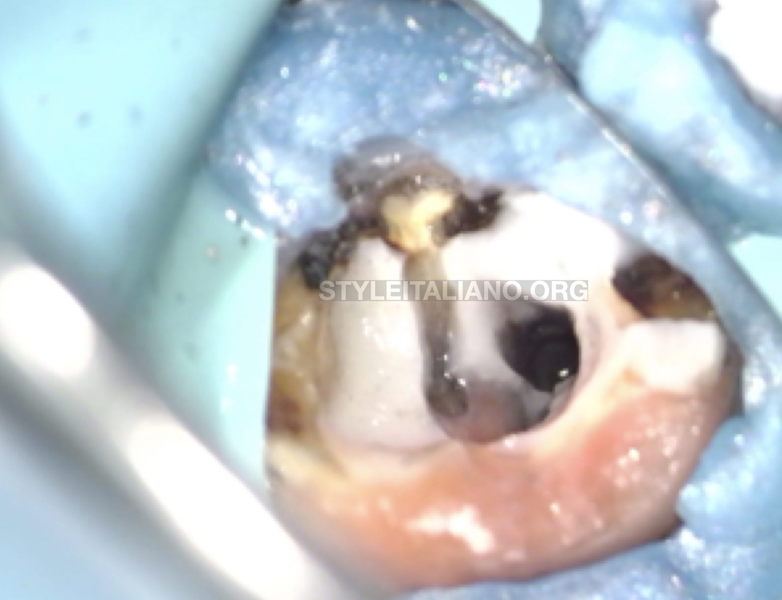
Fig. 7
After removing the cast post and core, remaining cement should be removed from the pulp chamber using an ultrasonic tip, the ET20, and the decay cleaned.
Cement remnants cleaned with ultrasonic tip under coolant water irrigation.
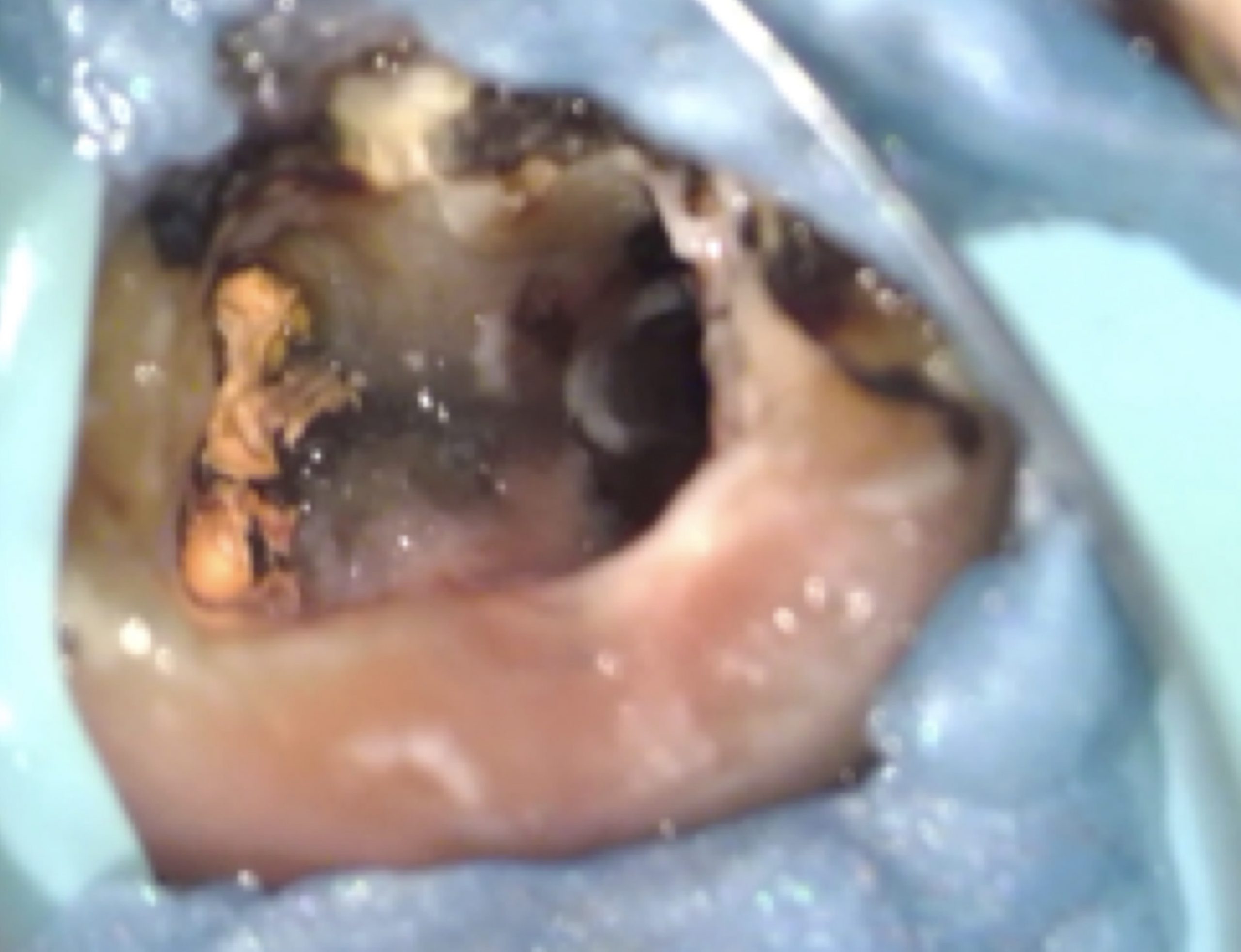
Fig. 8
After cleaning the cement, caries control should be performed specially on the lingual wall before starting the retreatment procedure.
Filling material is then retrieved from all the canals using a mechanical technique without dispensing any solvent in that particular case. The video shows as example the removal of the gutta-percha in the coronal third from the mesio-buccal canal with a rotary instrument.
The video shows an exemple of the shaping procedure in the mesio-buccal canal with a Ni-Ti instrument.
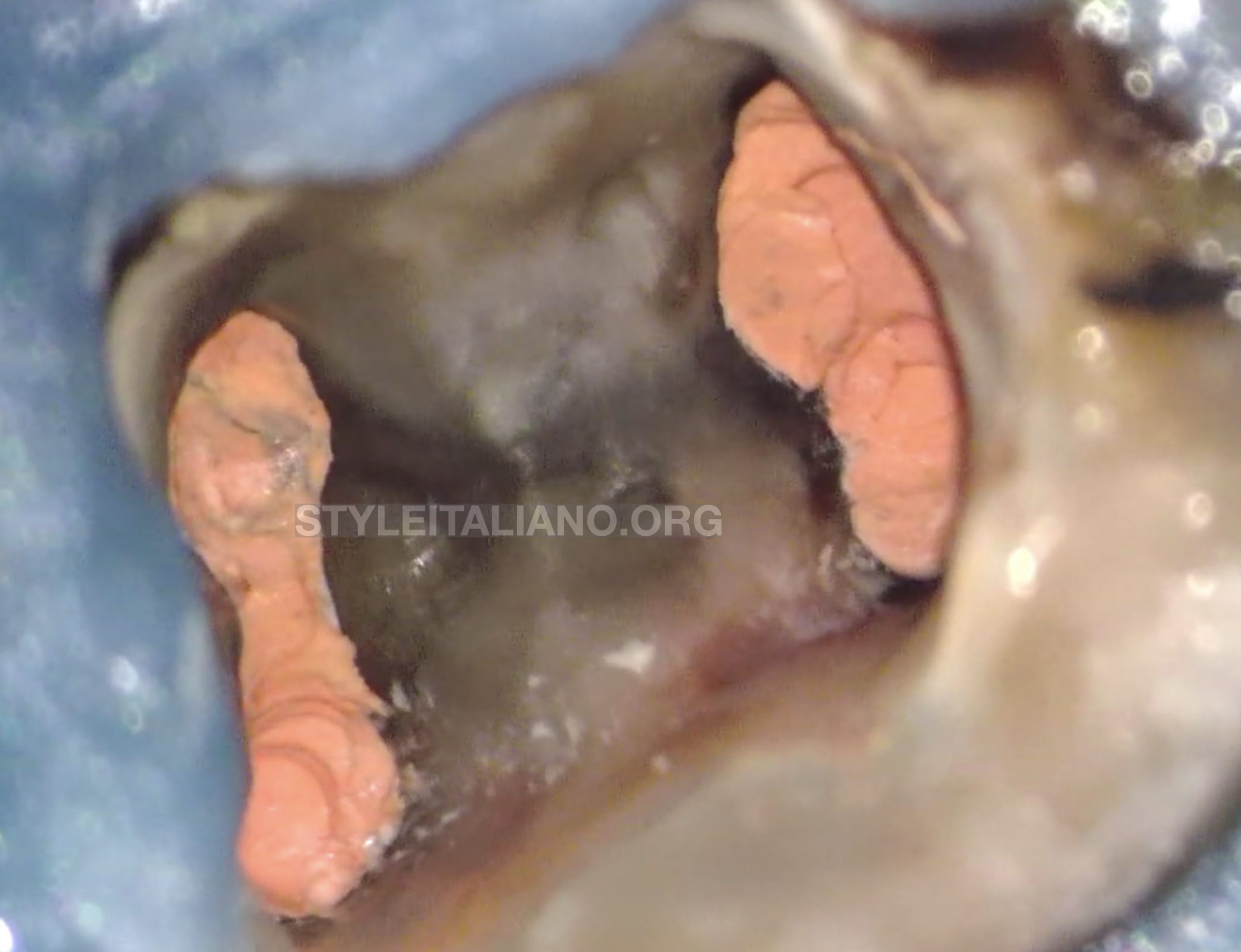
Fig. 9
The pulp chamber aspect after obturation.
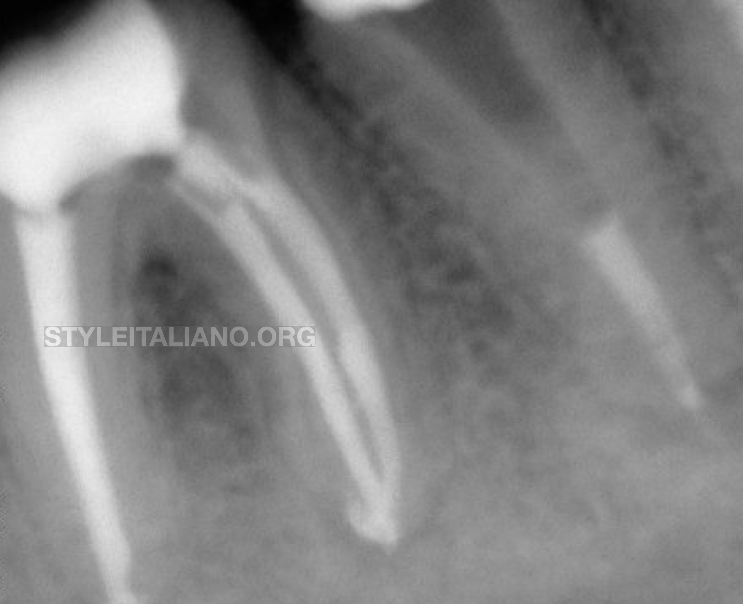
Fig. 10
final Xray after 3D filling with the warm vertical compaction and backpack using the injecting technique.
Conclusions
Post removal is mandatory in many cases specially when a non surgical retreatment is indicated with an inadequate coronal restoration. In fact, surgical procedure can only be performed when the coronal restoration is satisfying. Posts can be drilled with burs, pulled out using several devices or vibrated with ultrasonic appliances. In this case we opted for a combination of grinding for separation and vibration for retrieval. Studies showed that the smaller was the core structure the better was the transmission of the vibrating technique all along the post. By separating the core in 2 parts, this concept was followed because the distal part of the core containing the long post became smaller. On the other hand the risks of over enlargement of the post space, or removal of healthy surrounding dentine was avoided by exclusively working on the post thanks to the better visibility given by the high magnification and light provided by the operatory microscope.
Bibliography
- Abbott PV. Incidence of Root Fractures and Methods Used for Post Removal. Int Endod J. 2002 Jan;35(1):63-7.
- Dominici JT, Clark S, Scheetz J, Eleazer PD. Analysis of Heat Generation Using Ultrasonic Vibration for Post Removal. J Endod. 2005 Apr;31(4): 301-3).
- Garrido ADB, Oliveira AG, Osório JE, Silva-Sousa YT, Sousa-Neto MD. Evaluation of Several Protocols for the Application of Ultrasound During the Removal of Cast Intraradicular Posts Cemented With Zinc Phosphate Cement. Int Endod J. 2009 Jul;42(7):609-13.
- Garça IAA, Sponchiado Jùnior EC, Marques AAF, de Moura Martins L, Garrido ADB. Assessment of a Cavity to Optimize Ultrasonic Efficiency to Remove Intraradicular Posts. J Endod. 2017 Aug;43(8):1350-3.
- Kim JJ, Alapati S, Knoernschild KL, Jeong YH, Kim DG, Lee DJ. Micro-Computed Tomography of Tooth Volume Changes Following Post Removal. J Prosthodont 2017 Aug;26(6):522-528.